. 2014 Sep;63(9):3009-21.
doi: 10.2337/db13-1607. Epub 2014 Apr 16.

ADCY5 couples glucose to insulin secretion in human islets

Affiliations

ADCY5 couples glucose to insulin secretion in human islets

David J Hodson et al. Diabetes. 2014 Sep.

Abstract

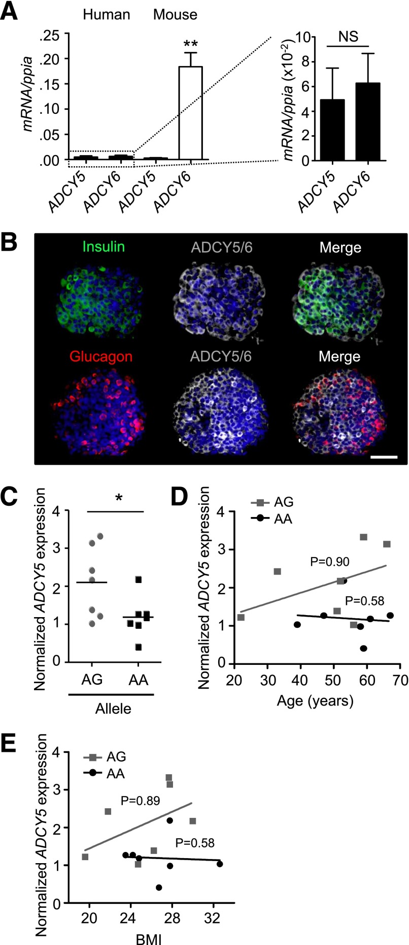
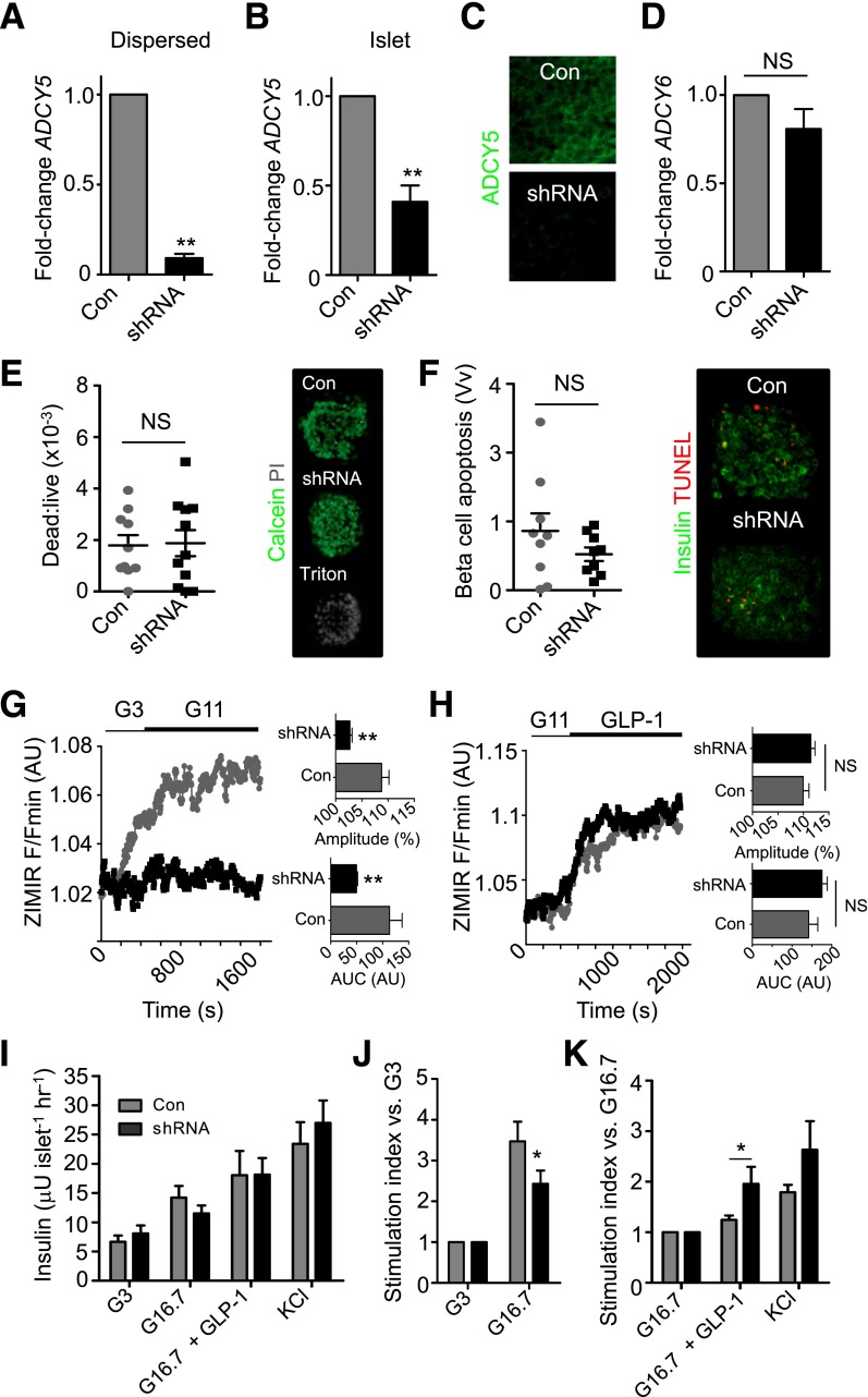
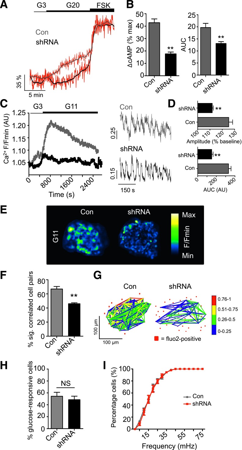
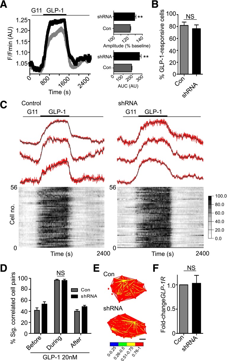
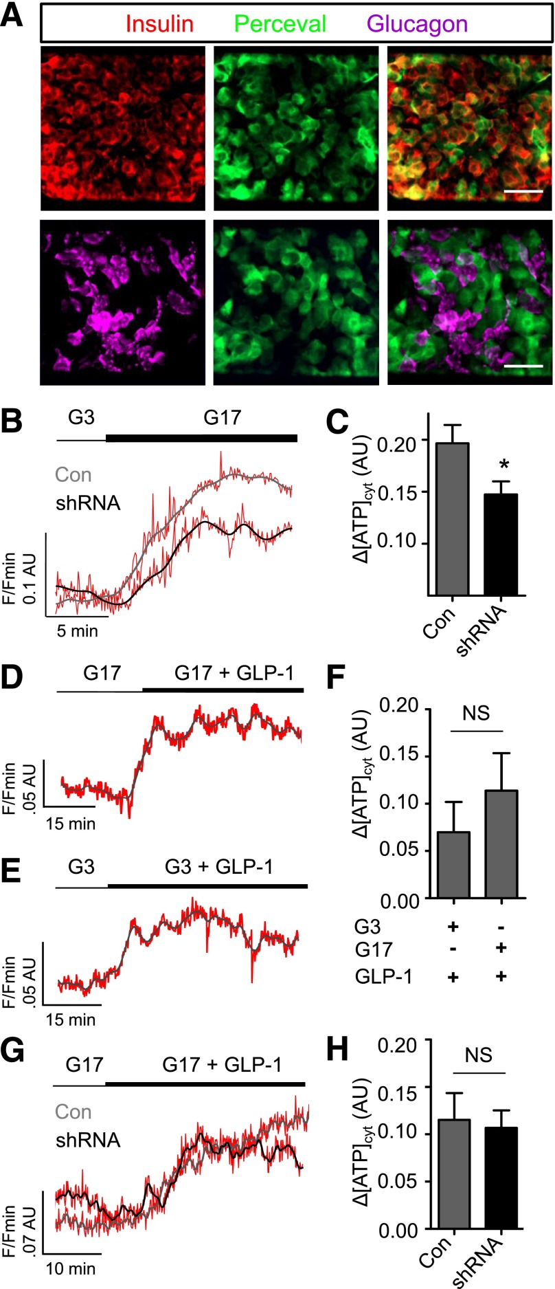
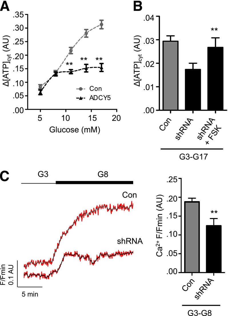
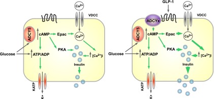
Single nucleotide polymorphisms (SNPs) within the ADCY5 gene, encoding adenylate cyclase 5, are associated with elevated fasting glucose and increased type 2 diabetes (T2D) risk. Despite this, the mechanisms underlying the effects of these polymorphic variants at the level of pancreatic β-cells remain unclear. Here, we show firstly that ADCY5 mRNA expression in islets is lowered by the possession of risk alleles at rs11708067. Next, we demonstrate that ADCY5 is indispensable for coupling glucose, but not GLP-1, to insulin secretion in human islets. Assessed by in situ imaging of recombinant probes, ADCY5 silencing impaired glucose-induced cAMP increases and blocked glucose metabolism toward ATP at concentrations of the sugar >8 mmol/L. However, calcium transient generation and functional connectivity between individual human β-cells were sharply inhibited at all glucose concentrations tested, implying additional, metabolism-independent roles for ADCY5. In contrast, calcium rises were unaffected in ADCY5-depleted islets exposed to GLP-1. Alterations in β-cell ADCY5 expression and impaired glucose signaling thus provide a likely route through which ADCY5 gene polymorphisms influence fasting glucose levels and T2D risk, while exerting more minor effects on incretin action.


Figures

Figure 1
Figure 1
ADCY5 is expressed in isolated human islets and affected by T2D risk alleles. A: ADCY5 and ADCY6 are expressed at similar levels in human islets (n = 4 donors). Conversely, Adcy6 mRNA expression is ∼40-fold more abundant than that of Adcy5, which is barely detectable in mouse islets (n = 3 female and 3 male animals; **P < 0.01 vs. ADCY5, Student t test). B: Immunostaining using an anti-ADCY5 immunoglobulin, with some reported cross-reactivity to ADCY6, reveals the cytoplasmic distribution of both proteins throughout the human α- and β-cell populations (DAPI, blue; scale bar, 60 µm). C: Scatter plot showing reduced ADCY5 mRNA abundance in islets from males <70 years of age who are carriers of the AA risk allele at rs11708067 (*P < 0.043 vs. AG; one-way ANOVA; n = 7 donors for each allele). Values represent mean ± SEM. D: Age of donors is not significantly correlated with ADCY5 mRNA expression (R2 = 0.21 and R2 = 0.01 for AG and AA, respectively; linear regression) (P values shown on graph). E: As for D, but BMI (R2 = 0.23 and R2 = 0.003, AG vs. AA, respectively; linear regression) (P values shown on graph).
Figure 2
Figure 2
ADCY5 silencing inhibits glucose- but not GLP-1–stimulated insulin secretion. A and B: Lentivirus harboring shRNA against ADCY5 reduces expression by >50% in both dispersed and intact islets (**P < 0.01 vs. Con; Student paired t test; n = 3–4 donors). C: ADCY5/6 protein expression is markedly reduced in the first few cell layers of intact islets, as determined using confocal imaging (n = 6 islets from two donors). D: ADCY6 mRNA expression is unaffected by ADCY5 silencing (NS, nonsignificant vs. Con; Student paired t test; n = 4 donors). E: Dead:live cell ratio is similar in Con and shRNA-treated islets (positive Con, Triton X-100; NS, nonsignificant vs.; Mann-Whitney U test; n = 10–11 islets from three donors). F: As for E, but TUNEL assay for apoptosis (n = 9 islets from three donors). The proportion of apoptotic β-cells was expressed as a fraction area versus nonapoptotic insulin-positive cell mass (Vv). G: ADCY5 knockdown suppresses glucose-induced insulin secretory dynamics, as shown by bar graphs of AUC and amplitude of ZIMIR responses (mean traces, left panel; n = 8–9 islets from four donors) (**P < 0.01 vs. Con; Mann-Whitney U test). H: GLP-1–stimulated insulin secretory dynamics are subtly improved after ADCY5 depletion (mean traces, left panel; AUC and amplitude, right panel; n = 7 islets from three donors) (NS, nonsignificant). I: Glucose-stimulated insulin release into static culture is impaired in shRNA-treated islets, as determined using radioimmunoassay (G3 and G16.7, 3 mmol/L and 16.7 mmol/L glucose, respectively) (n = 4–8 donors). KCl 30 mmol/L was added as a Con. J: As for I, but stimulation index vs. 3 mmol/L glucose to better account for the variation between islet preparations (*P < 0.02 vs. Con at 16.7 mmol/L glucose; Mann-Whitney U test). K: As for J, but vs. 16.7 mmol/L glucose (G16.7) (*P < 0.01 vs. 16.7 mmol/L glucose for each group; Mann-Whitney U test). Values represent mean ± SEM. hr, hour.
Figure 3
Figure 3
ADCY5 depletion suppresses glucose-induced increases in cytosolic free Ca2+. A: ADCY5 silencing decreases cystosolic cAMP levels, as determined using the recombinant probe Epac2-camps (representative traces shown; gray/black, smoothed; red, raw). B: As for A, but summary data showing a reduction in measured FRET signal versus maximal stimulation with FSK (%), as well as decreased AUC (**P < 0.01 vs. Con; Student t test; n = 12 recordings from three donors). C: ADCY5 knockdown suppresses 11 mmol/L glucose (G11)-evoked cytosolic Ca2+ rises (left panel: mean traces) (right panel: zoom-in of Ca2+ oscillations). D: AUC and amplitude of Ca2+ rises are reduced in shRNA-treated islets (right panel, **P < 0.01 vs. Con; Mann-Whitney U test; n = 10 islets from three donors). E: Pseudocolored Con and shRNA-treated human islets during exposure to 11 mmol/L glucose (recording time = 40 min; image cropped to display a single islet). F: ADCY5 is required for long-term evolutions in coordinated cell activity after exposure to elevated glucose (**P < 0.01 vs. Con; Mann-Whitney U test; n = 9–10 islets from three donors) (correlation calculated over 20–30 min; sig, significantly). G: Representative functional connectivity map depicting location, number, and strength (color-coded; 0 [blue] = lowest, 1 [red] = highest) of significantly correlated cell pairs (Pearson R coefficient, P < 0.05). Note that ADCY5 silencing decreases both the number and strength of correlations. H: Gene silencing does not significantly alter the percentage (%) of glucose (11 mmol/L)-responsive cells (NS, nonsignificant vs. Con; Mann-Whitney U test). I: The cumulative distribution of Ca2+-spiking frequencies remains similar in Con and shRNA-treated islets. Values represent mean ± SEM.
Figure 4
Figure 4
ADCY5 does not mediate GLP-1–stimulated cytosolic Ca2+ increases. A: ADCY5 knockdown subtly improves GLP-1 responses (left panel: mean traces), as evidenced by increased AUC and amplitude of cytosolic Ca2+ rises in shRNA-treated islets (right panel) (**P < 0.01 vs. Con; Mann-Whitney U test; n = 10 islets from three donors). B: The proportion of GLP-1–responsive cells is similar in Con and shRNA-treated islets (NS, nonsignificant vs. Con; Mann-Whitney U test). C: ADCY5 silencing does not affect coordinated β-cell responses to 11 mmol/L glucose plus GLP-1 (representative Ca2+ traces [top panel]; gray, smoothed; red, raw) (heat map depicting minimum–maximum for each cell [bottom panel]) (n = 10 islets from three donors; correlation measured using 5-min windows). D: Histogram showing mean % significantly correlated cell pairs in Con and shRNA-treated islets before, during, and after GLP-1 application (NS, nonsignificant; two-way ANOVA). E: Representative weighted graphs demonstrating large increases in β-cell connectivity after exposure to GLP-1 in both normal and ADCY5-depleted islets (scale bar, 50 µm). F: Gene silencing does not alter GLP-1R mRNA expression (NS, nonsignificant vs. Con; Student paired t test; n = 3 donors). Values represent mean ± SEM.
Figure 5
Figure 5
ADCY5 alters β-cell energetics. A: Expression of the ATP/ADP probe Perceval is predominantly restricted to β-cells, as shown using immunohistochemistry with antibodies against insulin and glucagon (scale bar, 25 µm [top panels] and 20 µm [bottom panels]). B: Glucose (17 mmol/L) (G17) still induces increases in ATP-to-ADP ratio after ADCY5 silencing (representative traces shown; gray/black, smoothed; red, raw). C: Bar graph showing a significant effect of shRNA treatment on the amplitude of cytosolic (cyt) ATP/ADP rises in response to 17 mmol/L glucose (*P < 0.05 vs. Con; Mann-Whitney U test; n = 9–10 islets from three donors). D: GLP-1 increases ATP/ADP in the presence of permissive (17 mmol/L) glucose concentrations (representative traces shown; gray/black, smoothed; red, raw). E: As for D, but in the presence of nonpermissive (3 mmol/L) glucose concentrations. F: Summary statistics demonstrate similar effects of GLP-1 on ATP/ADP in islets exposed to 3 or 17 mmol/L glucose (NS, nonsignificant vs. Con; Mann-Whitney U test; n = 10–13 recordings from five donors). G: ATP/ADP responses to GLP-1 are similar in Con and ADCY5 shRNA-treated islets (representative traces shown; gray/black, smoothed; red, raw). H: Summary statistics demonstrate no significant effect of gene silencing on GLP-1–induced ATP/ADP rises (NS, nonsignificant vs. Con; Mann-Whitney U test; n = 6 recordings from two donors). Values represent mean ± SEM.
Figure 6
Figure 6
ADCY5 targets nonmetabolic processes to alter Ca2+ responses. A: Impaired ATP/ADP responses are only present in ADCY5-silenced islets at glucose concentrations of ≥11 mmol/L (**P < 0.01 vs. Con; two-way ANOVA; n = 9 recordings from three donors). B: Elevation of cAMP using FSK rescues ATP/ADP rises in ADCY5-silenced islets after transition from 3 mmol/L to 17 mmol/L glucose (G3-G17) (**P < 0.01 vs. shRNA; one-way ANOVA; n = 9 recordings from three donors; n = 4–5 recordings). C: ADCY5 knockdown suppresses β-cell Ca2+ responses after transition from 3 mmol/L to 8 mmol/L glucose (G3-G8) (representative traces [left panel]; gray/black, smoothed; red, raw]), as evidenced by reduced amplitude (right panel) (**P < 0.01 vs. shRNA; Mann-Whitney U test; n = 8–9 recordings from three donors).
Figure 7
Figure 7
Schematic of ADCY5 function in human β-cells. Glucose-stimulated insulin secretion relies on KATP-dependent and -independent signals. The latter include cAMP generation, and this likely requires ADCY5 activation by the sugar to increase ATP generation, Ca2+ influx, and exocytosis (left panel). By contrast, incretins such as GLP-1, believed primarily to engage cAMP-signaling pathways, may potentiate insulin secretion via other ADCY isoforms (right panel). PKA, protein kinase A.

Similar articles

Cited by

References

    1. Scully T. Diabetes in numbers. Nature 2012;485:S2–S3 - PubMed
    1. Smith U, Gale EA. Cancer and diabetes: are we ready for prime time? Diabetologia 2010;53:1541–1544 - PubMed
    1. DeFronzo RA, Abdul-Ghani MA. Preservation of β-cell function: the key to diabetes prevention. J Clin Endocrinol Metab 2011;96:2354–2366 - PubMed
    1. Rutter GA, Parton LE. The beta-cell in type 2 diabetes and in obesity. Front Horm Res 2008;36:118–134 - PubMed
    1. McCulloch LJ, van de Bunt M, Braun M, Frayn KN, Clark A, Gloyn AL. GLUT2 (SLC2A2) is not the principal glucose transporter in human pancreatic beta cells: implications for understanding genetic association signals at this locus. Mol Genet Metab 2011;104:648–653 - PubMed

Publication types