. 2014 May 8;54(3):472-84.
doi: 10.1016/j.molcel.2014.03.014. Epub 2014 Apr 10.

Mouse SLX4 is a tumor suppressor that stimulates the activity of the nuclease XPF-ERCC1 in DNA crosslink repair

Affiliations

Mouse SLX4 is a tumor suppressor that stimulates the activity of the nuclease XPF-ERCC1 in DNA crosslink repair

Michael R G Hodskinson et al. Mol Cell. .

Abstract

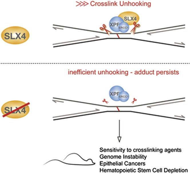
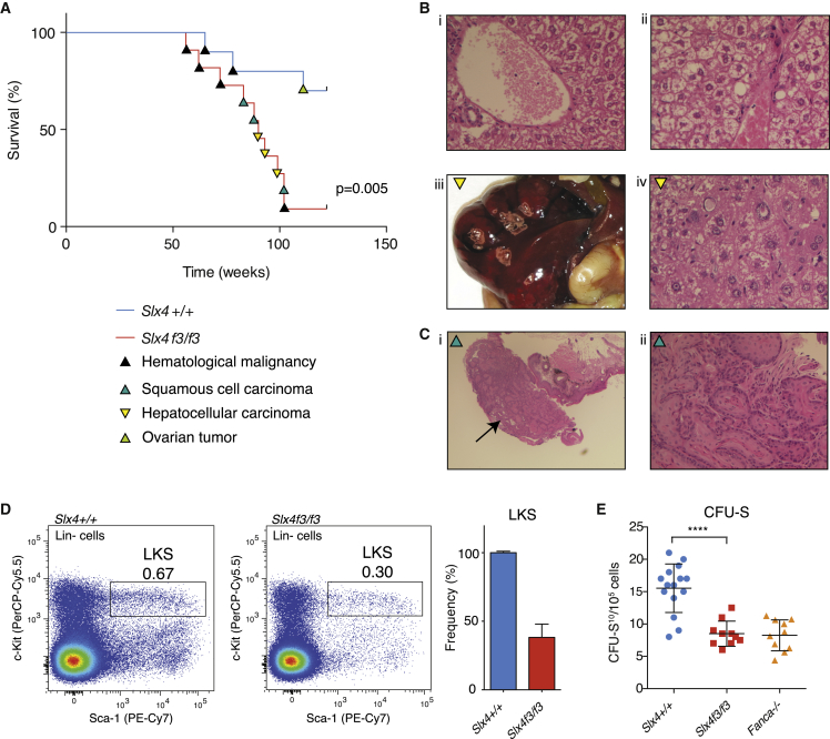
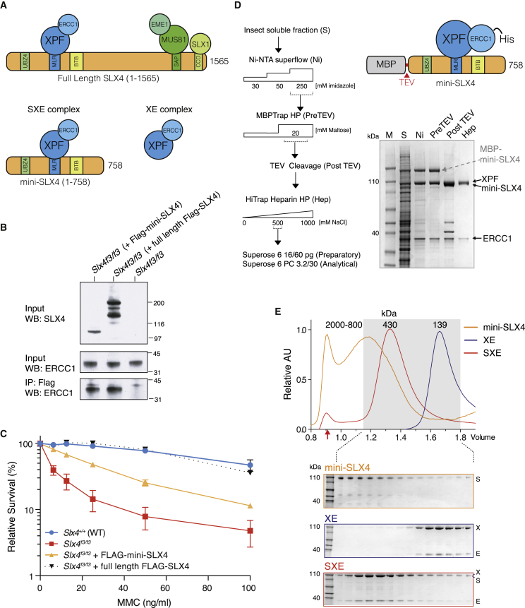
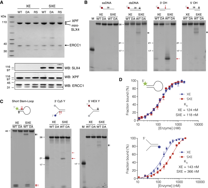
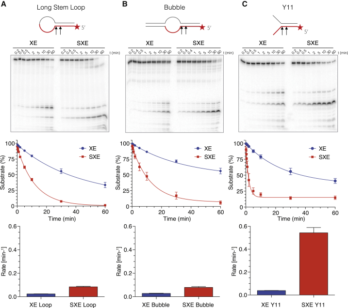
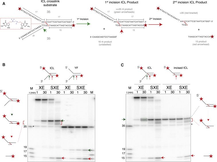
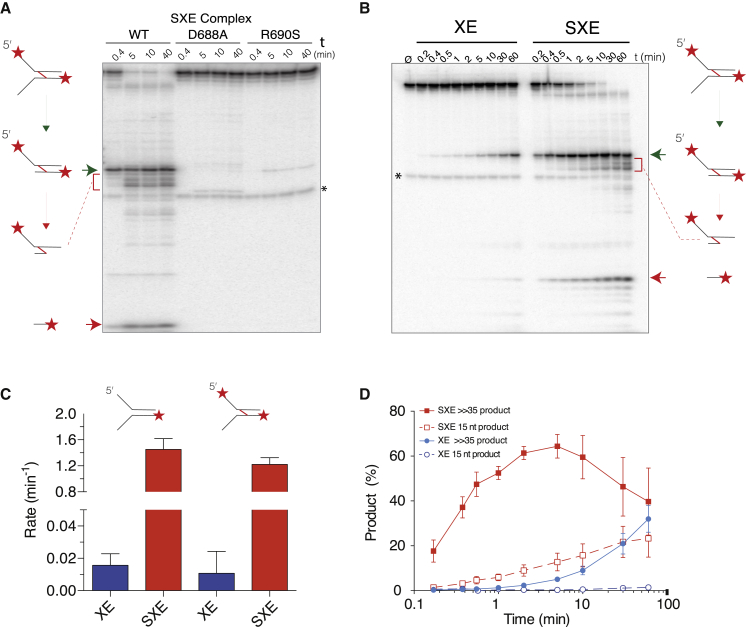
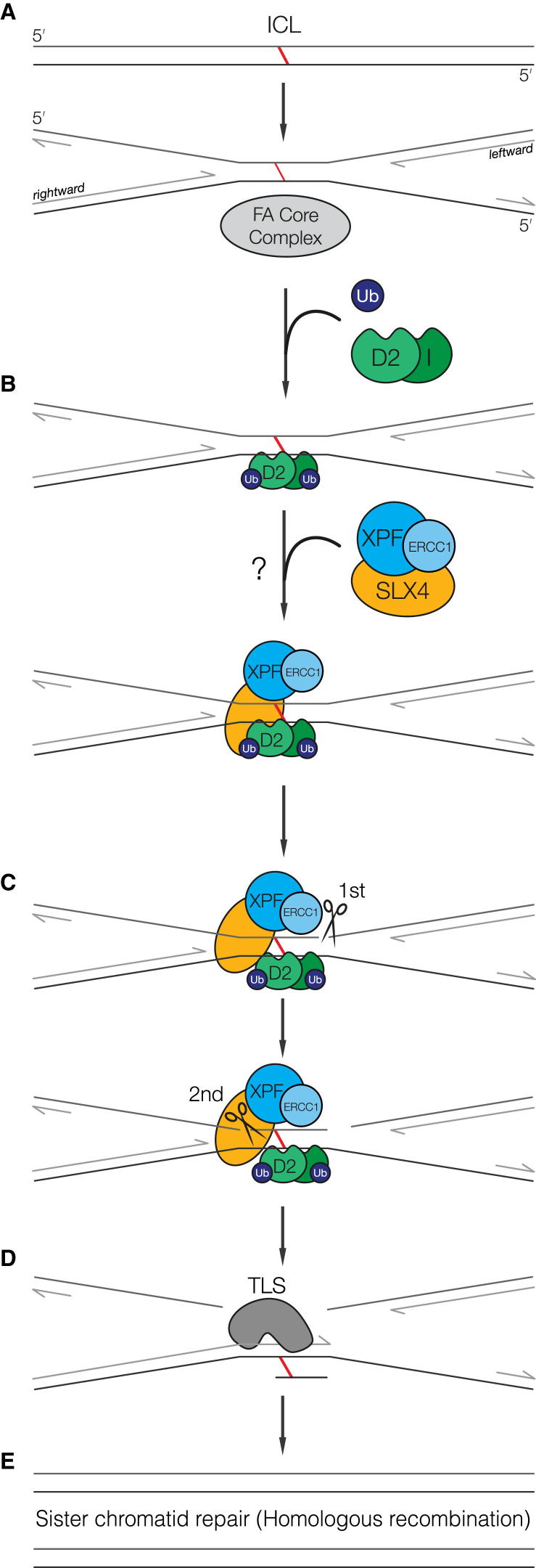
SLX4 binds to three nucleases (XPF-ERCC1, MUS81-EME1, and SLX1), and its deficiency leads to genomic instability, sensitivity to DNA crosslinking agents, and Fanconi anemia. However, it is not understood how SLX4 and its associated nucleases act in DNA crosslink repair. Here, we uncover consequences of mouse Slx4 deficiency and reveal its function in DNA crosslink repair. Slx4-deficient mice develop epithelial cancers and have a contracted hematopoietic stem cell pool. The N-terminal domain of SLX4 (mini-SLX4) that only binds to XPF-ERCC1 is sufficient to confer resistance to DNA crosslinking agents. Recombinant mini-SLX4 enhances XPF-ERCC1 nuclease activity up to 100-fold, directing specificity toward DNA forks. Mini-SLX4-XPF-ERCC1 also vigorously stimulates dual incisions around a DNA crosslink embedded in a synthetic replication fork, an essential step in the repair of this lesion. These observations define vertebrate SLX4 as a tumor suppressor, which activates XPF-ERCC1 nuclease specificity in DNA crosslink repair.


Figures

None
Graphical abstract
Figure 1
Figure 1
Slx4-Deficient Mice Are Cancer Prone and Have a Compromised HSPC Pool (A) Kaplan-Meier curve showing the tumor-free survival of our cohort of aged Slx4f3/f3 C57BL/6NTac mice (n = 28) and congenic controls (n = 28). (B) Hematoxylin and eosin staining of sections of liver in (1) 8-week-old and (2) 24-week-old Slx4f3/f3 mice, revealing karyomegaly and steatosis. (3) Gross pathology of a typical hepatic mass in Slx4f3/f3. (4) Histology of Slx4f3/f3 hepatic mass, showing a primary hepatocellular cancer. (C) (1) Low-power magnification of an anal mass (black arrow), and (2) higher magnification shows features of a typical squamous cell carcinoma with keratin whorls of the rectum. (D) Flow cytometry analysis of total bone marrow from Slx4+/+ and Slx4f3/f3 mice stained with hematopoietic stem and progenitor cell markers (Linagec-kit+Sca1+: LKS box). (E) Spleen colony forming assay (CFU-S10) was performed in lethally irradiated recipients revealing a reduction in the Slx4f3/f3 bone marrow. Error bars represent SEM. ∗∗∗∗p < 0.0001.
Figure 2
Figure 2
SLX4 1-758 Partially Complements Crosslinker Sensitivity and Can Be Purified in a Complex with XPF-ERCC1 (A) Cartoon depicts the SLX4 polypeptide (1–1565), domains, and interactions with the three nucleases: XPF-ERCC1, MUS81-EME1, and SLX1. A truncated SLX4 1-758 (mini-SLX4) contains the region that interacts with XPF-ERCC1. (B) Full-length FLAG-tagged SLX4 or FLAG-tagged mini-SLX4 was expressed in Slx4f3/f3 MEFs. Anti-FLAG immunoprecipitation shows that the ectopically expressed SLX4 polypeptides can be copurified with XPF-ERCC1. Note: ectopically expressed full-length SLX4 is prone to degradation/aggregation, accounting for the three bands seen by western blot (WB). (C) MTS viability of Slx4f3/f3 MEFs stably expressing full-length FLAG-SLX4 or FLAG-mini-SLX4, exposed to varying doses of Mitomycin C (MMC) for 4 days. (D) Expression and purification of a recombinant mini-SLX4 (1–758) in complex with XPF-ERCC1 (SXE) from insect cells. The purification scheme is shown next to a Coomassie gel depicting the various stages of purification. (E) Analytical gel filtration and Coomassie gels of purified mini-SLX4, XPF-ERCC1 (XE), and SXE complexes. Italicized letters correspond to the proteins shown on the Commassie gel. The shaded box represents those fractions loaded on SDS gels (below). A red arrow denotes the column void volume (∼2 MDa). Error bars represent SEM.
Figure 3
Figure 3
Comparison of the Activities of XE and SXE and Nuclease-Dead Mutants on Various Synthetic DNA Substrates (A) Coomassie gel (top) and western blot (WB) analysis (bottom) of the purified SXE, XE complexes with WT XPF, or catalytically dead mutant D688A (DA) used in the following assays and XPF Fanconi mutation R690S (RS). ERCC1 forms a doublet on WB, owing to a proteolytic site in the N terminus, shifting its mass by ∼2 kDa (). (B) Activity of WT SXE/XE or DA SXE/XE on DNA structures; single-stranded (ssDNA), double-stranded (dsDNA), 3′ overhang (3′ OH), and 5′ overhang (5′ OH). SXE shows enhanced activity toward 3′ overhangs (red arrow) and also low double-strand nicking activity (black arrow). The colored symbols denote fluorophore-labeled nucleotides. Red arrow marks structure-specific activity. (C) Activity of WT SXE/XE or DA SXE/XE on more complex splayed arms (5′ HEX Y, 3′ Cy5 Y) and stem-loop structures. SXE shows an induction in cleaving the 3′ end of Y-shaped substrate, cleaving near the ss/ds junction (21 nt marker). (D) Fluorescence anisotropy assay to determine binding of SXE and XE to either short stem-loop or splayed arms. Mini-SLX4 does not enhance the binding of XE to either short stem-loop or splayed arms. Normalized and averaged anisotropy ± SEM.
Figure 4
Figure 4
Mini-SLX4 Specifically Enhances XPF-ERCC1 Activity toward Y-Structured DNA (A–C) XE and SXE (5 nM) were reacted with different radiolabeled DNA substrates (∼1.5 pM), over a time course (A, long stem-loop; B, bubble; C, fork-structured DNA [Y11]). Substrates had identical primary sequence around the ss/ds bifurcation (depicted in red). The reaction products were separated by 12% denaturing PAGE gel (top panel), and the decay of the substrate band (S) was quantified and expressed as a percentage of initial substrate (middle panel). Data were fitted using single exponential decay in order to calculate reaction rates (bottom panel). XE data are plotted in blue; SXE data are plotted in red. SXE shows a modest stimulation of activity compared to XE toward stem-loop and bubble substrates and a pronounced induction of activity toward forked DNA (Y11). Error bars represent SEM.
Figure 5
Figure 5
SLX4 Promotes Unhooking of an ICL by XPF-ERCC1 (A) Outline of forked substrate containing a single nitrogen mustard-like interstrand crosslink (ICL) and its predicted reaction products. The substrate was generated from two oligonucleotides (each 35 nucleotides in length) with a crosslink between adjacent guanines, close to the ss/ds junction (red boxed inset). Sequential unhooking of the crosslinked DNA should result in an intermediate product (≫35 nt), followed by final products >35 and ≤15 nt (illustrated with green and red arrowheads). (B) The forked ICL substrate or an identical, but noncrosslinked, control (YF) were radiolabeled at the 5′ end and reacted with XE or SXE enzyme complexes and analyzed by denaturing PAGE. Cleavage sites and reaction products corresponding to those illustrated in (A) are shown as arrows and brackets (the equivalent products from YF migrate at 19 and 15 nt). Comparison of ICL and YF digestion reveals the first ≫35 nt product is most likely to result from an incision at the ss/ds boundary (corresponding to 19 nt product of noncrosslinked YF fork). This was confirmed with 3′-end labeling (Figure S5). (C) The primary reaction product (≫35 nt, green box) from the ICL substrate was purified as a substrate in a second reaction (“incised ICL”) to test whether the ICL was cleaved again (unhooked). The 15 and >35 nt product (red arrowhead and brackets) correspond to cleavage 5′ of the adducted guanine. All reactions contained 5 nM enzyme complex and ∼1.5 pM substrate. An asterisk denotes a low abundance, background band (a contaminant noncrosslinked oligonucleotide). Representative gels depict experiments performed at least three times.
Figure 6
Figure 6
SLX4 Increases the Efficiency of XPF-ERCC1 ICL Unhooking (A) WT SXE or SXE harboring either XPF D678A or R690S mutations were incubated with the ICL substrate labeled at the 5′ end. ICL cleavage products are clearly seen with WT SXE, whereas SXE R690S (associated with human FA) shows very weak activity. The cleavage products are illustrated with a green arrow, red bracket, and red arrow (as described in Figure 5). A noncrosslinked oligonucleotide contaminant is marked with an asterisk. (B) Representative time course, comparing reaction of ICL substrate with either XE or SXE. (C) Rates of substrate turnover for ICL or equivalent noncrosslinked control, calculated from data presented in (B). (D) Graph representing the ICL product formation for XE (blue) and SXE (red) enzyme complexes. Filled symbols mark the first incision product (shown in B above as a green arrow), open symbols depict 15 nt product (B, red arrow). The accumulation of the 15 nt product is dependent on the first product and marks the “unhooking” of the crosslink. Assays were performed with 5 nM enzyme complex and ∼1.5 pM labeled substrate, incubated for the time indicated, quenched, and separated by 12% denaturing PAGE gel. Data in (C) and (D) are plotted from a minimum of three independent experiments; error bars represent SEM.
Figure 7
Figure 7
Model for the Role of SXE in ICL Repair (A) Monoubiquitylation of FANCD2 and FANCI (ID) by the FA core complex is required for interstrand crosslink recognition. (B) Ubiquitylated ID recruits SLX4 in complex with XPF-ERCC1 either directly or via an unidentified intermediary protein(s). SXE preference for a 3′ single-stranded arm suggests the molecular recognition of the crosslink is triggered by the convergence of both replication forks at the ICL. (C) The presence of the leftward fork would trigger SXE cutting first 3′ and possibly then 5′, unhooking the ICL. (D) The intact (adducted) parental strand could then serve as a template for the rightward fork extension by translesion synthesis. (E) The adducted base can then be removed by a combination of nucleotide excision repair and the newly synthesized chromatid used to repair the resulting DSB. SLX4 involvement in this process may additionally require the action of MUS81-EME1 and/or SLX1.

Similar articles

Cited by

References

    1. Al-Minawi A.Z., Saleh-Gohari N., Helleday T. The ERCC1/XPF endonuclease is required for efficient single-strand annealing and gene conversion in mammalian cells. Nucleic Acids Res. 2008;36:1–9. - PMC - PubMed
    1. Andersen S.L., Bergstralh D.T., Kohl K.P., LaRocque J.R., Moore C.B., Sekelsky J. Drosophila MUS312 and the vertebrate ortholog BTBD12 interact with DNA structure-specific endonucleases in DNA repair and recombination. Mol. Cell. 2009;35:128–135. - PMC - PubMed
    1. Angelov T., Guainazzi A., Schärer O.D. Generation of DNA interstrand cross-links by post-synthetic reductive amination. Org. Lett. 2009;11:661–664. - PMC - PubMed
    1. Auerbach A.D., Wolman S.R. Susceptibility of Fanconi’s anaemia fibroblasts to chromosome damage by carcinogens. Nature. 1976;261:494–496. - PubMed
    1. Barnes D.E., Lindahl T. Repair and genetic consequences of endogenous DNA base damage in mammalian cells. Annu. Rev. Genet. 2004;38:445–476. - PubMed

Publication types