Neutrophil depletion suppresses pulmonary vascular hyperpermeability and occurrence of pulmonary edema caused by hantavirus infection in C.B-17 SCID mice
- PMID: 24719427
- PMCID: PMC4054411
- DOI: 10.1128/JVI.00254-14
Neutrophil depletion suppresses pulmonary vascular hyperpermeability and occurrence of pulmonary edema caused by hantavirus infection in C.B-17 SCID mice
Abstract
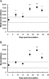
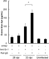
Hantavirus infections are characterized by vascular hyperpermeability and neutrophilia. However, the pathogenesis of this disease is poorly understood. Here, we demonstrate for the first time that pulmonary vascular permeability is increased by Hantaan virus infection and results in the development of pulmonary edema in C.B-17 severe combined immunodeficiency (SCID) mice lacking functional T cells and B cells. Increases in neutrophils in the lung and blood were observed when pulmonary edema began to be observed in the infected SCID mice. The occurrence of pulmonary edema was inhibited by neutrophil depletion. Moreover, the pulmonary vascular permeability was also significantly suppressed by neutrophil depletion in the infected mice. Taken together, the results suggest that neutrophils play an important role in pulmonary vascular hyperpermeability and the occurrence of pulmonary edema after hantavirus infection in SCID mice.
Importance: Although hantavirus infections are characterized by the occurrence of pulmonary edema, the pathogenic mechanism remains largely unknown. In this study, we demonstrated for the first time in vivo that hantavirus infection increases pulmonary vascular permeability and results in the development of pulmonary edema in SCID mice. This novel mouse model for human hantavirus infection will be a valuable tool and will contribute to elucidation of the pathogenetic mechanisms. Although the involvement of neutrophils in the pathogenesis of hantavirus infection has largely been ignored, the results of this study using the mouse model suggest that neutrophils are involved in the vascular hyperpermeability and development of pulmonary edema in hantavirus infection. Further study of the mechanisms could lead to the development of specific treatment for hantavirus infection.
Copyright © 2014, American Society for Microbiology. All Rights Reserved.
Figures



Similar articles
-
Hantavirus infection in SCID mice.J Vet Med Sci. 1997 Oct;59(10):863-8. doi: 10.1292/jvms.59.863. J Vet Med Sci. 1997. PMID: 9362032
-
Depletion of Alveolar Macrophages Does Not Prevent Hantavirus Disease Pathogenesis in Golden Syrian Hamsters.J Virol. 2016 Jun 24;90(14):6200-6215. doi: 10.1128/JVI.00304-16. Print 2016 Jul 15. J Virol. 2016. PMID: 27099308 Free PMC article.
-
The adaptive immune response does not influence hantavirus disease or persistence in the Syrian hamster.Immunology. 2013 Oct;140(2):168-78. doi: 10.1111/imm.12116. Immunology. 2013. PMID: 23600567 Free PMC article.
-
Hantavirus regulation of endothelial cell functions.Thromb Haemost. 2009 Dec;102(6):1030-41. doi: 10.1160/TH09-09-0640. Thromb Haemost. 2009. PMID: 19967132 Review.
-
Uncovering the mysteries of hantavirus infections.Nat Rev Microbiol. 2013 Aug;11(8):539-50. doi: 10.1038/nrmicro3066. Nat Rev Microbiol. 2013. PMID: 24020072 Review.
Cited by
-
Pathological Studies on Hantaan Virus-Infected Mice Simulating Severe Hemorrhagic Fever with Renal Syndrome.Viruses. 2022 Oct 13;14(10):2247. doi: 10.3390/v14102247. Viruses. 2022. PMID: 36298802 Free PMC article.
-
Deletions in Genes Participating in Innate Immune Response Modify the Clinical Course of Andes Orthohantavirus Infection.Viruses. 2019 Jul 25;11(8):680. doi: 10.3390/v11080680. Viruses. 2019. PMID: 31349540 Free PMC article.
-
Csf2ra deletion attenuates acute lung injuries induced by intratracheal inoculation of aerosolized ricin in mice.Front Immunol. 2022 Sep 20;13:900755. doi: 10.3389/fimmu.2022.900755. eCollection 2022. Front Immunol. 2022. PMID: 36203597 Free PMC article.
-
Hantavirus-induced disruption of the endothelial barrier: neutrophils are on the payroll.Front Microbiol. 2015 Mar 25;6:222. doi: 10.3389/fmicb.2015.00222. eCollection 2015. Front Microbiol. 2015. PMID: 25859243 Free PMC article.
-
Innate lymphoid cells are activated in HFRS, and their function can be modulated by hantavirus-induced type I interferons.PLoS Pathog. 2024 Jul 22;20(7):e1012390. doi: 10.1371/journal.ppat.1012390. eCollection 2024 Jul. PLoS Pathog. 2024. PMID: 39038044 Free PMC article.
References
Publication types
MeSH terms
Substances
LinkOut - more resources
Full Text Sources
Other Literature Sources
Medical

