. 2014 Apr;141(8):1649-59.
doi: 10.1242/dev.100842.

Early forming label-retaining muscle stem cells require p27kip1 for maintenance of the primitive state

Affiliations

Early forming label-retaining muscle stem cells require p27kip1 for maintenance of the primitive state

Joe V Chakkalakal et al. Development. 2014 Apr.

Abstract

Across different niches, subsets of highly functional stem cells are maintained in a relatively dormant rather than proliferative state. Our understanding of proliferative dynamics in tissue-specific stem cells during conditions of increased tissue turnover remains limited. Using a TetO-H2B-GFP reporter of proliferative history, we identify skeletal muscle stem cell, or satellite cells, that retain (LRC) or lose (nonLRC) the H2B-GFP label. We show in mice that LRCs and nonLRCs are formed at birth and persist during postnatal growth and adult muscle repair. Functionally, LRCs and nonLRCs are born equivalent and transition during postnatal maturation into distinct and hierarchically organized subsets. Adult LRCs give rise to LRCs and nonLRCs; the former are able to self-renew, whereas the latter are restricted to differentiation. Expression analysis revealed the CIP/KIP family members p21(cip1) (Cdkn1a) and p27(kip1) (Cdkn1b) to be expressed at higher levels in LRCs. In accordance with a crucial role in LRC fate, loss of p27(kip1) promoted proliferation and differentiation of LRCs in vitro and impaired satellite cell self-renewal after muscle injury. By contrast, loss of p21(cip1) only affected nonLRCs, in which myogenic commitment was inhibited. Our results provide evidence that restriction of self-renewal potential to LRCs is established early in life and is maintained during increased tissue turnover through the cell cycle inhibitor p27(kip1). They also reveal the differential role of CIP/KIP family members at discrete steps within the stem cell hierarchy.

Keywords: Mouse; Muscle; Quiescence; Regeneration; Satellite cell; Self-renewal; Stem cell.


Figures
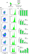
Fig. 1.
Fig. 1.
H2B-GFP labeling reveals the formation of LRC and nonLRCs at birth. (A) Timecourse of Dox feeding (E10.5-16) and chase and SC harvest periods of TetO-H2B-GFP mice. (B) SC sort profile and distribution of H2B-GFP intensity at different chase time points (E16.5 represents no chase). Vehicle-treated TetO-H2B-GFP control is in gray. (C) Percentage of sorted LRCs and nonLRCs expressing Pax7, Ki67, MyoD or Myf5 at different neonatal and postnatal stages. (D) Percentage of sorted p27kip1+ LRCs and nonLRCs at different neonatal and postnatal stages. (C,D) Data were averaged (n=3-4 mice per group) and expressed as mean±s.e.m. (t-tests, *P<0.05, LRC versus nonLRC).
Fig. 2.
Fig. 2.
H2B-GFP labeling reveals the re-establishment of LRCs and nonLRCs in response to injury. (A) Dox feeding and injury paradigm with adult TetO-H2B-GFP mice. (B) Representative SC sort profile of 6-week pulsed or 30-day post-injury muscle. (C) Representative distribution of H2B-GFP intensity from sorted SCs harvested 30 days post-injury (red) or from uninjured contralateral muscle (green). No-chase H2B-GFP profile isolated from Dox-fed TetO-H2B-GFP mice (black). H2B-GFP intensity profile from vehicle-fed TetO-H2B-GFP mice (gray filled line). Two discrete populations (LRC and nonLRC) of SCs form after injury. To determine the fraction of LRCs and nonLRCs within FACS isolated SCs, we created positive selection gates at the boundaries where the cell numbers reach a minimum across the total H2B-GFP intensity. The fraction of the total population within each gate was categorized as LRC or nonLRC (see Materials and Methods for more detail). (D) Transverse sections (top) and single fibers (middle) from Dox-fed no-chase TetO-H2B-GFP mice show GFP expression in Pax7+ SCs. H2B-GFP was not detected in Pax7+ cells from vehicle-treated TetO-H2B-GFP mice (bottom row). (E) H2B-GFP label retention in Pax7+ cells from single fibers in uninjured and regenerated muscle (30 days after injury). (F) Profile of H2B-GFP expression in uninjured (black) or 30-day regenerated (green) single muscle fiber-associated SCs; vehicle-treated H2B-GFP provided a negative control (gray). H2B-GFP profiles were collected from 6-8 mice per group.
Fig. 3.
Fig. 3.
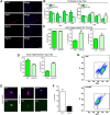
Proliferative history and cell fate become coupled during postnatal maturation. (A) Representative Pax7, Myf5, MyoD and MyoG immunostaining alone or in combination with DAPI staining (merge) of sorted LRCs and nonLRCs from TetO-H2B-GFP mice cultured for 4 days in plating medium. Scale bar: 50 µm. (B) Percentage of sorted LRCs or nonLRCs isolated throughout ontogeny expressing Pax7/MyoG after 4 days in culture. (C,D) Percentage of sorted LRCs or nonLRCs isolated from 30-day regenerated adult muscle and plated for 8 h (C, priming) or 4 days (D, fate) expressing Pax7, Myf5, MyoD or MyoG. (B-D) Data were averaged (n=3 mice per group) and expressed as mean±s.e.m. (t-tests, *P<0.05, LRC versus nonLRC). (E) LRCs stained with anti-Pax7 and anti-MyoG after 0 or 4 days in culture. Scale bar: 20 μm. (F) Percentage of Pax7+ or MyoG+ cells that retained GFP label after 4 days in culture. Data were averaged (n=3 mice per group) and expressed as mean±s.e.m. (P<0.05). (G) H2B-GFP intensity of sorted adult LRCs and nonLRCs that had been plated, treated with Dox on day 1 and cultured in plating medium for 9 days. LRC cultures form LRC and nonLRC populations, whereas cultured nonLRCs divide as a single population.
Fig. 4.
Fig. 4.
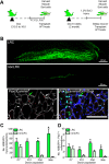
LRCs and nonLRCs are hierarchically organized and functionally distinct. (A) Schematic depicts isolation of 2000 LRCs and 2000 nonLRCs for transplant. (B) Representative whole-mount of regenerated (30 days post-transplantation) host TA muscle after engraftment of LRCs (top) or nonLRCs (bottom). Transverse sections of host muscle stained for Pax7 or laminin or with DAPI. Muscles contain donor (GFP+, green arrow) and host-derived (GFP, white arrow) Pax7+ cells underneath basal lamina and sublaminar myonuclei. Scale bars: 200 μm. (C,D) Number of donor-derived GFP+ myonuclei (C) and Pax7+/GFP+ (D) sublaminar cells. GFP+ cell numbers were summed over 200 μm of regenerated muscle. Data were averaged (n=3 mice per group) and expressed as mean±s.e.m. (t-tests, *P<0.05).
Fig. 5.
Fig. 5.
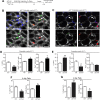
SC proliferative history during mdx disease progression. (A) Representative histological stains of TA muscles from 14-month control and mdx mice. Scale bars: 50 μm. (B) Number of Pax7+ SCs per single muscle fiber from 14-month control and mdx mice. (C) Percentage of sorted SCs from 14-month control and mdx mice expressing Pax7 or MyoG after 4 days in culture. (D) Dox feeding and chase strategy in H2B control and H2B mdx mice. Representative FACS profiles of H2B-GFP intensity in SCs from 14-month chased control (green) and mdx (red) mice. (E) Percentage of sorted LRCs and nonLRCs expressing Pax7 or MyoG+ after 4 days in culture. (F) Schematic and number of donor-derived GFP+ myonuclei (left) and Pax7+/GFP+ (right) sublaminar cells in host 14-month-old mdx muscle. GFP+ cell numbers were summed over 200 μm of regenerated muscle. (C,E,F) Data were averaged (n=3 mice per group) and expressed as mean±s.e.m. (t-tests, *P<0.05).
Fig. 6.
Fig. 6.
p27kip1 is enriched in LRCs throughout growth and regeneration. Expression of myogenic fate markers (A, Pax7; B, Myf5) and cell cycle inhibitors (C, p21cip1; D, p27kip1) in LRCs and nonLRCs during ontogeny or after injury. Expression values were normalized to the corresponding gene expression level in LRCs at the P7 time point. The normalization within the P7 time point was obtained by setting the first of four replicates to 1. Data were averaged (n=3-4 mice per group) and expressed as mean±s.e.m. (t-tests, *P<0.05).
Fig. 7.
Fig. 7.
p27kip1 is required for the maintenance of self-renewing LRCs. (A) Outline of experiments to assess BrdU/EdU label retention. (B) Representative images of Pax7+ and Ki67+ cells from postnatal p27 null muscle. Arrowhead and arrow indicate Pax7+/Ki67 and Pax7+/Ki67+ SCs, respectively. Scale bar: 20 μm. (C) Representative images of Pax7+ and EdU+ cells from postnatal p21 null muscles. Arrow indicates Pax7+/EdU+ SCs. Scale bar: 100 μm. (D) Postnatal (P17) p27 null and Ctrl muscles and (E) postnatal (P17) p21 null and Ctrl muscle analyzed for label-retaining (BrdU+/EdU+) SCs (left), the percentage of Ki67+ SCs (middle) and the number of SCs per serial muscle section (right). (F) Percentage of Pax7+/MyoG+ sorted SCs from p27 null and Ctrl muscles and (G) p21 null and Ctrl muscles after 4 days in culture. (D-G) Data were averaged (n=3-4 mice per group) and expressed as mean±s.e.m. (t-tests, *P<0.05, **P<0.01).
Fig. 8.
Fig. 8.
p27kip1 function is restricted to LRCs. Lentiviral shRNA strategy to knockdown p27kip1 (p27KD) in postnatal (P10) LRCs or nonLRCs (A) or to knockdown p21cip1 (p21KD) or p27kip1 in adult LRCs or nonLRCs (B). (C) Percentage of Pax7+ or MyoG+ LRCs and nonLRCs from postnatal (P10) muscle after p27KD (left), from adult muscle after p27KD (middle), and from adult muscle after p21KD (right). Data were averaged (n=3 mice per group) and expressed as mean±s.e.m. (t-tests, *P<0.05). (D) Muscle injury strategy. (E,F) Quantification of Pax7+ cells per single muscle fiber (E) and average muscle fiber size (F) from control or p27 null uninjured (UI), primary injured (1°) or secondary injured (2°) muscle. (G,H) Quantification of Pax7+ cells per mm2 (G) and average muscle fiber size (H) from control or p21 null uninjured (UI), primary injured (1°) or secondary injured (2°) muscle. (E-H) Data were averaged (n=3 mice per group) and expressed as mean±s.e.m. (t-tests, *P<0.05). (I) Control and p27 null uninjured and 2° injured muscle stained for laminin, pan myosin heavy chain and with DAPI to quantify average muscle fiber size. Scale bar: 200 μm.

Similar articles

Cited by

References

    1. Biressi S., Bjornson C. R. R., Carlig P. M. M., Nishijo K., Keller C., Rando T. A. (2013). Myf5 expression during fetal myogenesis defines the developmental progenitors of adult satellite cells. Dev. Biol. 379, 195-207 10.1016/j.ydbio.2013.04.021 - DOI - PMC - PubMed
    1. Blanpain C., Lowry W. E., Geoghegan A., Polak L., Fuchs E. (2004). Self-renewal, multipotency, and the existence of two cell populations within an epithelial stem cell niche. Cell 118, 635-648 10.1016/j.cell.2004.08.012 - DOI - PubMed
    1. Boldrin L., Zammit P. S., Muntoni F., Morgan J. E. (2009). Mature adult dystrophic mouse muscle environment does not impede efficient engrafted satellite cell regeneration and self-renewal. Stem Cells 27, 2478-2487 10.1002/stem.162 - DOI - PMC - PubMed
    1. Bröhl D., Vasyutina E., Czajkowski M. T., Griger J., Rassek C., Rahn H.-P., Purfürst B., Wende H., Birchmeier C. (2012). Colonization of the satellite cell niche by skeletal muscle progenitor cells depends on notch signals. Dev. Cell 23, 469-481 10.1016/j.devcel.2012.07.014 - DOI - PubMed
    1. Cerletti M., Jurga S., Witczak C. A., Hirshman M. F., Shadrach J. L., Goodyear L. J., Wagers A. J. (2008). Highly efficient, functional engraftment of skeletal muscle stem cells in dystrophic muscles. Cell 134, 37-47 10.1016/j.cell.2008.05.049 - DOI - PMC - PubMed

Publication types

Substances