. 2014 Apr 8:11:29.
doi: 10.1186/1742-4690-11-29.

Nuclear import of SAMHD1 is mediated by a classical karyopherin α/β1 dependent pathway and confers sensitivity to VpxMAC induced ubiquitination and proteasomal degradation

Affiliations

Nuclear import of SAMHD1 is mediated by a classical karyopherin α/β1 dependent pathway and confers sensitivity to VpxMAC induced ubiquitination and proteasomal degradation

Torsten Schaller et al. Retrovirology. .

Abstract

Background: The deoxynucleotide-triphosphate (dNTP) hydrolase sterile alpha motif domain and HD domain 1 (SAMHD1) is a nuclear protein that inhibits HIV-1 infection in myeloid cells as well as quiescent CD4 T-cells, by decreasing the intracellular dNTP concentration below a level that is required for efficient reverse transcription. The Vpx proteins of the SIVSMM/HIV-2 lineage of lentiviruses bind SAMHD1 and recruit an ubiquitin ligase, leading to polyubiquitination and proteasomal degradation.

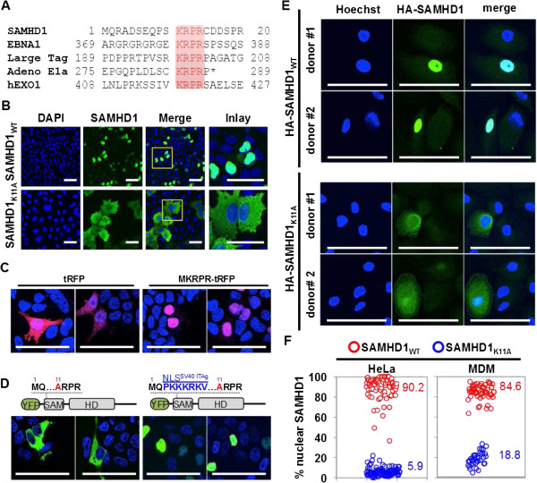
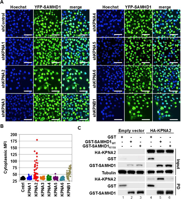
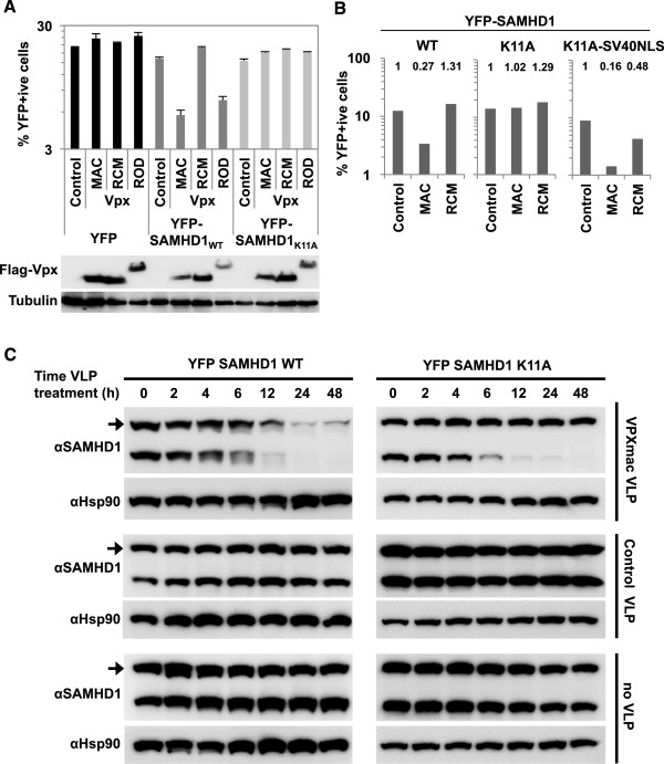
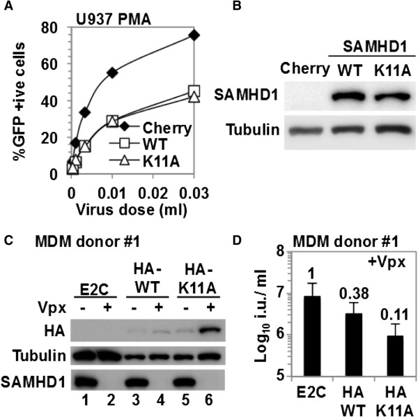
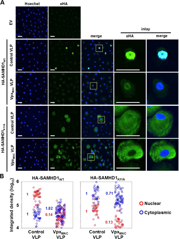
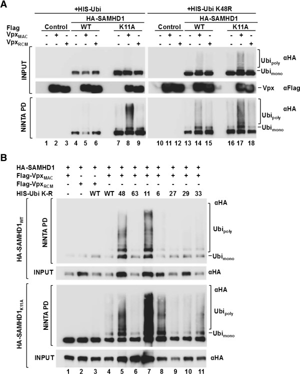
Results: Here, we have investigated the importance of nuclear localization for SAMHD1's antiviral function as well as its sensitivity to the Vpx protein of SIVMAC. Using GST pull down assays, as well as RNA silencing approaches, we show that SAMHD1 preferentially uses karyopherin α2 (KPNA2) and a classical N-terminal nuclear localization signal (14KRPR17) to enter the nucleus. Reduction of karyopherin β1 (KPNB1) or KPNA2 by RNAi also led to cytoplasmic re-distribution of SAMHD1. Using primary human monocyte-derived macrophages (MDM), a cell type in which SAMHD1 is naturally expressed to high levels, we demonstrate that nuclear localization is not required for its antiviral activity. Cytoplasmic SAMHD1 still binds to VpxMAC, is efficiently polyubiquitinated, but is not degraded. We also find that VpxMAC-induced SAMHD1 degradation was partially reversed by ubiquitin carrying the K48R or K11R substitution mutations, suggesting involvement of K48 and K11 linkages in SAMHD1 polyubiquitination. Using ubiquitin K-R mutants also revealed differences in the ubiquitin linkages between wild type and cytoplasmic forms of SAMHD1, suggesting a potential association with the resistance of cytoplasmic SAMHD1 to VpxMAC induced degradation.

Conclusions: Our work extends published observations on SAMHD1 nuclear localization to a natural cell type for HIV-1 infection, identifies KPNA2/KPNB1 as cellular proteins important for SAMHD1 nuclear import, and indicates that components of the nuclear proteasomal degradation machinery are required for SAMHD1 degradation.


Figures

Figure 1
Figure 1
Determination of the SAMHD1 nuclear localization signal (NLS). A) Comparison of the SAMHD1 amino acid sequence with known nuclear localization signals (NLS) from EBNA1, polyoma virus large T antigen, adenovirus E1a and human EXO1 identifies an N-terminal four amino acid stretch as a putative NLS. B) Substitution of a critical lysine residue K11 with alanine abrogates SAMHD1 nuclear localization and causes cytoplasmic accumulation. HeLa cells transduced with HA-SAMHD1WT or HA-SAMHD1K11A expressing MLV vectors were subjected to immunofluorescence using a HA-specific antibody and nuclei were stained with DAPI. C) Expression of turbo red fluorescence protein (tRFP) fused to amino-terminal KRPR sequence in 293 T cells causes redistribution of the protein from a pan-cellular to a nuclear localization. Nuclei were stained with DAPI and tRFP was detected by autofluorescence. D) Fusion of the SV40 large T antigen NLS is sufficient to rescue nuclear localization of YFP-SAMHD1K11A. 293 T cells were transfected with YFP-SAMHD1K11A (left panels) or YFP-SAMHD1K11A fused to the NLS of the SV40 large T antigen (right panels) and autofluorescence was measured 48 h after transfection. Nuclei were stained with DAPI. E) Primary monocyte-derived macrophages from two independent donors were transduced with HA-tagged wild type or K11A mutant SAMHD1 expressing HIV-1 lentiviral vectors and HA-immunostaining was performed to detect SAMHD1 three days later. F) Quantification of the percentage of nuclear SAMHD1 for HA-SAMHD1WT and HA-SAMHD1K11A expressing cells comparing HeLa cells with primary monocyte derived macrophages (MDM). Each circle represents one cell and numbers indicate average percentages of cells shown. Scale bars, 50 μm.
Figure 2
Figure 2
SAMHD1 nuclear import is mediated by KPNA2 and KPNB1. A) HeLa cells stably transduced with an MLV-vector encoding YFP-tagged wild type SAMHD1 were transiently transduced with Retro-SIREN-Q (Clontech) shRNA encoding vectors expressing two independent shRNAs for each karyopherin or shControl and subjected to autofluorescence microscopy 48 h after transduction. Nuclei were stained with Hoechst. All pictures were generated using the same microscope settings and analyzed using the same conditions. Scale bars, 50 μm. B) Cytoplasmic fluorescence was determined using software ImageJ for thirty cells for each slide shown in A) in an area of 535 pixels (total image size 1024x1024 pixels) of cytoplasm in proximity of the nucleus for each cell. Each dot represents measurement of cytoplasmic mean fluorescence intensities (MFIs) for one cell. Each cell was only counted once. C) GST-tagged wild type or SAMHD1K11A encoding plasmids or the GST control were co-transfected with empty vector or HA-KPNA2 encoding vector in 293 T cells and GST-pull down (PD) was performed 48 h later. Tubulin served as the input loading control.
Figure 3
Figure 3
SAMHD1WT and SAMHD1K11A bind VpxMAC with similar efficiencies. 293 T cells were co-transfected with plasmids expressing GST, GST-SAMHD1 or GST-SAMHD1K11A along with plasmids expressing Flag-tagged Vpx derived from HIV-2ROD, SIVMAC or SIVRCM. GST-pull down was performed and samples were separated using 15% SDS-PAGE gels. Vpx proteins were detected using an anti-Flag antibody, and SAMHD1 was detected using an anti-GST antibody. Tubulin served as a loading control for the input.
Figure 4
Figure 4
SAMHD1K11A has reduced sensitivity to VpxMAC mediated degradation. A) 293 T cells were transfected with plasmids encoding YFP, YFP-SAMHD1WT, or YFP-SAMHD1K11A along with plasmids encoding for Flag-tagged Vpx proteins from HIV-2ROD, SIVMAC or SIVRCM as indicated and percentages of YFP-positive cells were determined 24 h after transfection. A parallel sample was used for western blot analysis using an anti-Flag antibody (lower panel). Tubulin served as a loading control. B) YFP-tagged SAMHD1WT, SAMHD1K11A or SAMHD1K11A-SV40NLS were co-transfected in parallel with Flag-Vpx expression plasmids as in A) and percentages of YFP + cells were measured 24 h post transfection. Fold changes are indicated above the bars. C) Time course of VpxMAC VLP or control VLP treatment of THP-1 cells expressing YFP-SAMHD1WT or YFP-SAMHD1K11A. Western blot analysis was performed with an anti-SAMHD1 antibody detecting endogenous SAMHD1 as well as ectopically expressed YFP-SAMHD1 (arrow). Hsp90 served as loading control.
Figure 5
Figure 5
Wild type SAMHD1 as well as SAMHD1K11A inhibit HIV-1 infection in cell lines and primary monocyte derived macrophages. A) U937 cells transduced with lentiviral vectors expressing wild type or K11A mutant SAMHD1, or mCherry respectively, were differentiated with 100 ng/ml PMA for 2 days and then infected with an HIV-1 YFP reporter vector at different doses. Infected cells were enumerated by flow cytometry 48 h after infection. A representative of at least three independent experiments is shown. B) A parallel sample to A) was used for western blotting using a SAMHD1 specific antibody. Tubulin served as a loading control. C) Monocyte-derived macrophages were transduced with lentiviral vectors encoding HA-tagged SAMHD1WT or SAMHD1K11A in the presence or absence of virus like particles delivering VpxMAC and cells were used for western blotting using anti-HA, anti-SAMHD1 as well as anti-tubulin antibodies. Cells transduced with a lentivirus vector expressing E2-Crimson (E2C) served as a control. D) A parallel sample of C) was used to test the infectivity of an HIV-1 GFP reporter virus which was measured 48 h post infection by flow cytometry. Infectious titers (i.u./ml) were calculated from three independent viral doses. Numbers above bars present fold changes compared to the E2C control.
Figure 6
Figure 6
The nuclear fraction of SAMHD1K11A in MDM is sensitive to VpxMAC induced degradation. A) Monocyte-derived macrophages were transduced with lentiviral vectors encoding HA-tagged SAMHD1WT or SAMHD1K11A in the presence of virus like particles (VLPs) delivering VpxMAC or control VLPs and cells were fixed for HA-immunostaining 72 h later. Scale bars, 30 μm. Shown are representative pictures from one of four independent donors. B) The integrated MFI densities were calculated on MDM expressing HA-SAMHD1WT or HA-SAMHD1K11A for nuclear and cytoplasmic areas using ImageJ software. Numbers within the graphs show fold changes of the average integrated densities normalized to control VLP treated cells.
Figure 7
Figure 7
SAMHD1K11A is ubiquitinated in the presence of VpxMAC. A) 293 T cells were co-transfected with plasmids encoding HA-tagged wild type or K11A mutant SAMHD1 along with plasmids expressing VpxMAC or VpxRCM and plasmids encoding either HIS-tagged wild type ubiquitin (HIS-Ubi) or K48R mutant ubiquitin (HIS-UbiK48R) for 24 h before cells were lysed and subjected to NiNTA pull down. Cell lysates (Input) or pulled down samples (NiNTA PD) were subjected to SDS-PAGE using 10% gels and western blotting using an anti-HA antibody. Mono- and polyubiquitinated SAMHD1 are indicated. B) Similar experiment as shown in A) but using ubiquitin mutants HIS-UbiK6R, K11R, K27R, K29R, K33R, K48R or K63R.

Similar articles

Cited by

References

    1. Sayah DM, Sokolskaja E, Berthoux L, Luban J. Cyclophilin A retrotransposition into TRIM5 explains owl monkey resistance to HIV-1. Nature. 2004;430:569–573. doi: 10.1038/nature02777. - DOI - PubMed
    1. Stremlau M, Owens CM, Perron MJ, Kiessling M, Autissier P, Sodroski J. The cytoplasmic body component TRIM5alpha restricts HIV-1 infection in old world monkeys. Nature. 2004;427:848–853. doi: 10.1038/nature02343. - DOI - PubMed
    1. Sheehy AM, Gaddis NC, Malim MH. The antiretroviral enzyme APOBEC3G is degraded by the proteasome in response to HIV-1 Vif. Nat Med. 2003;9:1404–1407. doi: 10.1038/nm945. - DOI - PubMed
    1. Goldstone DC, Ennis-Adeniran V, Hedden JJ, Groom HC, Rice GI, Christodoulou E, Walker PA, Kelly G, Haire LF, Yap MW, de Carvalho LP, Stoye JP, Crow YJ, Taylor IA, Webb M. HIV-1 restriction factor SAMHD1 is a deoxynucleoside triphosphate triphosphohydrolase. Nature. 2011;480:379–382. doi: 10.1038/nature10623. - DOI - PubMed
    1. Hrecka K, Hao C, Gierszewska M, Swanson SK, Kesik-Brodacka M, Srivastava S, Florens L, Washburn MP, Skowronski J. Vpx relieves inhibition of HIV-1 infection of macrophages mediated by the SAMHD1 protein. Nature. 2011;474:658–661. doi: 10.1038/nature10195. - DOI - PMC - PubMed

Publication types

MeSH terms

Substances