Review
. 2014 Apr;21(4):336-45.
doi: 10.1038/nsmb.2787.

Dynamic regulation of macroautophagy by distinctive ubiquitin-like proteins

Affiliations
Review

Dynamic regulation of macroautophagy by distinctive ubiquitin-like proteins

Daniel J Klionsky et al. Nat Struct Mol Biol. 2014 Apr.

Abstract

Autophagy complements the ubiquitin-proteasome system in mediating protein turnover. Whereas the proteasome degrades individual proteins modified with ubiquitin chains, autophagy degrades many proteins and organelles en masse. Macromolecules destined for autophagic degradation are 'selected' through sequestration within a specialized double-membrane compartment termed the phagophore, the precursor to an autophagosome, and then are hydrolyzed in a lysosome- or vacuole-dependent manner. Notably, a pair of distinctive ubiquitin-like proteins (UBLs), Atg8 and Atg12, regulate degradation by autophagy in unique ways by controlling autophagosome biogenesis and recruitment of specific cargos during selective autophagy. Here we review structural mechanisms underlying the functions and conjugation of these UBLs that are specialized to provide interaction platforms linked to phagophore membranes.


Figures
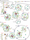
Figure 1
Figure 1
Morphology of macroautophagy and location/functions of UBL Atg proteins. The key morphological intermediates of macroautophagy are shown for mammals (a) and yeast (b). In either case, the process begins with the nucleation of the phagophore, the initial sequestering compartment (for simplicity, an omegasome is not depicted). This step and the subsequent expansion of the phagophore require the function of Atg8/LC3. Upon completion the phagophore becomes a double-membrane autophagosome. In mammals the autophagosome may fuse with a late endosome to form an amphisome, or may fuse directly with the lysosome to generate an autolysosome. In yeast, the autophagosome fuses with the vacuole, releasing the inner vesicle that is termed an autophagic body. The inner vesicle of the autophagosome is lysed and the cargoes are degraded, with the breakdown products being released into the cytosol for reuse. Atg8/LC3-II is present on both sides of the phagophore, and on the early autophagosome, but is released from the outer membrane by Atg4/ATG4B-dependent deconjugation during autophagosome maturation. Regulatory kinases including protein kinase A and TOR inhibit Atg1/ULK1 and Atg1/ATG13 during normal growth. See the text for details on the functions of the proteins involved in the UBL-protein conjugation systems.
Figure 2
Figure 2
Cargo recognition and packaging in selective macroautophagy. Different types of selective autophagy are illustrated. The yeast cytoplasm-to-vacuole targeting (Cvt) pathway is a biosynthetic process that delivers certain resident hydrolases to the vacuole. The propeptide of precursor aminopeptidase I (prApe1) is the key ligand that binds the Atg19 receptor. The latter interacts with the scaffold Atg11, and also contains an AIM motif for binding Atg8, connecting the cargo with the phagophore. A similar process occurs during mitophagy and pexophagy, where Atg32 and Atg36 act as receptors that bind both Atg11 and Atg8. During xenophagy in mammalian cells invasive bacteria may first be recognized by LGALS8 and subsequently by ubiquitin, which act as ligands. Receptors, including CALCOCO2/NDP52, SQSTM1 and OPTN bind the ligands and one or more of the Atg8 orthologs. Ams1, α-mannosidase; GBRP, GABARAP; PAS, phagophore assembly site.
Figure 3
Figure 3
Structural basis for Atg8 interacting motif (AIM)/LC3 interacting region (LIR) binding to ubiquitin-like protein Atg8 and orthologs. UBLs are shown in yellow, AIM or LIR peptides or proteins in green, and key interacting residues are shown in sticks. (a)Ubiquitin from a complex with E1. (b) Atg8 from a complex with E1, with distinctive N-terminal helices colored in orange,. (c) Overall similar modes of interactions with Atg8, LC3, and GABARAP as in an Atg8 complex with the AIM peptide from Atg19. (d) LC3B complex with the LIR peptide from SQSTM1/p62,. (e) GABARAPL1 complex with the LIR peptide from NBR1. (f) LC3C complex with the CLIR from CALCOCO2/NDP52. (g) LC3B complex with the phosphor-LIR from OPTN. (h) GABARAP complex with a synthetic high-affinity inverse LIR peptide. (i) Close-up views highlighting selected variations in LIR binding to LC3 and GABARAP family members from structures of LC3B bound to a conventional LIR (left), LC3C bound to the CLIR (middle), and LC3B bound to phosphor-LIR (right).
Figure 4
Figure 4
Structural basis for Atg8/LC3 processing and deconjugation. Top: Crystal structure of unprocessed rat LC3B (yellow) in a complex with human ATG4B (blue) harboring a Ser substitution in place of the catalytic Cys74 (here noted as Cys). The left view shows LC3B in the same orientation as in Figure 3, and the right shows ATG4B with a transparent surface, rotated 90° about the x-axis to provide a close-up view. The C-terminal LC3B residues including the penultimate Phe119, neo C-terminal Gly120, and pro sequence are shown in sticks, as are ATG4B's Cys-Asp-His of the catalytic triad (here the Cys is substituted with Ser) and Trp clamping down the LC3 C terminus. Bottom: schematic view of Atg4 functions, as illustrated with the S. cerevisiae Atg8 C-terminal sequence. Atg4 orthologs from other organisms display overall similar functions, including with Atg8 orthologs displaying different C-terminal sequences. The Atg12– Atg5-Atg16 conjugate is shown in its role as an E3 ligase for Atg8.
Figure 5
Figure 5
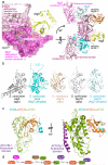
Selected structures of autophagy UBL ligation enzymes. (a) Structural insights into autophagy UBL activation by the E1, Atg7. Left, Close-up view showing one of two Atg8 molecules (yellow) in a complex with the homodimeric adenylation/catalytic cysteine domain of yeast Atg7 with the two subunits shown in magenta and pink,. The catalytic Cys and Asp residues are shown in sticks. The magnesium ion is shown as a gray sphere, with ATP in sticks. The Atg8 molecule bound to protomer 2 (pink) is not visible in this view. Right, Model of an (Atg7-Atg8-Atg3)2 intermediate, generated by superimposing the structure on the left and the structure of an Atg7-Atg3 complex. A key feature is that an autophagy UBL (Atg8 shown here) is transferred in trans from the catalytic Cys of one Atg7 protomer to an E2 (Atg3 shown here) recruited to the amino terminal domain of the opposite monomer of the Atg7 homodimer. (b) Conformational variability of the active site region of autophagy E2 enzymes. For Atg3 and Atg10 from S. cerevisiae, the active site and other structural regions are dramatically reoriented upon binding to Atg7, although the crystal structure of isolated Atg10 from K. marxianus appears closer to the active conformation,,-. (c) Crystal structure of human ATG3 (cyan)-ATG12 (orange)–ATG5 (green)-ATG16L1 N-terminal domain (purple)1. Note that the overall structures of the Atg12–Atg5-Atg16 subcomplexes are similar between human and S. cerevisiae,. The isopeptide linkage between the ATG12 C terminus and ATG5 is shown in sticks, and the ATG12 region implicated in binding to the E2, ATG3, is indicated. (d) Schematic illustration of Atg12–Atg5-Atg16 conjugate formation.

Similar articles

Cited by

References

    1. Yang Z, Klionsky DJ. Eaten alive: a history of macroautophagy. Nat Cell Biol. 2010;12:814–822. doi:10.1038/ncb0910-814. - PMC - PubMed
    1. Reggiori F, Klionsky DJ. Autophagic processes in yeast: mechanism, machinery and regulation. Genetics. 2013;194:341–361. doi:10.1534/genetics.112.149013. - PMC - PubMed
    1. Mijaljica D, et al. Receptor protein complexes are in control of autophagy. Autophagy. 2012;8:1701–1705. doi:10.4161/auto.21332. - PMC - PubMed
    1. Oku M, Sakai Y. Peroxisomes as dynamic organelles: autophagic degradation. Febs J. 2010;277:3289–3294. doi:10.1111/j.1742-4658.2010.07741.x. - PubMed
    1. Kanki T, Klionsky DJ, Okamoto K. Mitochondria autophagy in yeast. Antioxid Redox Signal. 2011;14:1989–2001. doi:10.1089/ars.2010.3762. - PMC - PubMed

Publication types

MeSH terms