. 2014 May 29;123(22):3512-23.
doi: 10.1182/blood-2013-12-545335. Epub 2014 Apr 2.

Siglec-G-CD24 axis controls the severity of graft-versus-host disease in mice

Affiliations

Siglec-G-CD24 axis controls the severity of graft-versus-host disease in mice

Tomomi Toubai et al. Blood. .

Abstract

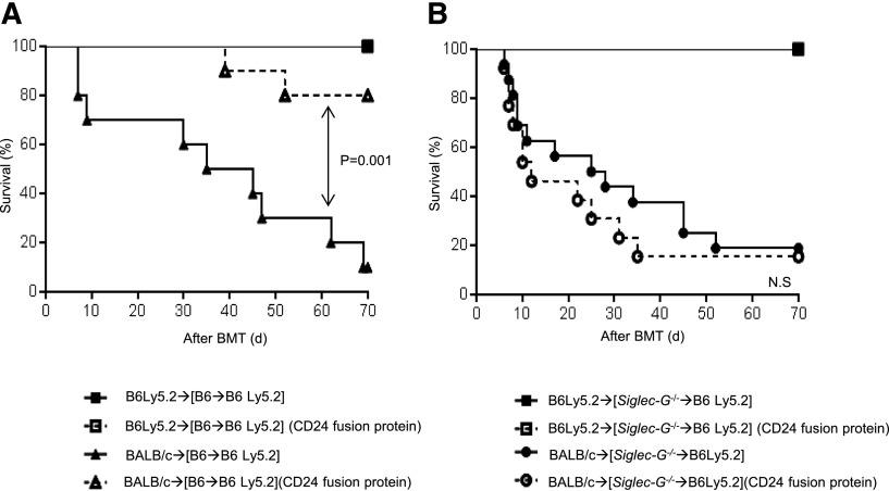
Activation of sialic-acid-binding immunoglobulin-like lectin-G (Siglec-G) by noninfectious damage-associated molecular patterns controls innate immune responses. However, whether it also regulates T-cell-mediated adaptive immune responses is not known. Graft-versus-host reaction is a robust adaptive immune response caused by allogeneic hematopoietic cell transplantation that have been activated by antigen-presenting cells (APCs) in the context of damaged host tissues following allogeneic hematopoietic cell transplantation. The role of infectious and noninfectious pattern recognition receptor-mediated activation in the induction and aggravation of graft-versus-host disease (GVHD) is being increasingly appreciated. But the role of pathways that control innate immune responses to noninfectious stimuli in modulating GVHD has heretofore not been recognized. We report that Siglec-G expression on host APCs, specifically on hematopoietic cells, negatively regulates GVHD in multiple clinically relevant murine models. Mechanistic studies with various relevant Siglec-G and CD24 knockout mice and chimeric animals, along with rescue experiments with novel CD24 fusion protein demonstrate that enhancing the interaction between Siglec-G on host APCs with CD24 on donor T cells attenuates GVHD. Taken together, our data demonstrate that Siglec-G-CD24 axis, controls the severity of GVHD and suggest that enhancing this interaction may represent a novel strategy for mitigating GVHD.


Figures

Figure 1
Figure 1
The exacerbation of GVHD in Siglec-G–deficient hosts is dependent on the intensity of conditioning in experimental MHC-mismatched allo-HCT. (A) Siglec-G expression on splenic DCs tested by FACS with antiSiglec-G antibody in naïve mouse and at 24 hours after TBI; the representative histogram (upper) and summarized data from 3 experiments (lower) (n = 2 to 4 per group). (B) The absence of Siglec-G decreased phosphorylated and total of SHP-1. Immunoblot analysis of phosphorylated(p)- and total SHP-1 protein in lysates of splenocytes from WT-B6 or Siglec-G−/− animals after irradiation. The bar graph shows the ratio pSHP-1/total SHP-1. (C-H) Siglec-G deficiency exacerbates GVHD. WT-B6 and Siglec-G−/− mice were lethally irradiated with 13 Gy and infused with 2 to 2.5 × 10⁶ CD90+ T cells along with 5 × 10⁶ TCD-BM cells from either syngeneic B6 or allogeneic MHC-mismatched BALB/c donors. (C) Survival following 13 Gy TBI (n = 6 to 11 per group, pooled from 2 experiments); P = .007 when B6 vs Siglec-G−/− B6 mice were used as recipients. (D) Histopathologic analysis of GI tracts (small and large intestine) by hematoxylin and eosin stain (left) and GVHD score on days 7 and 14 after allo-HCT (n = 3 to 6 per group, pooled from 2 experiments). Original magnification ×200. (E) Donor T-cell (H-2kd+CD4+CD8+) expansion in the spleen on days 7 and 14 after allo-HCT (n = 4 to 6 per group, pooled from 2 experiments). (F-H) Serum HMGB-1, IFN-γ, and TNF-α levels on day 7 after allo-HCT (n = 3 to 6 per group, pooled from 2 experiments). The bar shows the mean ± standard error of the mean (SEM).
Figure 2
Figure 2
The absence of Siglec-G expression on host APCs enhances GVHD. To make BM chimeras, WT-B6 Ly5.1, WT-B6 Ly5.2, and Siglec-G−/− animals were lethally irradiated with 11 Gy and infused with 5 × 10⁶ BM cells and 5 × 10⁶ splenocytes from syngeneic WT-B6 Ly5.2, WT-B6 Ly5.1, or Siglec-G−/− donors. Four months later, [B6 Ly5.2→B6 Ly5.1], [B6 Ly5.1→B6Ly5.2], [Siglec-G−/−→B6Ly5.2], or [B6 Ly5.2→Siglec-G−/−] animals were irradiated with 9 Gy and received 2.5 × 10⁶ CD90+ T cells along with 5 × 10⁶ TCD-BM cells from either syngeneic B6 or allogeneic MHC-mismatched BALB/c donors, or received 1.5 × 10⁶ CD90+ T cells along with 5 × 10⁶ TCD-BM cells from either syngeneic B6 or allogeneic MHC-matched, multiple MiHA-mismatched, C3H.sw donors. (A) Survival (left) and clinical GVHD score (right) (n = 6 to 18 per group, pooled from 2 experiments) in MHC-mismatched BALB/c→B6 model. (B) Survival (left) and clinical GVHD score (right) (n = 8 to 12 per group, pooled from 2 experiments) in MHC-matched, multiple MiHA-mismatched C3H.sw→B6 model. (C) Donor T-cell (H-2kd+CD4+CD8+) expansion in the spleen on day 7 after allo-HCT (n = 3 to 6 per group, pooled from 2 experiments) in MHC-mismatched BALB/c→B6 model. (D) Donor T-cell (CD229.1+CD4+CD8+) expansion in the spleen on day 14 after allo-HCT (n = 3 to 6 per group, pooled from 2 experiments). (E) Donor T-cell (H-2d+CD3+) expansion in the spleen and MLNs on day 1 after allo-HCT (n = 3 per group, pooled from 2 experiments). (F) Serum TNF-α levels on day 1 after allo-HCT (n = 3 to 6 per group, pooled from 2 experiments). (G) Donor T-cell (H-2d+CD3+) expansion in the spleen and intraepitherial lymphocytes on day 3 after allo-HCT (n = 3 per group, pooled from 2 experiments). (H) Donor IFN-γ+CD3+ T-cell expansion in the spleen on day 3 after allo-HCT (n = 3 per group, pooled from 2 experiments). (I) Serum IFN-γ levels on day 3 after allo-HCT (n = 3 to 6 per group, pooled from 2 experiments). (D-I) The bars show the mean ± SEM.
Figure 3
Figure 3
Siglec-G deficiency enhances DAMP-induced inflammatory responses from hematopoietic APCs. (A-B) Both B6 and Siglec-G−/− BMDCs, (C) peritoneal macrophages, and the GI tract DCs were harvested and stimulated with HMGB-1 (5 μg/ml) for 16 hours. The supernatants were analyzed for TNF-α (A,C-D), and IL-6 (B). (E) TLR-4 expression on BMDCs. The data are representative of 3 independent experiments and show the mean ± SEM.
Figure 4
Figure 4
Siglec-G–deficient DCs show enhanced stimulation of allogeneic T cells. BMDCs from WT-B6 and Siglec-G−/− mice were used as stimulators in an MLR with T cells from either BALB/c (allogeneic) or C57BL/6 (syngeneic) mice and analyzed for (A) T-cell proliferation based on 3H-thymidine incorporation at 96 hours. The data are the mean ± SEM of quadruplicate cultures and are representative of 3 independent experiments. (B) Supernatants from the cultures were collected at 80 hours and analyzed for IFN-γ by ELISA. The data are the mean ± SEM of quadruplicate cultures and are from 1 of 3 similar experiments. (C) Representative figures for CFSE, and (D) Annexin-V staining of cells gated on CD90.2+ T cells after the MLR. CFSE-labeled splenic CD90+ T cells from BALB/c mice were cultured for 96 hours with BMDCs from either WT-B6 or Siglec-G−/− mice. The cells were then harvested and analyzed. The right column shows the collective data of 3 independent experiments. N.S., no significant differences between B6 and Siglec-G−/− DCs.
Figure 5
Figure 5
Disruption of the CD24–Siglec-G interaction enhances allogeneic T-cell responses. BMDCs from WT-B6 and Siglec-G−/− mice were used as stimulators in an MLR with T cells from either CD24−/− BALB/c (allogeneic) or C57BL/6 (syngeneic) mice and analyzed for (A) T-cell proliferation based on 3H-thymidine incorporation at 96 hours. The data are the mean ± SEM of quadruplicate cultures and are from 1 of 3 similar experiments. (B-C) Supernatants from the cultures were collected at 80 hours and analyzed for IFN-γ (B) and IL-2 (C) by ELISA. The data are the mean ± SEM of quadruplicate cultures and are from 1 of 3 similar experiments. (D) Survival. WT-B6 mice were lethally irradiated with 13 Gy and infused with 2.5 × 10⁶ CD90+ T cells along with 5 × 10⁶ TCD-BM cells from either syngeneic B6 or allogeneic MHC-mismatched BALB/c or CD24−/− donors (n = 4 to 15 per group, pooled from 2 experiments). The bar shows the mean ± SEM. (E) Clinical GVHD score. For ▲ vs ●, **P < .01 and *P < .05. (F) Survival. WT-B6 mice were lethally irradiated with 13 Gy and infused with 2.5 × 10⁶ CD90+ T cells along with 5 × 10⁶ TCD-BM cells from either syngeneic B6 or allogeneic MHC-mismatched CD24−/− donors. The recipients were injected with the CD24 Fc protein on day −1 (5 mg/kg) or diluent control on day −1 before allo-HCT (n = 4 to 10 per group, pooled from 2 experiments). For ♦ vs ◊, P = .02. The bar shows the mean ± SEM.
Figure 6
Figure 6
Enhanced Siglec-G interaction with the CD24 fusion protein mitigates GVHD. (A) Survival. WT-B6 mice were lethally irradiated with 13 Gy and infused with 2.5 × 10⁶ CD90+ T cells along with 5 × 10⁶ TCD-BM cells from either syngeneic B6 or allogeneic MHC-mismatched BALB/c donors. The recipients were injected with the CD24 Fc protein on day −1 (5 mg/kg) or diluent control on day −1 before allo-HCT (n = 4 to 25 per group, pooled from 3 experiments). For ▲ vs △, P = .03. The bar shows the mean ± SEM. (B) Siglec-G−/− mice were lethally irradiated with 13 Gy and infused with 2.5 × 10⁶ CD90+ T cells along with 5 × 10⁶ TCD-BM cells from either syngeneic B6 or allogeneic MHC-mismatched BALB/c donors. The recipients were injected with the CD24 Fc protein on day −1 (5 mg/kg) or diluent control on day −1 before allo-HCT (n = 4 to 12 per group, pooled from 2 experiments). The bar shows the mean ± SEM.
Figure 7
Figure 7
Enhanced Siglec-G interaction with the CD24 fusion protein mitigates GVHD in WT but not Siglec-G−/− mice. (A-B) Survival. [WT-B6→B6Ly5.2] and [Siglec-G−/−→B6 Ly5.2] chimeras were lethally irradiated with 9 Gy and infused with 2.5 × 10⁶ CD90+ T cells along with 5 × 10⁶ TCD-BM cells from either syngeneic B6 or allogeneic MHC-mismatched BALB/c donors. The recipients were injected with the CD24 Fc protein on day −1 (5 mg/kg) or diluent control on day −1 before allo-HCT (n = 5 to 17 per group, pooled from 2 experiments). For ▲ vs △, P = .001. (B). The bar shows the mean ± SEM.

Comment in

  • Siglec-G signaling DAMPens GVHD.
    Zeiser R. Zeiser R. Blood. 2014 May 29;123(22):3376-7. doi: 10.1182/blood-2014-04-568832. Blood. 2014. PMID: 24876528 No abstract available.

Similar articles

Cited by

References

    1. Iwasaki A, Medzhitov R. Regulation of adaptive immunity by the innate immune system. Science. 2010;327(5963):291–295. - PMC - PubMed
    1. Crocker PR, Paulson JC, Varki A. Siglecs and their roles in the immune system. Nat Rev Immunol. 2007;7(4):255–266. - PubMed
    1. Ravetch JV, Lanier LL. Immune inhibitory receptors. Science. 2000;290(5489):84–89. - PubMed
    1. Chen GY, Tang J, Zheng P, Liu Y. CD24 and Siglec-10 selectively repress tissue damage-induced immune responses. Science. 2009;323(5922):1722–1725. - PMC - PubMed
    1. Chen GY, Chen X, King S, et al. Amelioration of sepsis by inhibiting sialidase-mediated disruption of the CD24-SiglecG interaction. Nat Biotechnol. 2011;29(5):428–435. - PMC - PubMed

Publication types

MeSH terms