. 2014 Apr 3:4:4576.
doi: 10.1038/srep04576.

ATP and potassium ions: a deadly combination for astrocytes

Affiliations

ATP and potassium ions: a deadly combination for astrocytes

David G Jackson et al. Sci Rep. .

Abstract

The ATP release channel Pannexin1 (Panx1) is self-regulated, i.e. the permeant ATP inhibits the channel from the extracellular space. The affinity of the ATP binding site is lower than that of the purinergic P2X7 receptor allowing a transient activation of Panx1 by ATP through P2X7R. Here we show that the inhibition of Panx1 by ATP is abrogated by increased extracellular potassium ion concentration ([K(+)]o) in a dose-dependent manner. Since increased [K(+)]o is also a stimulus for Panx1 channels, it can be expected that a combination of ATP and increased [K(+)]o would be deadly for cells. Indeed, astrocytes did not survive exposure to these combined stimuli. The death mechanism, although involving P2X7R, does not appear to strictly follow a pyroptotic pathway. Instead, caspase-3 was activated, a process inhibited by Panx1 inhibitors. These data suggest that Panx1 plays an early role in the cell death signaling pathway involving ATP and K(+) ions. Additionally, Panx1 may play a second role once cells are committed to apoptosis, since Panx1 is also a substrate of caspase-3.


Figures
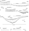
Figure 1
Figure 1. Extracellular K+ attenuates the inhibitory effect of BzATP on Panx1 channel currents in Xenopus oocytes.
(a) BzATP (30 µM) applied to oocytes expressing Panx1 and repeatedly pulsed from a holding potential of −60 mV to +60 mV to open the channels significantly attenuated the currents. Application of 40 mM [K+]o to the same cell held at −50 mV and pulsed to −40 mV induced an inward current, indicating opening of Panx1 channels. The currents were slightly affected by 30 µM BzATP but were diminished by 100 µM carbenoxolone (CBX). (b) At 300 µM, BzATP inhibited both voltage- and K+-induced currents. (c) With 75 mM [K+] as stimulus, neither 30 nor 300 µM BzATP had a discernible inhibitory effect. (d) In uninjected control oocytes, the pulse protocol induced small currents that were not inhibited by 300 µM BzATP. As reported earlier, increased extracellular K+ (40 mM) induced a small current of unknown origin in these cells. This K+-induced current was also not affected by BzATP (300 µM).
Figure 2
Figure 2. Quantitative analysis of inhibition of Panx1 currents by ATP or BzATP at different K+ concentrations.
(a) In normal Ringer solution (1 mM K+), 500 µM ATP inhibited voltage-induced currents. With the same ATP concentration, currents induced by K+ were minimally affected (10 mM [K+]o) or not at all (42.5 mM [K+]o). BzATP (300 µM) was effectively inhibiting at 40 mM [K+]o, but not at 75 mM. (b) Competition between BzATP and K+. At 40 mM [K+]o, increasing the BzATP concentration restored the inhibitory capacity of this compound. Increasing the [K+]o to 75 mM eliminated the inhibitory effect of even 300 µM BzATP.
Figure 3
Figure 3. Inhibition of K+-induced currents by different Panx1 inhibitors as a function of [K+]o.
With the exception of BB FCF and Fast green FCF, inhibition of Panx1 currents was independent of the [K+]o. Only at 75 mM [K+], a slight attenuation was observed. The inhibition by BB FCF and Fast Green FCF was attenuated at 40 mM and not detectable at 75 mM [K+].
Figure 4
Figure 4. Pannexin truncation and cell survival.
The truncation mutants Panx1 359stop and Panx1 408stop form channels similar to wtPanx1. (a) Oocytes expressing Panx1 408stop were voltage clamped, held at -50 mV and stepped to +50 mV at a rate of 0.1 Hz. The voltage steps induced large currents that were sensitive to the Panx1 inhibitor carbenoxolone (CBX). The truncation mutant Panx1 378stop forms a constitutively open channel. (b) Voltage ramps from −100 to +100 mV were applied to uninjected oocytes (green, control) and oocytes expressing wtPanx1 (blue) or Panx1 378stop (red). Recordings were taken 48 hours after mRNA injection of wt Panx1 or 9 hours after injection of Panx1 378stop. Survival of Panx1 378stop expressing cells was limited and thus did not allow for longer incubation times, hence some currents with smaller amplitude than wtPanx1. The rectification typical for wtPanx1 was absent for Panx1 378stop. Furthermore, the reversal potential was shifted to the left for the mutant. (c) The Panx1 inhibitors probenecid and carbenoxolone (CBX) reversibly inhibited currents through Panx1 378stop channels. (d) Oocytes expressing wtPanx1 were viable 48 hours after mRNA injection (left), while oocytes expressing Panx1 378stop had undergone cell death already 36 hours after mRNA injection despite of incubation in Ringer's solution supplemented with 100 µM CBX.
Figure 5
Figure 5. Panx1 mediates astrocyte cell death.
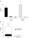
(A) Means ± s.e.m. values of fold changes in LDH released from WT (black), Panx1 KO (red) and P2X7R KO (green) astrocytes exposed for 1 hr to 50 mM K+-ACSF relative to control condition (2.5 mM K+ ACSF; white bar). Note that mefloquine (MFQ) at 100 nM blocked LDH released from WT cells. (B) LDH released from astrocytes exposed for 1 hr to 25 mM K+-ACSF is enhanced by 30 µM BzATP and blocked by 300 µM BzATP. ***P < 0.001 (ANOVA followed by Tukey multiple comparison test). In parentheses are the number of samples.
Figure 6
Figure 6. Panx1-dependent caspase-3 activation.
(A) Representative epifluorescence image showing cleaved caspase-3 (green) in nuclei (red) of WT astrocytes exposed to ACSF containing 2.5 and 50 mM [K+]. (B) Histograms of the mean ± s.e.m values of the fraction of WT (black bars), Panx1 KO (red bars) and P2X7 KO (green) astrocytes displaying cleaved caspase-3 following exposure to 10, 25 and 50 mM K+-ACSF. ***P < 0.001 (ANOVA followed by Tukey multiple comparison test. Minimum of 4 independent experiments). (C) Bar histograms of the mean ± s.e.m. values of caspase-3 positive cells (relative to control) exposed to elevated extracellular [K+] in the absence and presence of BzATP. (D) Effect of BBG (5 μM) and BB FCF (100 μM) on caspase-3 activation induced by 50mM K+-ACSF in WT (black symbols) and Panx1 KO (red symbols). Note that in Panx1 KO astrocytes high [K+] did not lead to caspase-3 activation. ***P<0.001 (ANOVA followed by Tukey' multiple comparison test). Mean+ s.e.m. are from 9–11 fields obtained from 3–4 different cultures.
Figure 7
Figure 7. AnnexinV staining in astrocytes.
(Top) Examples of epifluorescence images of cultured WT astrocytes treated for 1–2 hr with 2.5 and 10 mM K+- ACSF showing staining for Annexin V (green) and Dapi (red). (Bottom) Mean ± s.e.m. values of Annexin V fluorescence per total number of nuclei (left) and the mean ± s.e.m. fold changes in Annexin V (right) obtained for WT and Panx1 KO exposed to 2.5 mM K+-ACSF and to 10 mM K+-ACSF in the absence and presence of 300 µM BzATP. In parentheses are the number of images used for measurements that were obtained from 2 Panx1KO and 3 WT mice. Note that although not significant, there was a slight increase in Annexin V as measured in WT astrocytes exposed to 10 mM K+-ACSF. To avoid any bias, we measured total fluorescence and divided by cell number. This procedure results in an underestimation of K+-induced cell death because of a few dead cells in the control condition that were highly fluorescent.
Figure 8
Figure 8. In situ blockade of Panx1 channels by BzATP.
Means ± s.e.m. of the relative values (test/control) of YoPro fluorescence changes induced by 50 mM K+-ACSF in the absence and presence of 300 µM BzATP recorded from strata pyramidale (left) and radiatum (right) of hippocampi of wild-type (black symbols), Panx1 KO (red symbols), P2X7R KO (green symbols) and dKO (yellow symbols) mice. Data for each genotype were normalized to their corresponding values recorded at 2.5 mM K+-ACSF (open circles). ***P < 0.001, **P < 0.01 (ANOVA followed by Dunnet's test). Each symbol correspond to values obtained from a single hippocampal slice from a minimum of three mice per genotype.
Figure 9
Figure 9. Early signaling events in cell undergoing secondary cell death.
In the central nervous system, damaged cells (top) release ATP, glutamate and K+ into a narrow extracellular space and thus can reach concentrations approximating those in the cytoplasm. Extracellular ATP binds to purinergic receptors, including P2X7R, activating it and also activating the Panx1 channel through intracellular signaling cascade. ATP efflux through the Panx1 channel then provides a positive feedback for P2X7R activation. Under normal circumstances this positive feedback would be interrupted by the inhibition of the Panx1 channel by extracellular ATP. Increased [K+]o renders this negative feedback loop ineffective as shown in the present study. In addition, [K+]o stimulates Panx1 directly. P2X7R and/or Panx1 signal to the inflammasome and thereby activate caspase-1. In addition, caspase-3 gets activated through an unknown pathway (present study). Panx1 is a substrate of caspase-3 and its cleavage results irreversibly in constitutive channel activity. Cell death thus is a combination of apoptotic events combined with the rundown of all membrane gradients for small molecules due to the permanently active Panx1 channel.

Similar articles

Cited by

References

    1. Zheng L. M., Zychlinsky A., Liu C. C., Ojcius D. M. & Young J. D. Extracellular ATP as a trigger for apoptosis or programmed cell death. J Cell Biol 112, 279–288 (1991). - PMC - PubMed
    1. Di Virgilio F., Pizzo P., Zanovello P., Bronte V. & Collavo D. Extracellular ATP as a possible mediator of cell-mediated cytotoxicity. Immunol Today 11, 274–277 (1990). - PubMed
    1. Ferrari D. et al. ATP-mediated cytotoxicity in microglial cells. Neuropharmacology 36, 1295–1301 (1997). - PubMed
    1. Burnstock G. Purinergic signalling and disorders of the central nervous system. Nat Rev Drug Discov 7, 575–590 (2008). - PubMed
    1. Qu Y., Franchi L., Nunez G. & Dubyak G. R. Nonclassical IL-1 beta secretion stimulated by P2X7 receptors is dependent on inflammasome activation and correlated with exosome release in murine macrophages. J Immunol 179, 1913–1925 (2007). - PubMed

Publication types

MeSH terms