. 2014 May;62(5):790-803.
doi: 10.1002/glia.22642.

Age-related changes in astrocytic and ependymal cells of the subventricular zone

Age-related changes in astrocytic and ependymal cells of the subventricular zone

Vivian Capilla-Gonzalez et al. Glia. 2014 May.

Abstract

Neurogenesis persists in the adult subventricular zone (SVZ) of the mammalian brain. During aging, the SVZ neurogenic capacity undergoes a progressive decline, which is attributed to a decrease in the population of neural stem cells (NSCs). However, the behavior of the NSCs that remain in the aged brain is not fully understood. Here we performed a comparative ultrastructural study of the SVZ niche of 2-month-old and 24-month-old male C57BL/6 mice, focusing on the NSC population. Using thymidine-labeling, we showed that residual NSCs in the aged SVZ divide less frequently than those in young mice. We also provided evidence that ependymal cells are not newly generated during senescence, as others studies suggest. Remarkably, both astrocytes and ependymal cells accumulated a high number of intermediate filaments and dense bodies during aging, resembling reactive cells. A better understanding of the changes occurring in the neurogenic niche during aging will allow us to develop new strategies for fighting neurological disorders linked to senescence.


Figures
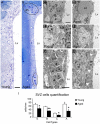
FIGURE 1
FIGURE 1
Aging reduces the main cellular components of the SVZ niche. Light and transmission electron microscopy analysis of the SVZ. (A) Typical organization of the lateral wall in young mice with SVZ cells homogeneously distributed. (B) The aged SVZ showed a loss of cells. Remaining cells appeared forming groups (black circles) and were mainly located in the dorsal horn. (C) The young SVZ showed small lipid droplets in the ependymal layer. (D) Detail of a large lipid droplet in the ependymal layer of the aged SVZ. (E) The young mice typically presented neurons in the neuropil next to the SVZ. (F) Detail of a neuron close to the ventricle wall in the aged SVZ. (G) Typical neurogenic niche showing the SVZ cells next to the ependymal layer in a young mouse. Note the presence of myelin axons (white arrowhead) in the neuropil. (H) Lateral ventricle wall of an aged mouse displaying a depletion of SVZ cells. Note the presence of myelin axons (white arrowheads) next to the ependymal layer. (I) Cell quantification using electron microscopy showed a number reduction for type E, B, A, and C cells. a, neuroblast; b, astrocyte; c, type C cell; dh, dorsal horn; e, ependymal cell; Lp, lipid droplet; Lv, lateral ventricle; n, neuron. Scale bar: A–B 20 μm, C–F 2 μm, G–H 20 μm. *P<0.05, **P<0.01. [Color figure can be viewed in the online issue, which is available at wileyonlinelibrary.com.]
FIGURE 2
FIGURE 2
The molecular markers characteristic of the main SVZ cell types are altered during aging. Immunoassay against Nestin (white), GFAP (green), DCX (red), and S100 (orange) in the dorsal horn of the lateral ventricles. (A) Nestin expression was reduced in the aged SVZ. (B) GFAP expression was reduced in aged mice, but remaining GFAP+ cells were more intensely labeled, compared to the young brain. (C) DCX expression was diminished during aging. (D) Cells expressing S100 marker showed flattened morphology in the aged SVZ, compared to the young SVZ. Some S100+ cells located in the ependymal layer of aged mice co-expressed the GFAP marker (inset in D). Lv, lateral ventricle. Scale bar 20 μm. [Color figure can be viewed in the online issue, which is available at wileyonlinelibrary.com.]
FIGURE 3
FIGURE 3
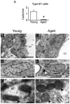
Aging alters the ultrastructure of the SVZ astrocytes. Electron microscopy analysis of the astrocytes within the SVZ. (A) Cell quantification of B1 astrocytes revealed a reduction of this subpopulation of cells in the aged SVZ. (B) Both B1 astrocytes (NSCs) and B2 astrocytes (non-neurogenic astrocytes) were frequently found in the SVZ of the young brain. (C) Astrocytes in the aged SVZ were mainly close to the underlying striatal parenchyma, corresponding to B2 astrocytes. (D) Astrocyte with light cytoplasm in the SVZ of young mice showed perinuclear mitochondria. (E) Astrocyte in the aged SVZ presented mitochondria distributed in a dispersed manner and dense bodies (white arrowhead) in the cytosol. (F) Detail of mitochondria (black arrowheads) in the cytoplasm of an astrocyte in a young mouse, with increased number of cristae and dense matrix. (G) Detail of mitochondria (black arrowheads) in the cytosol of an astrocyte in the aged SVZ with less developed cristae. Note the presence of abundant intermediate filaments (arrows). b, astrocyte; b1, B1 astrocyte; b2, B2 astrocyte; e, ependymal cell; Lv, lateral ventricle. Scale bar: B–C 5 μm, D–E 1 μm, F–G 500 nm. *P<0.01.
FIGURE 4
FIGURE 4
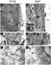
Aging alters the ultrastructure of the ependymal cells. Electron microscopy analysis of ependymal cells in the ventricle wall. (A) Typical ependymal cell in the young SVZ with cubical morphology and multiple cilia in the apical surface. (B) Elongated ependymal cell in the aged SVZ with dense bodies (box) in the cytosol. (C) Detail of cell junctions (black arrowhead) between two ependymal cells (asterisks), which constitute the monolayer. (D) Thin multilayer constituted by 4 different ependymal processes (asterisks), which were connected by cell junctions (black arrowheads). (E) Detail of an ependymal cytosol in the young mouse showing intermediate filaments (arrows). (F) Detail of an ependymal cytoplasm in the aged SVZ showing an increase in intermediate filaments (arrows). e, ependymal cell; Lv, lateral ventricle. Scale bar: A–B 1 μm, C–D 500 nm, E–F 200 nm.
FIGURE 5
FIGURE 5
The ventricular wall presents cilia-devoid patches during aging. (A–E) Scanning electron microscopy images. (A) In young mice, the ventricular wall was completely covered by cilia of ependymal cells. (B) Cilia-devoid patches were abundant in the aged brain. (C) Detail of tangled cilia in an ependymal cell of aged mice. (D) Ventricular surface in the aged brain showed an extensive network of structures resembling axons (white arrows). (E) Detail of structure-like axons observed over the lateral wall of the aged brain. (F) Detail of structure-like axons observed over the lateral wall of the aged brain using transmission electron microscopy. Scale bar A–B 10μm, C, E, F 1 μm, D 5 μm.
Figure 6
Figure 6
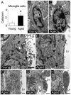
The cytoarchitecture of the SVZ microenvironment changes during aging. (A) Cell quantification of microglia cells in the SVZ. (B) Microglia cell in the young SVZ showing few dense bodies (arrow) in the cytoplasm. (C) Microglia cell in the aged SVZ showing round shape and abundant dense bodies (arrow) in the cytoplasm. (D) Small fractons (arrowhead) located between ependymal cells in the young SVZ. (E) Large penetrative fractons (arrowheads) located between ependymal cells in the aged SVZ. (F) Neuron-like cytosol with a high number of mitochondria, vesicles, and microtubules within the aged SVZ. (G) Neuron-like cytosol through the ependymal layer in the aged SVZ. (H) Neuron-like cytosol on the ventricle wall of the aged brain. e; ependymal cell, Lv; lateral ventricle, Mg; microglia cell. Scale bar: B,C 1 μm, D,E 10 μm, F, H 1μm, G 500 nm. *P<0.05.
Figure 7
Figure 7
Aged astrocytes preserve their proliferative capacity but divide less frequently. (A) Timeline for the BrdU administration protocol. Animals received a single dose of BrdU 2 h before euthanize. (B) Bar graph depicting a significant reduction of BrdU+ cells in the aged SVZ. (C,D) Immunoassay against BrdU in coronal brain sections. (C) Young mice presented a high number of BrdU+ cells in the dorsal horn of the SVZ. (D) Aged mice presented a drastic reduction in proliferating cells. (E–G) Immunoassay against GFAP (green), S100 (red), and BrdU (white) in coronal brain sections. BrdU+ cells coexpressed GFAP, but not S100 in both young (E) and aged (F) mice. (G) Detail of a BrdU/GFAP+ cells close to ependymal layer in aged mice. (H) Timeline for the 3H-Thy administration protocol. Animals received 10 doses of 3H-Thy (1 dose per day) 6 weeks before euthanized. (I) Bar graph depicting a trend to decrease the number of 3H-Thy+ cells in the aged SVZ. (J–N) Ultrastructural analysis of SVZ cells incorporating 3H-Thy. (J) Labeled astrocytes (arrowhead) were identified by the presence of intermediate filaments (arrows) in the cytosol. (K,L) The young SVZ showed astrocytes with low intensity of the 3H-Thy marker. (M,N) In the aged brain, astrocytes displayed an intense 3H-Thy labeling. Lv, lateral ventricle; sac, sacrifice. Scale bar A,B 50 μm, C,D 20 μm, J–N 5 μm, detail in J 500 nm. *P<0.01. [Color figure can be viewed in the online issue, which is available at wileyonlinelibrary.com.]

Similar articles

Cited by

References

    1. Achanta P, Capilla-Gonzalez V, Purger D, Reyes J, Sailor K, Song H, Garcia-Verdugo JM, Gonzalez-Perez O, Ford E, Quinones-Hinojosa A. Subventricular zone localized irradiation affects the generation of proliferating neural precursor cells and the migration of neuroblasts. Stem Cells. 2012;30:2548–2560. - PMC - PubMed
    1. Ahlenius H, Visan V, Kokaia M, Lindvall O, Kokaia Z. Neural stem and progenitor cells retain their potential for proliferation and differentiation into functional neurons despite lower number in aged brain. J Neurosci. 2009;29:4408–4419. - PMC - PubMed
    1. Alfaro-Cervello C, Soriano-Navarro M, Mirzadeh Z, Alvarez-Buylla A, Garcia-Verdugo JM. Biciliated ependymal cell proliferation contributes to spinal cord growth. J Comp Neurol. 2012;520:3528–3552. - PMC - PubMed
    1. Alvarez-Buylla A, Garcia-Verdugo JM. Neurogenesis in adult subventricular zone. J Neurosci. 2002;22:629–634. - PMC - PubMed
    1. Batiz LF, Jimenez AJ, Guerra M, Rodriguez-Perez LM, Toledo CD, Vio K, Paez P, Perez-Figares JM, Rodriguez EM. New ependymal cells are born postnatally in two discrete regions of the mouse brain and support ven tricular enlargement in hydrocephalus. Acta Neuropathol. 2011;121:721–735. - PubMed

Publication types