

Alveolar macrophage depletion increases the severity of acute inflammation following nonlethal unilateral lung contusion in mice
- PMID: 24662861
- PMCID: PMC4543300
- DOI: 10.1097/TA.0000000000000163
Alveolar macrophage depletion increases the severity of acute inflammation following nonlethal unilateral lung contusion in mice
Abstract
Background: Lung contusion (LC) is a common injury resulting from blunt thoracic trauma. LC is an important risk factor for the development acute lung injury, adult respiratory distress syndrome, and ventilator-associated pneumonia, all of which increase mortality from trauma. LC produces a nonspecific immune cellular response. Neutrophil recruitment is known to increase the severity of inflammation during LC. However, the exact role of macrophages in modulating the response to LC has not been well described.
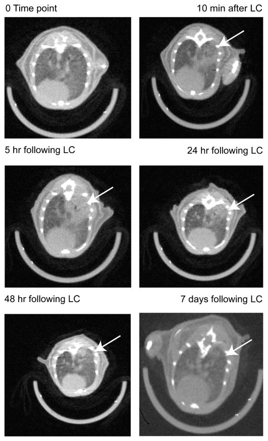
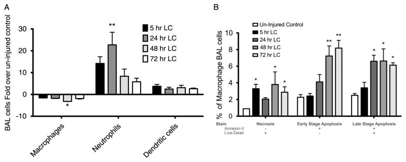
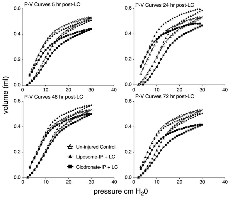
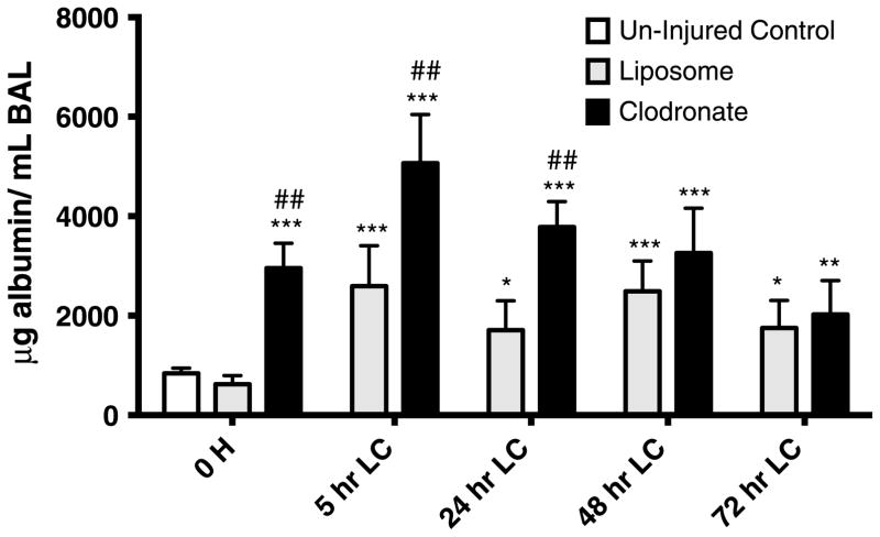
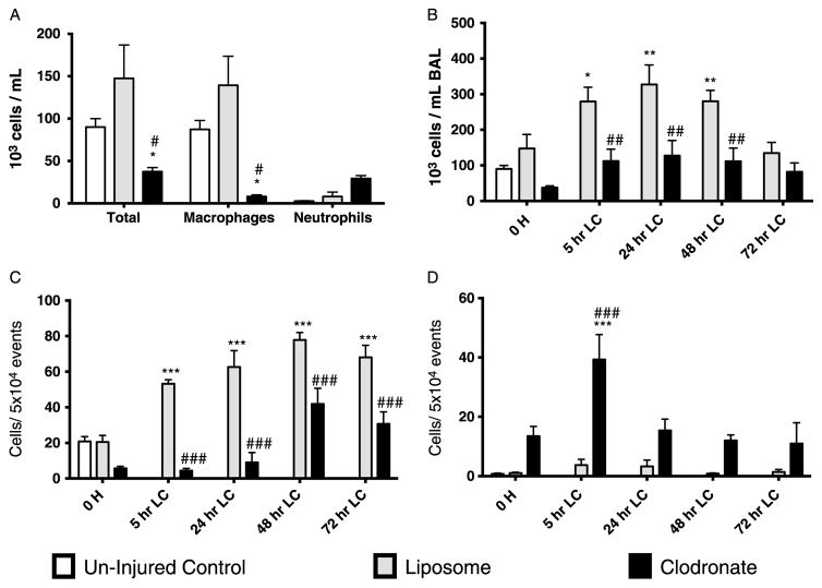
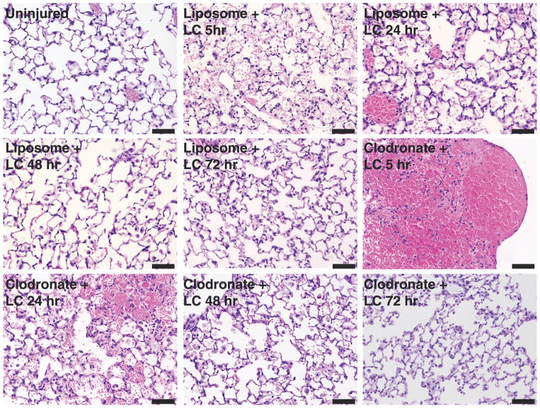
Methods: We used a cortical contusion impactor to induce unilateral LC in mice. Thoracic micro computed tomographic scans of these animals were obtained to document radiologic changes over time following LC. To understand the role of macrophages during LC, liposomal clodronate was used to deplete macrophage levels before traumatic insult. Acute inflammatory attributes after LC were assessed, by measuring pressure-volume mechanics; quantifying bronchial alveolar lavage levels of leukocytes, albumin, and cytokines; and finally examining lung specimen histopathology at 5, 24, 48, and 72 hours after injury.
Results: After LC, alveolar macrophage numbers were significantly reduced and exhibited slowed recovery. Simultaneously, there was a significant increase in bronchial alveolar lavage neutrophil counts. The loss of macrophages could be attributed to both cellular apoptosis and necrosis. Pretreatment with clodronate increased the severity of lung inflammation as measured by worsened pulmonary compliance, increased lung permeability, amplification of neutrophil recruitment, and increases in early proinflammatory cytokine levels.
Conclusion: The presence of regulatory alveolar macrophages plays an important role in the pathogenesis of acute inflammation following LC.
Figures
Similar articles
-
Toll-Like Receptor-9 (TLR9) is Requisite for Acute Inflammatory Response and Injury Following Lung Contusion.Shock. 2016 Oct;46(4):412-9. doi: 10.1097/SHK.0000000000000601. Shock. 2016. PMID: 26939039 Free PMC article.
-
Interleukin 10 overexpression alters survival in the setting of gram-negative pneumonia following lung contusion.Shock. 2014 Apr;41(4):301-10. doi: 10.1097/SHK.0000000000000123. Shock. 2014. PMID: 24430542 Free PMC article.
-
Contributing factors in the development of acute lung injury in a murine double hit model.Eur J Trauma Emerg Surg. 2020 Feb;46(1):21-30. doi: 10.1007/s00068-019-01121-5. Epub 2019 Apr 1. Eur J Trauma Emerg Surg. 2020. PMID: 30937460
-
Lung contusion: inflammatory mechanisms and interaction with other injuries.Shock. 2009 Aug;32(2):122-30. doi: 10.1097/SHK.0b013e31819c385c. Shock. 2009. PMID: 19174738 Free PMC article. Review.
-
Role of alveolar macrophages in the inflammatory response after trauma.Shock. 2014 Jul;42(1):3-10. doi: 10.1097/SHK.0000000000000167. Shock. 2014. PMID: 24667621 Review.
Cited by
-
The Role of Macrophages in the Pathogenesis of ALI/ARDS.Mediators Inflamm. 2018 May 13;2018:1264913. doi: 10.1155/2018/1264913. eCollection 2018. Mediators Inflamm. 2018. PMID: 29950923 Free PMC article. Review.
-
Macrophage and dendritic cell subset composition can distinguish endotypes in adjuvant-induced asthma mouse models.PLoS One. 2021 Jun 1;16(6):e0250533. doi: 10.1371/journal.pone.0250533. eCollection 2021. PLoS One. 2021. PMID: 34061861 Free PMC article.
-
Control of lung defence by mucins and macrophages: ancient defence mechanisms with modern functions.Eur Respir J. 2016 Oct;48(4):1201-1214. doi: 10.1183/13993003.00120-2015. Epub 2016 Sep 1. Eur Respir J. 2016. PMID: 27587549 Free PMC article. Review.
-
Pneumonia in severely injured patients with thoracic trauma: results of a retrospective observational multi-centre study.Scand J Trauma Resusc Emerg Med. 2019 Mar 14;27(1):31. doi: 10.1186/s13049-019-0608-4. Scand J Trauma Resusc Emerg Med. 2019. PMID: 30871601 Free PMC article.
-
Alpha-defensins inhibit ERK/STAT3 signaling during monocyte-macrophage differentiation and impede macrophage function.Respir Res. 2023 Dec 11;24(1):309. doi: 10.1186/s12931-023-02605-0. Respir Res. 2023. PMID: 38082274 Free PMC article.
References
-
- Miller PR, Croce MA, Kilgo PD, Scott J, Fabian TC. Acute respiratory distress syndrome in blunt trauma: identification of independent risk factors. Am Surg. 2002;68:845–850. discussion 850–851. - PubMed
-
- Cohn SM. Pulmonary contusion: review of the clinical entity. J Trauma. 1997;42:973–979. - PubMed
-
- Kollmorgen DR, Murray KA, Sullivan JJ, Mone MC, Barton RG. Predictors of mortality in pulmonary contusion. Am J Surg. 1994;168:659–663. discussion 663–664. - PubMed
Publication types
MeSH terms
Grants and funding
LinkOut - more resources
Full Text Sources
Other Literature Sources
Medical
