. 2014 Feb 28;9(2):e90669.
doi: 10.1371/journal.pone.0090669. eCollection 2014.

Identification of binding sites in Huntingtin for the Huntingtin Interacting Proteins HIP14 and HIP14L

Affiliations

Identification of binding sites in Huntingtin for the Huntingtin Interacting Proteins HIP14 and HIP14L

Shaun S Sanders et al. PLoS One. .

Abstract

Huntington disease is an adult onset neurodegenerative disease characterized by motor, cognitive, and psychiatric dysfunction, caused by a CAG expansion in the HTT gene. Huntingtin Interacting Protein 14 (HIP14) and Huntingtin Interacting Protein 14-like (HIP14L) are palmitoyl acyltransferases (PATs), enzymes that mediate the post-translational addition of long chain fatty acids to proteins in a process called palmitoylation. HIP14 and HIP14L interact with and palmitoylate HTT and are unique among PATs as they are the only two that have an ankyrin repeat domain, which mediates the interaction between HIP14 and HTT. These enzymes show reduced interaction with and palmitoylation of mutant HTT, leading to increased mutant HTT inclusion formation and toxicity. The interaction between HIP14 and HTT goes beyond that of only an enzyme-substrate interaction as HTT is essential for the full enzymatic activity of HIP14. It is important to further understand and characterize the interactions of HTT with HIP14 and HIP14L to guide future efforts to target and enhance this interaction and increase enzyme activity to remediate palmitoylation of HTT and their substrates, as well as to understand the relationship between the three proteins. HIP14 and HIP14L have been previously shown to interact with HTT amino acids 1-548. Here the interaction of HIP14 and HIP14L with N- and C-terminal HTT 1-548 deletion mutations was assessed. We show that HTT amino acids 1-548 were sufficient for full interaction of HTT with HIP14 and HIP14L, but partial interaction was also possible with HTT 1-427 and HTT 224-548. To further characterize the binding domain we assessed the interaction of HIP14-GFP and HIP14L-GFP with 15Q HTT 1-548Δ257-315. Both enzymes showed reduced but not abolished interaction with 15Q HTT 1-548Δ257-315. This suggests that two potential binding domains exist, one around residues 224 and the other around 427, for the PAT enzymes HIP14 and HIP14L.


Conflict of interest statement

Competing Interests: The authors have declared that no competing interests exist.

Figures

Figure 1
Figure 1. Overview schematics of the domain organization of HTT (A), HIP14 (B), and HIP14L (C).
The domain organization of HTT is shown in (A) with the poly-glutamine domains of WT (15Q) and mutant (128Q) HTT (NP_002102) are shown in grey and black rectangles, respectively, the proline rich repeat is shown in a hatched rectangle, and the H1 alpha-rod domain is shown in a dotted rectangle with the amino acids indicated above. (B) The domain organization of HIP14 (NP_056151) is shown in (B) and of HIP14L (NP_061901) in (C) with the 7 ankyrin repeats making up the ankyrin repeat domain shown in numbered solid grey rectangles, the transmembrane domains shown in hatched rectangles labeled TM1-6, and the DHHC cysteine-rich domain shown in solid black rectangles labeled DHHC. The amino acids corresponding to the appropriate domains are indicated below.
Figure 2
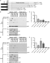
Figure 2. HIP14 and HIP14L interaction with C-terminal deletion mutants of HTT 1-548.
(A) A schematic diagram of the HTT 1–548 C-terminal deletion mutants used in co-immunoprecipitation experiments with HIP14-GFP and HIP14L-GFP showing the 15Q or 128Q poly-Q domains, the proline rich region (PRR), and the H1 alpha-rod domain. (B) A representative image (top two panels) of the co-immunoprecipitation between these C-terminal deletion mutants and HIP14-GFP where GFP was immunoprecipitated and the resulting blots were probed for HTT (top panel) and GFP (bottom panel) showing less 15Q and 128Q HTT 1–427 co-immunoprecipitated with HIP14-GFP. On the right is a beads alone (no antibody) control showing no non-specific binding of the proteins to the beads. The bottom two images show the expression of the HTT deletion mutants (top panel) and of HIP14-GFP (bottom panel). (C) Quantification of three independent co-immunoprecipitation experiments where the % HTT interaction with HIP14 is the indicated HTT band intensity as a percentage of the HIP14-GFP band intensity from the same sample, normalized to 15Q HTT 1–548. (D) A representative image (top two panels) of the co-immunoprecipitation between the HTT 1–548 C-terminal deletion mutants and HIP14L-GFP where GFP was immunoprecipitated and the resulting blots were probed for HTT (top panel) and GFP (bottom panel). Less 15Q and 128Q HTT 1–427 co-immunoprecipitated with HIP14L-GFP. On the right is a beads alone (no antibody) control showing no non-specific binding of the proteins to the beads. The bottom two panels show the expression of the HTT deletion mutants (top panel) and of HIP14L-GFP (bottom panel). (E) Quantification of three independent co-immunoprecipitation experiments where the % HTT interaction with HIP14L-GFP is the indicated HTT band intensity as a percentage of the HIP14L-GFP band intensity from the same sample, normalized to 15Q HTT 1–548.
Figure 3
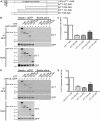
Figure 3. HIP14 and HIP14L interaction with N-terminal deletion mutants of HTT 1–548.
(A) A diagram of the HTT 1–548 N-terminal deletion mutants used in co-immunoprecipitation experiments with HIP14-GFP and HIP14L-GFP showing the 15Q poly-Q domains, the proline rich region (PRR), and the H1 alpha-rod domain. (B) A representative image (top two panels) of the co-immunoprecipitation between these N-terminal deletion mutants and HIP14-GFP where GFP was immunoprecipitated and the resulting blots were probed for HTT (top panel) and GFP (bottom panel) showing less HTT 88–548, HTT 151–548, and HTT 224–548 co-immunoprecipitated with HIP14-GFP and no HTT 427–548 was co-immunoprecipitated with HIP14. On the right is a beads alone control showing no non-specific binding of the proteins to the beads. The bottom two images show the expression of the HTT deletion mutants (top panel) and of HIP14-GFP (bottom panel). (C) Quantification of three co-immunoprecipitation experiments where the % HTT interaction with HIP14 is the indicated HTT band intensity as a percentage of the HIP14-GFP band intensity from the same sample, normalized to 15Q HTT 1–548. (D) A representative image (top two panels) of the co-immunoprecipitation between the HTT 1–548 N-terminal deletion mutants and HIP14L-GFP where GFP was immunoprecipitated and the resulting blots were probed for HTT (top panel) and GFP (bottom panel). Less HTT 88–548, HTT 151–548, and HTT 224–548 and no HTT 427–548 was co-immunoprecipitated with HIP14L-GFP. On the right is a beads alone control showing no non-specific binding of the proteins to the beads. The bottom two panels show the expression of the HTT deletion mutants (top panel) and of HIP14L-GFP (bottom panel). (E) Quantification of three co-immunoprecipitation experiments where the % HTT interaction with HIP14L-GFP is the indicated HTT band intensity as a percentage of the HIP14L-GFP band intensity from the same sample, normalized to 15Q HTT 1–548.
Figure 4
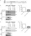
Figure 4. HIP14 and HIP14L interaction with 15Q HTT 1-548Δ257-315.
(A) A diagram of the 15Q HTT 1-548Δ257-315 deletion mutant used in co-immunoprecipitation experiments with HIP14-GFP and HIP14L-GFP showing the 15Q poly-Q domains, the proline rich region (PRR), and the H1 alpha-rod domain. (B) A representative image (top two panels) of the co-immunoprecipitation between 15Q HTT 1-548Δ257-315 deletion mutant and HIP14-GFP where GFP was immunoprecipitated and the resulting blots were probed for HTT (top panel) and GFP (bottom panel) showing less of the 15Q HTT 1-548Δ257-315 deletion mutant co-immunoprecipitated with HIP14-GFP and compared to 15Q HTT 1–548. On the right is a beads alone (no antibody) control showing no non-specific binding of the proteins to the beads. The bottom two images show the expression of the 15Q HTT 1-548Δ257-315 deletion mutant (top panel) and of HIP14-GFP (bottom panel). (C) Quantification of three independent co-immunoprecipitation experiments where the % HTT interaction with HIP14 is the indicated HTT band intensity as a percentage of the HIP14-GFP band intensity from the same sample, normalized to 15Q HTT 1–548. (D) A representative image (top two panels) of the co-immunoprecipitation between the 15Q HTT 1-548Δ257-315 deletion mutant and HIP14L-GFP where GFP was immunoprecipitated and the resulting blots were probed for HTT (top panel) and GFP (bottom panel). Less 15Q HTT 1-548Δ257-315 deletion mutant was co-immunoprecipitated with HIP14L-GFP. On the right is a beads alone (no antibody) control showing no non-specific binding of the proteins to the beads. The bottom two panels show the expression of the 15Q HTT 1-548Δ257-315 deletion mutant (top panel) and of HIP14L-GFP (bottom panel). (E) Quantification of three independent co-immunoprecipitation experiments where the % HTT interaction with HIP14L-GFP is the indicated HTT band intensity as a percentage of the HIP14L-GFP band intensity from the same sample, normalized to 15Q HTT 1–548.
Figure 5
Figure 5. A schematic diagram of the two hypothetical binding scenarios of HTT with HIP14 or HIP14L.
In both (A) and (B) for HIP14 or HIP14L the numbered, solid grey boxes are the seven ankyrin repeats that make up the ankyrin repeat domain, the six TMDs are in hatched boxes labeled TM1–TM6, and the DHHC-CR domain is a black box labeled DHHC. (A) In this first scenario, the HIP14 and HIP14L HTT binding site (solid pink box) is between amino acids 224–427 and this binding site interacts with the ankyrin repeat domain of HIP14 or HIP14L. (B) In an alternate scenario there are two binding sites (solid pink boxes), one between amino acids 1–427 and the other between amino acids 224–548, that both interact with the ankyrin repeat domain.

Similar articles

Cited by

References

    1. Roos RA (2010) Huntington's disease: a clinical review. Orphanet Journal of Rare Diseases 5: 40 10.1186/1750-1172-5-40 - DOI - PMC - PubMed
    1. Sturrock A, Leavitt BR (2010) The Clinical and Genetic Features of Huntington Disease. Journal of Geriatric Psychiatry and Neurology 23: 243–259 10.1177/0891988710383573 - DOI - PubMed
    1. The Huntington's disease collaborative research group (1993) A novel gene containing a trinucleotide repeat that is expanded and unstable on Huntington's disease chromosomes. Cell 72: 971–983. - PubMed
    1. Kalchman MA, Graham RK, Xia G, Koide HB, Hodgson JG, et al. (1996) Huntingtin is ubiquitinated and interacts with a specific ubiquitin-conjugating enzyme. J Biol Chem 271: 19385–19394. - PubMed
    1. Singaraja RR, Hadano S, Metzler M, Givan S, Wellington CL, et al. (2002) HIP14, a novel ankyrin domain-containing protein, links huntingtin to intracellular trafficking and endocytosis. Human Molecular Genetics 11: 2815–2828. - PubMed

Publication types

LinkOut - more resources