Inorganic nanovehicle targets tumor in an orthotopic breast cancer model
- PMID: 24651154
- PMCID: PMC3961742
- DOI: 10.1038/srep04430
Inorganic nanovehicle targets tumor in an orthotopic breast cancer model
Abstract
The clinical efficacy of conventional chemotherapeutic agent, methotrexate (MTX), can be limited by its very short plasma half-life, the drug resistance, and the high dosage required for cancer cell suppression. In this study, a new drug delivery system is proposed to overcome such limitations. To realize such a system, MTX was intercalated into layered double hydroxides (LDHs), inorganic drug delivery vehicle, through a co-precipitation route to produce a MTX-LDH nanohybrid with an average particle size of approximately 130 nm. Biodistribution studies in mice bearing orthotopic human breast tumors revealed that the tumor-to-liver ratio of MTX in the MTX-LDH-treated-group was 6-fold higher than that of MTX-treated-one after drug treatment for 2 hr. Moreover, MTX-LDH exhibited superior targeting effect resulting in high antitumor efficacy inducing a 74.3% reduction in tumor volume compared to MTX alone, and as a consequence, significant survival benefits. Annexin-V and propidium iodine dual staining and TUNEL analysis showed that MTX-LDH induced a greater degree of apoptosis than free MTX. Taken together, our data demonstrate that a new MTX-LDH nanohybrid exhibits a superior efficacy profile and improved distribution compared to MTX alone and has the potential to enhance therapeutic efficacy via inhibition of tumor proliferation and induction of apoptosis.
Figures


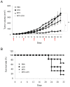
 ), LDH (
 ), MTX (
 ), and MTX-LDH (X) were administered via intraperitoneal injection on days 0, 7, 14, 21, and 28 (indicated by arrows), and tumor volume was measured using calipers every 2 days. (B) Survival rate of tumor-bearing mice treated as in (A). (*p < 0.05, **p < 0.01).

Similar articles
-
Anionic clay as the drug delivery vehicle: tumor targeting function of layered double hydroxide-methotrexate nanohybrid in C33A orthotopic cervical cancer model.Int J Nanomedicine. 2016 Jan 20;11:337-48. doi: 10.2147/IJN.S95611. eCollection 2016. Int J Nanomedicine. 2016. PMID: 26855572 Free PMC article.
-
Integration of phospholipid-hyaluronic acid-methotrexate nanocarrier assembly and amphiphilic drug-drug conjugate for synergistic targeted delivery and combinational tumor therapy.Biomater Sci. 2018 Jun 25;6(7):1818-1833. doi: 10.1039/c8bm00009c. Biomater Sci. 2018. PMID: 29785434
-
Anticancer drug-inorganic nanohybrid and its cellular interaction.J Nanosci Nanotechnol. 2007 Nov;7(11):3700-5. doi: 10.1166/jnn.2007.061. J Nanosci Nanotechnol. 2007. PMID: 18047040
-
In vivo pharmacological evaluation and efficacy study of methotrexate-encapsulated polymer-coated layered double hydroxide nanoparticles for possible application in the treatment of osteosarcoma.Drug Deliv Transl Res. 2017 Apr;7(2):259-275. doi: 10.1007/s13346-016-0351-6. Drug Deliv Transl Res. 2017. PMID: 28050892
-
A functionalized cell membrane biomimetic nanoformulation based on layered double hydroxide for combined tumor chemotherapy and sonodynamic therapy.J Mater Chem B. 2024 Jul 31;12(30):7429-7439. doi: 10.1039/d4tb00813h. J Mater Chem B. 2024. PMID: 38967310
Cited by
-
Development of polysaccharide-coated layered double hydroxide nanocomposites for enhanced oral insulin delivery.Drug Deliv Transl Res. 2024 Sep;14(9):2345-2355. doi: 10.1007/s13346-023-01504-7. Epub 2024 Jan 12. Drug Deliv Transl Res. 2024. PMID: 38214820 Free PMC article.
-
LD50 dose fixation of nanohybrid-layered double hydroxide-methotrexate.Indian J Pharmacol. 2022 Sep-Oct;54(5):379-380. doi: 10.4103/ijp.ijp_899_21. Indian J Pharmacol. 2022. PMID: 36537409 Free PMC article. No abstract available.
-
Homogeneous Incorporation of Gallium into Layered Double Hydroxide Lattice for Potential Radiodiagnostics: Proof-of-Concept.Nanomaterials (Basel). 2020 Dec 26;11(1):44. doi: 10.3390/nano11010044. Nanomaterials (Basel). 2020. PMID: 33375387 Free PMC article.
-
Effect of protocatechuic acid-layered double hydroxide nanoparticles on diethylnitrosamine/phenobarbital-induced hepatocellular carcinoma in mice.PLoS One. 2019 May 29;14(5):e0217009. doi: 10.1371/journal.pone.0217009. eCollection 2019. PLoS One. 2019. PMID: 31141523 Free PMC article.
-
Improvement in Therapeutic Efficacy and Reduction in Cellular Toxicity: Introduction of a Novel Anti-PSMA-Conjugated Hybrid Antiandrogen Nanoparticle.Mol Pharm. 2018 May 7;15(5):1778-1790. doi: 10.1021/acs.molpharmaceut.7b01024. Epub 2018 Apr 4. Mol Pharm. 2018. PMID: 29616555 Free PMC article.
References
-
- Choy J. H., Kwon S. J. & Park G. S. High-Tc superconductors in the two-dimensional limit: [(Py-CnH2n+1)2HgI4]-Bi2Sr2Cam−1CumOy (m = 1 and 2). Science 280, 1589–1592 (1998). - PubMed
-
- Xu Z. P. et al. Stable suspension of layered double hydroxide nanoparticles in aqueous solution. J. Am. Chem. Soc. 128, 36–37 (2006). - PubMed
-
- Xu Z. P. et al. Subcellular compartment targeting of layered double hydroxide nanoparticles. J. Control. Release 130, 86–94 (2008). - PubMed
-
- Wong Y. Y. et al. Efficient delivery of siRNA to cortical neurons using layered double hydroxide nanoparticles. Biomaterials 31, 8770–8779 (2010). - PubMed
-
- Bao H. F. et al. Synthesis of well-dispersed layered double hydroxide core@ordered mesoporous silica shell nanostructure (LDH@mSiO2) and its application in drug delivery. Nanoscale 3, 4069–4073 (2011). - PubMed
Publication types
MeSH terms
Substances
LinkOut - more resources
Full Text Sources
Other Literature Sources
Medical

