. 2014 Mar 19;9(3):e92140.
doi: 10.1371/journal.pone.0092140. eCollection 2014.

Up-regulation of the ATP-binding cassette transporter A1 inhibits hepatitis C virus infection

Affiliations

Up-regulation of the ATP-binding cassette transporter A1 inhibits hepatitis C virus infection

Simone Bocchetta et al. PLoS One. .

Abstract

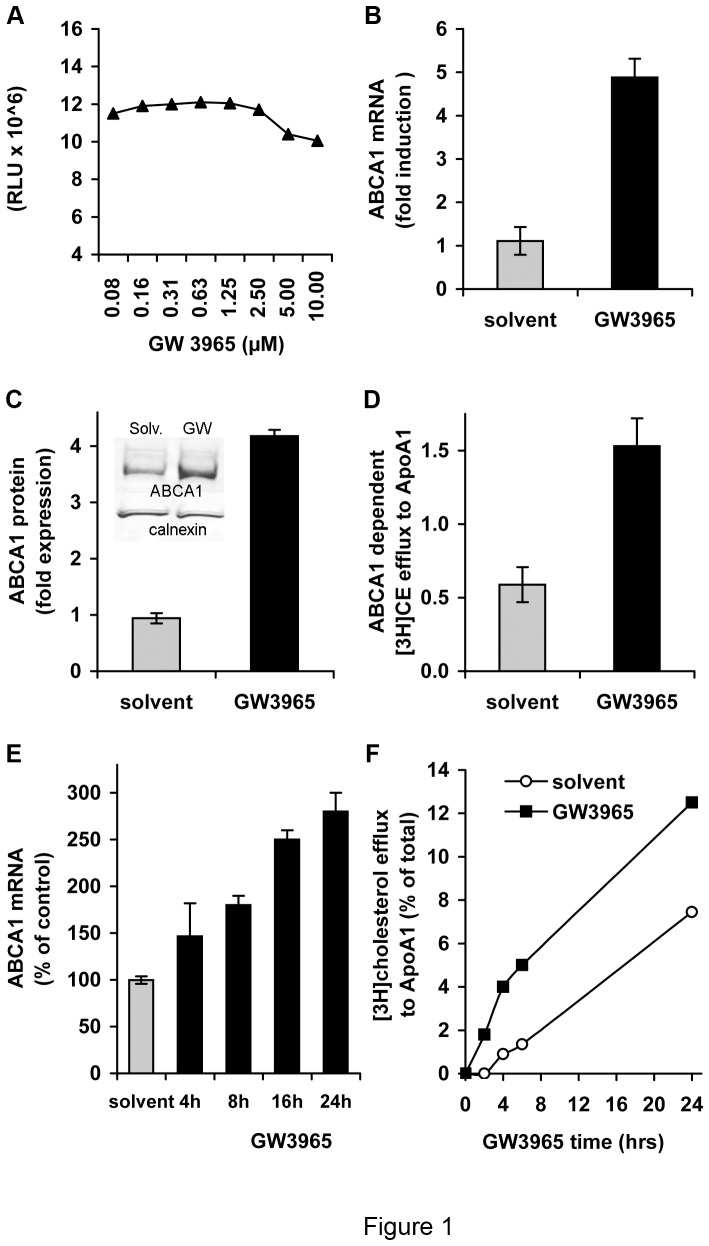
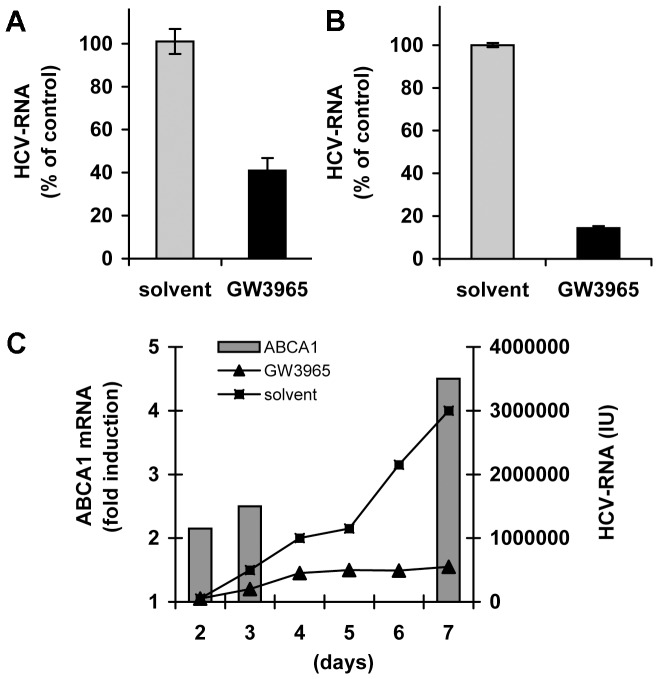
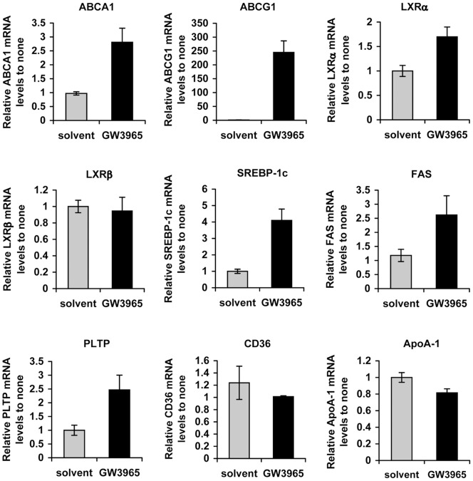
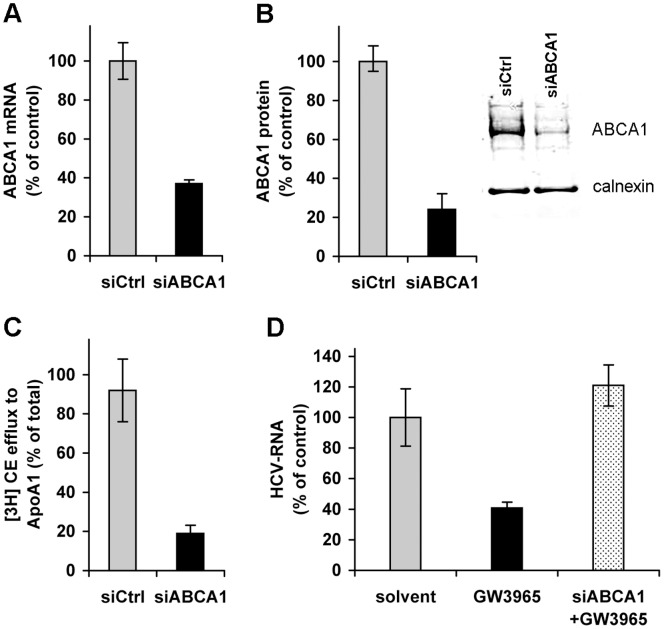
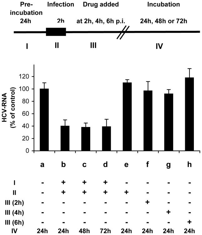
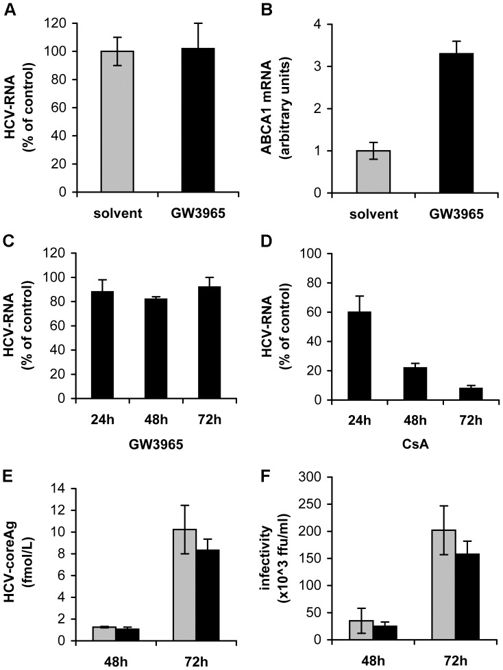
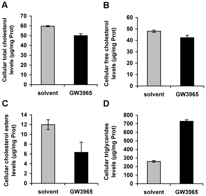
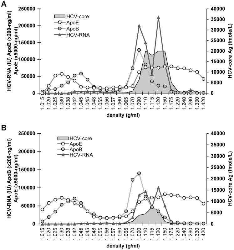
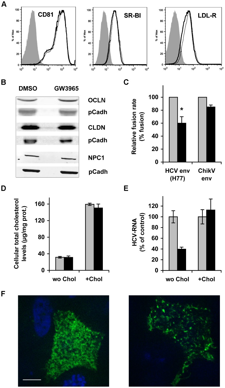
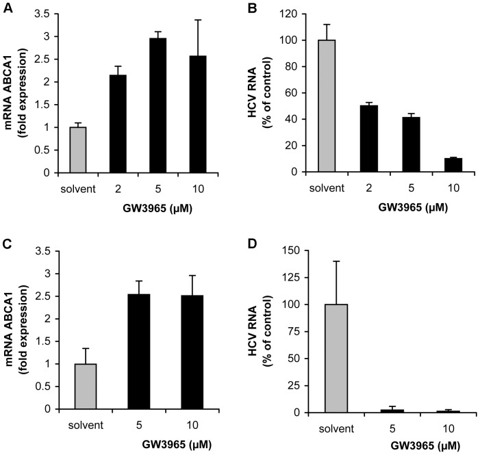
Hepatitis C virus (HCV) establishes infection using host lipid metabolism pathways that are thus considered potential targets for indirect anti-HCV strategies. HCV enters the cell via clathrin-dependent endocytosis, interacting with several receptors, and virus-cell fusion, which depends on acidic pH and the integrity of cholesterol-rich domains of the hepatocyte membrane. The ATP-binding Cassette Transporter A1 (ABCA1) mediates cholesterol efflux from hepatocytes to extracellular Apolipoprotein A1 and moves cholesterol within cell membranes. Furthermore, it generates high-density lipoprotein (HDL) particles. HDL protects against arteriosclerosis and cardiovascular disease. We show that the up-regulation of ABCA1 gene expression and its cholesterol efflux function in Huh7.5 hepatoma cells, using the liver X receptor (LXR) agonist GW3965, impairs HCV infection and decreases levels of virus produced. ABCA1-stimulation inhibited HCV cell entry, acting on virus-host cell fusion, but had no impact on virus attachment, replication, or assembly/secretion. It did not affect infectivity or properties of virus particles produced. Silencing of the ABCA1 gene and reduction of the specific cholesterol efflux function counteracted the inhibitory effect of the GW3965 on HCV infection, providing evidence for a key role of ABCA1 in this process. Impaired virus-cell entry correlated with the reorganisation of cholesterol-rich membrane microdomains (lipid rafts). The inhibitory effect could be reversed by an exogenous cholesterol supply, indicating that restriction of HCV infection was induced by changes of cholesterol content/distribution in membrane regions essential for virus-cell fusion. Stimulation of ABCA1 expression by GW3965 inhibited HCV infection of both human primary hepatocytes and isolated human liver slices. This study reveals that pharmacological stimulation of the ABCA1-dependent cholesterol efflux pathway disrupts membrane cholesterol homeostasis, leading to the inhibition of virus-cell fusion and thus HCV cell entry. Therefore besides other beneficial roles, ABCA1 might represent a potential target for HCV therapy.


Conflict of interest statement

Competing Interests: The authors have declared that no competing interests exist.

Figures

Figure 1
Figure 1. GW3965 treatment up-regulates ABCA1 expression and its cholesterol efflux function.
(A) Cell toxicity of GW3965. Huh7.5 cells were cultured in the presence of indicated concentrations of the drug for 24 h. The luminescent signal is expressed in luminescence units (RLU). (B) Up-regulation of ABCA1 mRNA expression by GW3695 treatment. Huh7.5 cells were treated for 24 h with 1 µM GW3695 or drug solvent (DMSO). Then ABCA1 mRNA was determined by qRT-PCR. (C) ABCA1 protein production in drug-stimulated Huh7.5 cells. Cells were treated for 24 h with 1 µM GW3965 and analysed by Western blot (shown in the insert). Protein content in the ABCA1 band (220 kDA) in GW3965-(GW), and DMSO-(solv) treated cells was quantified relative to the calnexin band using the Odyssey Infrared Imaging System. (D) GW3965 stimulation promotes ABCA1-mediated cholesterol efflux to ApoA1. Huh7.5 cells were labelled with [3H] cholesterol then incubated with GW3965 or drug solvent. ABCA1-dependent [3H] cholesterol efflux was assayed by comparing cell-associated and free radioactivity. (E) Kinetics of ABCA1 gene expression following stimulation of cells with GW3965. Huh7.5 cells were treated with 1 µM GW3965 for the indicated time and ABCA1 mRNA was determined by qRT-PCR. Results were expressed as relative values compared to ABCA1 expression in cells treated with drug solvent. (F) Kinetics of cholesterol efflux in cells stimulated with GW3965. Huh7.5 cells were labelled with [3H] cholesterol for 24 h, and incubated for an additional 16 h with 1 μM GW3965 or drug solvent. ABCA1-dependent [3H] cholesterol efflux was assayed in the presence of ApoA1 and either GW3965 or solvent for the indicated period of time.
Figure 2
Figure 2. Stimulation of ABCA1 inhibits HCV infection.
(A) Reduction of intracellular HCV RNA levels in cells that over-express ABCA1. Huh7.5 cells were pre-treated with 1 µM GW3965 then infected with HCV. Cells were grown for a further 24 h, total RNA was extracted and intracellular HCV RNA was determined by qRT-PCR. Results are expressed as the percentage of HCV RNA relative to that in cells treated with drug solvent prior to infection. (B) Decrease of HCV RNA levels in the supernatant collected from drug-stimulated cells. Huh7.5 cells were pre-treated with 1 µM GW3965 then infected with HCV. After a further 72 h, HCV-RNA in the culture medium was determined by qRT-PCR. Results are expressed as the percentage of HCV RNA secreted from drug-treated cells compared to solvent-treated cells. (C) Effect of GW3965 treatment on long-term HCV infection. Huh 7.5 cells were pre-treated with 1 µM GW3965, infected with HCV and grown for up to 7 days in the presence of the drug. ABCA1 mRNA was determined by qRT-PCR every 24 h and results are expressed as a fold-increase of ABCA1 mRNA compared to solvent-treated cells (grey bars). HCV RNA in the cell supernatant was measured at the same time points by qRT-PCR (line curves for GW3965 treated [filled triangles] or control [filled squares] cells) and is expressed in International Units (IU).
Figure 3
Figure 3. GW3695 treatment modulates expression of genes involved in lipid metabolism.
Huh7.5 cells were treated with 1 µM GW3695. Total RNA was extracted from cells and the mRNA levels corresponding to several genes regulating lipoprotein metabolism: ABCA1, ABCG1, nuclear LXRα and LXRβ receptors, a sterol regulatory element binding protein-1c (SREBP-1c), fatty acid synthase (FAS) and phospholipid transfer protein (PLTP), CD36 and ApoA1 were determined by qRT-PCR. The results were normalized to housekeeping genes and compared to the levels of corresponding mRNAs in solvent-treated cells.
Figure 4
Figure 4. ABCA1 plays a key role in the inhibition of HCV infection.
(A) Silencing of ABCA1. Huh7.5 cells were transfected with siRNA that targets ABCA1, or with a control siRNA. Total RNA was extracted after 48 h and ABCA1 mRNA determined by qRT-PCR. Results are expressed as the percentage of ABCA1 mRNA in cells transfected with siRNA targeting ABCA1 relative to mRNA levels in control cells. (B) Decreased ABCA1 protein synthesis in ABCA1-knocked-down cells. Huh7.5 cells were transfected with ABCA1-specific siRNA as above. Cell lysates were subjected to Western blot analysis. Staining of the ABCA1 protein with specific antibodies is shown in the insert. Results are expressed as the ABCA1 protein content in ABCA1-siRNA transfected cells relative to that in control siRNA transfected cells, normalized to calnexin (quantification using the Odyssey Infrared Imaging System). (C) Loss of cholesterol efflux function in ABCA1-silenced cells. Huh7.5 cells were transfected with siRNA that targets ABCA1 or with control siRNA. ABCA1-dependent [3H] cholesterol efflux was assayed in the presence of ApoA1. Results are expressed as percentage of cholesterol efflux to ApoA1 in ABCA1 knocked-down cells relative to control si-RNA transfected cells. (D) Silencing of ABCA1 antagonizes GW3965-mediated inhibition of HCV infection. The expression of ABCA1 was reduced by transfection with ABCA1-specific siRNA (as in A) and cells were treated with 1 µM GW3965, infected and grown for a further 24 h. HCV RNA was determined by qRT-PCR. Results are expressed as the percentage of HCV RNA in drug-treated (GW3965) cells or ABCA1-silenced and subsequently GW3965-treated cells (siABCA1+GW3965), compared to solvent-treated cells.
Figure 5
Figure 5. Up-regulation of ABCA1 inhibits HCV cell entry.
The effect of GW3965 on the HCV cell cycle was analysed by adding the drug at different time points. A flow-chart is depicted in the upper panel of the graph. RNA in Huh7.5 cells infected in the presence of DMSO is shown in (a); that in cells pre-treated for 24 h with 1 µM GW3965 and infected in the presence of the drug are shown in (b), (c) and (d); results for cells treated with GW3965 during virus inoculation without pre-treatment are shown in (e); those of assays where the drug was added at 2 h, 4 h, or 6 h post-infection are presented in (f), (g) and (h) respectively. For each experiment cells were incubated for the indicated time period after infection (IV). The efficiency of infection was expressed as intracellular HCV RNA measured by qRT-PCR as a per cent of the control (a).
Figure 6
Figure 6. Stimulation of ABCA1 has no impact on HCV cell attachment, HCV RNA replication, assembly/secretion or infectivity of virus particles.
(A–B) GW3965 treatment does not affect HCV cell attachment. Huh7.5 cells were pre-treated for 24 h with 1 µM GW3965, to raise ABCA1 levels and tested for their capacity to attach HCV using a “binding assay”. (A) HCV RNA attached to drug pre-treated cells (GW3965) is expressed as per cent relative to HCV RNA attached to cells treated with DMSO (solvent). (B) ABCA1 mRNA levels were determined by corresponding qRT-PCR and expressed in arbitrary units. (C) Stimulation of ABCA1 does not affect HCV RNA replication. Huh7 cells that express the sub-genomic replicon were incubated for 72 h with 1 μM GW3965 or with the equivalent concentration of drug solvent. HCV RNA was quantified by qRT-PCR in drug-treated cells relative to HCV RNA in control replicon cells grown in the presence of drug solvent. (D) HCV RNA replication is inhibited by CsA (control for C). Replicon cells were grown in medium containing 0.67 µM CsA or drug solvent (EtOH) for 72 h. HCV RNA was measured by qRT-PCR every 24 h and results were expressed as the percentage of HCV RNA in drug treated cells relative to HCV RNA content in cells grown in the presence of drug solvent. (E) ABCA1 up-regulation does not affect virus particle assembly/secretion. Huh7.5 cells were infected with HCV and 1 µM GW3965 was applied to cells 2 h post-infection. Every 24 h drug was replenished. HCV core antigen secreted to the cell supernatant was quantified at 48 h and 72 h post infection. Results were normalized with respect to the total protein content in the supernatant and are expressed in fmol/L. (F) Infectivity of virus particles secreted to the cell supernatant from cells treated according to the procedure described in (E) and determined using In Cell Western Blot assay. Results are expressed in ffu/ml.
Figure 7
Figure 7. Analysis of cellular lipids in GW3965-treated cells.
Huh7.5 cells were treated with 1 μM GW3965 (GW3965) or drug solvent (solvent). Cellular lipids were extracted and (A) cellular total cholesterol, (B) cell-free cholesterol, (C) cholesterol esters and (D) triglyceride levels were quantified and expressed relative to total protein levels.
Figure 8
Figure 8. Analysis of HCV particles secreted from cells that over-express ABCA1.
Physical properties of the nascent virus particles produced in cells stimulated or not with GW3965 were analysed by centrifugation in iodixanol gradient. Huh7.5 cells were pre-incubated with solvent (panel A) or 1 µM GW3965 (panel B) and the drug was maintained until 72 h post-infection when cell supernatants were collected, concentrated and subjected to gradient centrifugation. HCV RNA in gradient fractions was quantified by qRT-PCR and core antigen, ApoB and ApoE by ELISA assays.
Figure 9
Figure 9. GW3965 treatment does not change the expression of HCV receptors, but affects HCV-cell fusion and modifies lipid raft structure.
(A) Analysis of the expression of CD81, SR-BI and LDL-R in Huh7.5 cells treated with 1 μM GW3965 by Flow Cytometry using anti-receptor antibodies. GW3965-stimulated cells are shown by bold lines and non-treated cells by plain lines. Filled histograms represent cells stained with the secondary antibody only. (B) Western Blot assessment of the expression of OCLN, CLDN1 and NPC1 receptor in GW3965-treated cells, compared to solvent-treated cells. Pan Cadherin (pCadh) was used as a loading control. (C) GW3965 treatment inhibits HCV envelope-induced cell fusion. 293T cells that co-express a luciferase marker and the HCV E1–E2 envelope glycoproteins or the Chikungunya virus envelope glycoproteins were co-cultured with Huh7-Tat indicator cells. Co-cultured cells were incubated with 1 µM GW3965 or DMSO and exposed to pH 5. Luciferase activity was measured 72 h later. Data are presented as the fusion rate in the presence of the drug (black bars) relative to fusion in the absence of drug (gray bars), which was considered as 100%. The graph represents the average of 3 independent experiments (*P<0.03). (D–E). Cholesterol loading counteracts the inhibitory effect of ABCA1 over-expression on HCV infection. Huh7.5 cells were stimulated with 1 µM GW3965 to over-express ABCA1, and then incubated with 20 µM cholesterol/MβCD. In (D) the determination of total cellular cholesterol after replenishment of ABCA1 overexpressing cells with cholesterol/MβCD is shown. GW3965-treated cells (black bars) were compared to solvent-treated (grey bars). The infection of Huh7.5 cells after cholesterol supply is shown in (E). Intracellular HCV RNA was determined by qRT-PCR at 24 h post-infection, and is expressed as the percentage of HCV RNA in drug-treated (black bars) compared to solvent-treated (grey bars) cells, supplied (+Chol) or not (wo Chol) with cholesterol. (F) GW3965 treatment modifies plasma membrane organisation and thus the distribution of lipid raft-associated protein. Huh7.5 cells were transfected with DNA encoding the Glycosylphosphatidyl-inositol-anchor attachment signal of the folate receptor fused to GFP (GFP-FR). Cells were subsequently exposed to 1 μM GW3965 or drug solvent (control). Two days post-transfection cells were fixed and GFP-FR fluorescence was visualised using a Zeiss axioplan 2 microscope (x63 objective). Slices of 0.46 µm were acquired. The images shown are a Z projection of 5 slices of the cell surface that face the cell medium. The right panel represents GW3965-stimulated cells and the left control cells. The scale bar corresponds to 10 µm.
Figure 10
Figure 10. Over-expression of ABCA1 inhibits HCV infection of primary human hepatocytes and human liver slices.
(A–B) Inhibition of HCV infection of primary human hepatocytes. (A) Primary human hepatocytes were treated with 2–10 μM GW3965 (non-toxic concentrations for cells) or with drug solvent, prior to HCV infection. Twenty-four hours post-infection, ABCA1 mRNA was determined by qRT-PCR and expressed in arbitrary units, taking into account ABCA1 levels in liver cells pre-treated with the drug. (B) GW3965-treated and solvent-treated primary human hepatocytes were inoculated with HCV. After 24 h, intracellular HCV RNA was quantified by qRT-qPCR. The efficiency of infection in drug pre-treated cells was expressed as the percentage of infection compared to solvent-treated cells. (C–D) ABCA1 over-expression inhibits HCV infection of human liver slices. Human liver slices were cultured for 24 h, treated with 5 or 10 μM GW3965 or with DMSO before infection with HCVcc. At 24 h post-infection, total RNA was extracted and ABCA1 mRNA (C) and HCV RNA (D) were quantified by corresponding qRT-PCR assays and expressed as the percentage of RNA compared to the values obtained for solvent-treated cells.

Similar articles

Cited by

References

    1. Lange CM, Jacobson IM, Rice CM, Zeuzem S (2014) Emerging therapies for the treatment of hepatitis C. EMBO Mol Med. 6: 4–15. - PMC - PubMed
    1. Asselah T, Marcellin P (2012) Direct acting antivirals for the treatment of chronic hepatitis C: one pill a day for tomorrow. Liver International 32: 88–102. - PubMed
    1. Buhler S, Bartenschlager R (2012) New targets for antiviral therapy of chronic hepatitis C. Liver Int. 32 Suppl 19–16. - PubMed
    1. Bartenschlager R, Cosset FL, Lohmann V (2010) Hepatitis C virus replication cycle. J Hepatol 53: 583–585. - PubMed
    1. Ye J (2007) Reliance of host cholesterol metabolic pathways for the life cycle of hepatitis C virus. PLoS Pathog 3: e108. - PMC - PubMed

Publication types

MeSH terms

Grants and funding

SB was supported by the Doctoral Fellowship from the Italian Ministry of Science and Education and MY by the Postdoctoral Fellowship from the French Embassy in Japan. The funders had no role in study design, data collection and analysis, decision to publish, or preparation of the manuscript.