. 2014 Mar 17;204(6):931-45.
doi: 10.1083/jcb.201305148.

Oxidative stress-induced assembly of PML nuclear bodies controls sumoylation of partner proteins

Affiliations

Oxidative stress-induced assembly of PML nuclear bodies controls sumoylation of partner proteins

Umut Sahin et al. J Cell Biol. .

Abstract

The promyelocytic leukemia (PML) protein organizes PML nuclear bodies (NBs), which are stress-responsive domains where many partner proteins accumulate. Here, we clarify the basis for NB formation and identify stress-induced partner sumoylation as the primary NB function. NB nucleation does not rely primarily on intermolecular interactions between the PML SUMO-interacting motif (SIM) and SUMO, but instead results from oxidation-mediated PML multimerization. Oxidized PML spherical meshes recruit UBC9, which enhances PML sumoylation, allow partner recruitment through SIM interactions, and ultimately enhance partner sumoylation. Intermolecular SUMO-SIM interactions then enforce partner sequestration within the NB inner core. Accordingly, oxidative stress enhances NB formation and global sumoylation in vivo. Some NB-associated sumoylated partners also become polyubiquitinated by RNF4, precipitating their proteasomal degradation. As several partners are protein-modifying enzymes, NBs could act as sensors that facilitate and confer oxidative stress sensitivity not only to sumoylation but also to other post-translational modifications, thereby explaining alterations of stress response upon PML or NB loss.


Figures

Figure 1.
Figure 1.
PML NB nucleation does not depend on SUMO interactions with SIM. (A) Schematic representation of the current model, in which NB biogenesis relies on noncovalent intermolecular interactions between PML-attached SUMO and the PML SIM. (B) PML and SP100 immunolocalization showing morphologically normal NBs in pml−/− immortalized MEFs stably expressing PML or the indicated mutants (left), and in HeLa cells in which SUMO1 and SUMO2/3 were inactivated (right). Bar, 5 µm. Quantitative data are indicated in Fig. S1 A. (C) Super-resolution microscopy analysis showing NBs formed by PML WT, PML3KR, or PML3KRΔSIM in pml−/− MEFs. Bar, 1 µm. (D) Western blot analysis under nonreducing conditions of pml−/− MEFs stably expressing the indicated HA-tagged PML mutants. Lysates of total cells (T) or nuclear matrix (NM) fractions. Lamin B and RXRA are fractionation controls. The top arrow (PML2) points to covalently linked PML multimers; sumoylated PML (PML + Sn) and unmodified PML are indicated.
Figure 2.
Figure 2.
SIM and SUMOs are NB-targeting signals. (A) Prototypical PML partner with both a SIM and a SUMO conjugation site may interact with PML SUMO (1) and SIM (2) in an ordered manner. (B) Colocalization of GFP-SIM or SUMO1/2/3ΔGG-GFP fusions with NBs. (Top) As2O3 (arsenic)-enhanced NB recruitment of GFP-SIM fusions stably expressed in CHO-PML (arsenic: 10−6 M, 1 h). (Bottom) Recruitment to NBs of SUMO1, 2, or 3ΔGG-GFP fusions in CHO-PML but not in CHO cells. Insets show GFP labeling alone. (C) Quantification of GFP fusions’ recruitment on NBs: ratios of GFP intensities (in NBs versus in the nucleoplasm) were calculated cell by cell, and averaged from 20 cells. P-values are indicated. (D) FRAP analysis of NB-associated SUMOΔGG-GFPs or SIM-GFPs in CHO-PML cells (the graph represents means of five experiments); standard deviations are shown for GFP and S3ΔGG-GFPSIM. t1/2 recoveries after photo-bleaching are shown below (table). (E) Real-time diffusion analysis after Dendra-DAXX photoconversion performed on NBs or in the nucleoplasm of CHO-PML cells (means of five experiments are represented below). Error bars represent standard deviation. Half times of Dendra-DAXX diffusion after photoconversion are indicated below (t1/2). Insets show a representative green to red photoconverted ROI surrounding NB or in the nucleoplasm at t = 2.5 s, t = 9.5 s, and t = 45.3 s after switch. Bar, 0.5 µm. (F) Immunolocalization of PMLΔSIM or PMLK160R stably expressed in pml−/− MEFs or CHO cells, and of endogenous DAXX or transfected SP100, as indicated. (G) Immunolocalization of DAXX mutants stably expressed in CHO-PML cells. Bars, 5 µm.
Figure 3.
Figure 3.
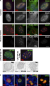
Topologically and biochemically distinct compartments in NBs. (A) In situ NM preparations from MRC5 cells, showing the matrix association of endogenous PML/SUMO2/3, in contrast to endogenous DAXX, SUMO1, or transfected FLAG-RNF4. Control, total cell labeling; nuclear matrix, in situ NM preparation; double PML/partner protein (top) and partner only (bottom) labeling are shown. Bar, 5 µm. (B) Confocal (top bar, 5 µm) and immunoelectron microscopy analysis (bottom bar, 0.5 µm) of stably transfected CHO-SP100 or CHO-SP100/PML cells. In the dual PML/SP100 labeling (far right), PML is revealed by large and SP100 by smaller gold particles. (C) Deconvoluted confocal analysis of IFNα-treated MRC5 primary fibroblasts stained with anti-PML (red), anti-SUMO1, anti-SUMO-2/3, anti-SP100, or anti-DAXX (green) antibodies. Bar, 1 µm.
Figure 4.
Figure 4.
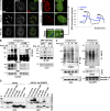
UBC9 recruitment into NBs favors hyper-sumoylation of partner proteins. (A, left) Confocal analysis of endogenous UBC9 localization in Triton X-100 pre-extracted H1299 cells, showing recruitment onto NBs after arsenic or IFNα exposure. Representative of two independent experiments, n ≥ 300 cells examined. Bar, 5 µm. (Right) Localization of UBC9-GFP stably expressed in CHO-PML cells before and after 2 h exposure to arsenic. Representative of three independent experiments, n ≥ 300 cells. Bar, 5 µm. Zooms show the regions (outlined above) used to quantify GFP fluorescence intensity (graph). Representative of three repeats. Bar, 2 µm. (B, left) Western blot analysis of endogenous SP100 profiles, performed on protein extracted 48 h after transfection with PML or control siRNAs. (Right) Sumoylation of hSP100A stably expressed in pml+/+ or pml−/− MEFs treated or not with IFNα for 24 h. Arrows and the bracket point to sumoylated species (see also Fig. S4, A, B, and D). (C) Western blot analysis of endogenous TDG (left) or HIPK2 (right) immunoprecipitated (IP) from H1299 cells treated as indicated. Inputs are shown. RanGAP1-S, sumoylated RanGAP1. (D) SUMO-GFP or SIM-GFP fusions are sumoylated. The indicated GFP fusions and (His)x6-SUMO2 were overexpressed in CHO-PML cells, purified over a Ni-NTA column and analyzed by Western blot using anti-GFP antibodies. Left, inputs; right, denaturing purification of sumoylated proteins. Dotted lines show sumoylated GFP fusions specifically purified from unspecific binding of GFP to the Ni-NTA column.
Figure 5.
Figure 5.
RNF4 recruitment into NBs results in sumoylation decay through partner ubiquitination. (A, top) PML–RNF4 interactions detected by PLA Duolink assay (red dots) and PML NB immunolocalization (green); Z-stack projections are shown. (Bottom) Quantification of the Duolink dots per cell and percentages of colocalization with NBs (means from 20 cells). (B) Western blot analysis of endogenous SP100 hyper-sumoylation (bracket) upon exposure to arsenic in H1299 transfected with the indicated siRNAs. (C) Western blot analysis of transduced hSP100A in MEFs treated as indicated, demonstrating arsenic-induced proteasomal degradation after 24 h. (D) SUMO and ubiquitin conjugation of SP100 upon exposure to arsenic. (Left) Endogenous SP100 immunoprecipitates from arsenic-treated HeLa cells probed with anti-SUMO1 and anti-SP100 antibodies. (Right) Nickel-purified His-ubiquitin conjugates from His-ubiquitin–overexpressing HeLa cells, probed with antibodies to SP100 and ubiquitin. (E) Polyubiquitination of indicated GFP fusions in CHO-PML cells overexpressing (His)x6-Ubiquitin, purified over nickel column and analyzed by Western blot using anti-GFP antibodies. Left, inputs; right, denaturing purification of ubiquitinated proteins.
Figure 6.
Figure 6.
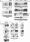
PML and RNF4 cooperate to regulate sumoylation in response to oxidative stress. (A) Western blot analysis of SUMO1 and -2/3 conjugates from H1299 cells treated as indicated with arsenic, and for 8 h with MG132. (B) Western blot analysis of arsenic-induced transient hyper-sumoylation in H1299 cells transfected with the indicated siRNAs. SUMO1 and -2/3 conjugates are shown along with PML, RNF4, and actin controls. Star, sumoylation of RanGAP1, a non–NB-associated protein, does not respond to arsenic; PML-S, sumoylated PML. (C) Western blot analysis of immunoprecipitated polyubiquitin conjugates (IP: Ub) from pml+/+ or pml−/− MEFs treated or not with arsenic and IFNα for 48 h. (D) Western blot analysis of sumoylation after transduction of RNF4 (R) or catalytically inactive RNF4-DN mutant in IFNα-treated pml+/+ or pml−/− MEFs. Brackets, HMW SUMO1 conjugates; asterisk, sumoylated RanGAP1. (E) Western blot analysis of global sumoylation in immortalized pml+/+ or pml−/− MEFs.
Figure 7.
Figure 7.
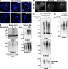
Induction of NB formation and sumoylation in oxidant-treated mice. (A) In vivo oxidative stress effects on NB formation: Immunofluorescence analysis of PML NBs on liver sections from mice exposed to APAP (acetaminophen: N-acetyl-p-aminophenol), As2O3, doxorubicin (Doxo), or paraquat for 2 h. Quantifications are shown in Fig. S5 C. n ≥ 50 cells examined. (B) In vivo sumoylation after exposure to oxidative stress. (Left) Western blot analysis of liver cells from mice treated with paraquat. (Right) Western blot analysis of bone marrow (BM) cells from mice treated with arsenic; two mice are shown for each from two independent experiments. (C) Immunofluorescence analysis of APL cells obtained after in vivo administration of arsenic or paraquat for the indicated period of time. Bar, 5 µm. (D) His-SUMO1–transduced mouse APL bone marrow (BM) cells were isolated after in vivo arsenic administration. His-conjugates were purified and probed with antibodies to SUMO1, SUMO2/3, and RARA. PML/RARA-S, poly-sumoylated PML/RARA. Representative data from six mice, three independent experiments. (E) Western blot analysis of bone marrow cells of APL mice after in vivo treatment with arsenic or paraquat.
Figure 8.
Figure 8.
NBs are redox-regulated hubs of sumoylation and SUMO-initiated, RNF4-mediated ubiquitination. Nucleation of the NB mesh relies solely on ROS-induced PML oxidation, while intermolecular PML SUMO–SIM interactions are not involved in this initial nucleation step. Stress-induced UBC9 recruitment results in subsequent PML sumoylation, critical for partners’ recruitment through their SIM. In situ sumoylation of partners secures partner–PML, partner–partner, and partner–RNF4 interactions in NBs, promoting their ubiquitination or other post-translational modifications (PTMs) like acetylation or phosphorylation.

Similar articles

Cited by

References

    1. Bernardi R., Pandolfi P.P. 2007. Structure, dynamics and functions of promyelocytic leukaemia nuclear bodies. Nat. Rev. Mol. Cell Biol. 8:1006–1016 10.1038/nrm2277 - DOI - PubMed
    1. Bischof O., Kirsh O., Pearson M., Itahana K., Pelicci P.G., Dejean A. 2002. Deconstructing PML-induced premature senescence. EMBO J. 21:3358–3369 10.1093/emboj/cdf341 - DOI - PMC - PubMed
    1. Bossis G., Melchior F. 2006. Regulation of SUMOylation by reversible oxidation of SUMO conjugating enzymes. Mol. Cell. 21:349–357 10.1016/j.molcel.2005.12.019 - DOI - PubMed
    1. Chiantore M.V., Vannucchi S., Accardi R., Tommasino M., Percario Z.A., Vaccari G., Affabris E., Fiorucci G., Romeo G. 2012. Interferon-β induces cellular senescence in cutaneous human papilloma virus-transformed human keratinocytes by affecting p53 transactivating activity. PLoS ONE. 7:e36909 10.1371/journal.pone.0036909 - DOI - PMC - PubMed
    1. Ching R.W., Ahmed K., Boutros P.C., Penn L.Z., Bazett-Jones D.P. 2013. Identifying gene locus associations with promyelocytic leukemia nuclear bodies using immuno-TRAP. J. Cell Biol. 201:325–335 10.1083/jcb.201211097 - DOI - PMC - PubMed

Publication types

MeSH terms