Review
. 2014 Feb 23:2:535-62.
doi: 10.1016/j.redox.2014.02.006. eCollection 2014.

Hydrogen peroxide sensing, signaling and regulation of transcription factors

Affiliations
Review

Hydrogen peroxide sensing, signaling and regulation of transcription factors

H Susana Marinho et al. Redox Biol. .

Abstract

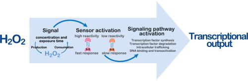
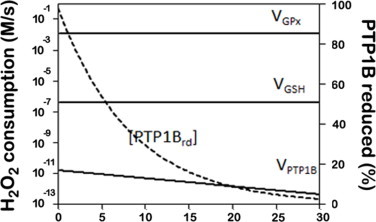
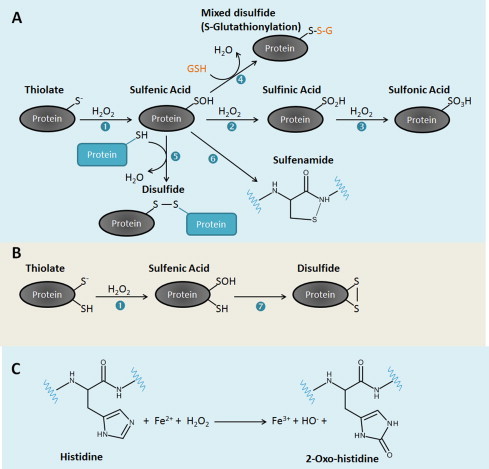
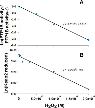
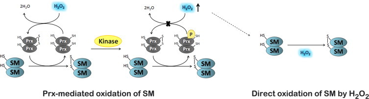
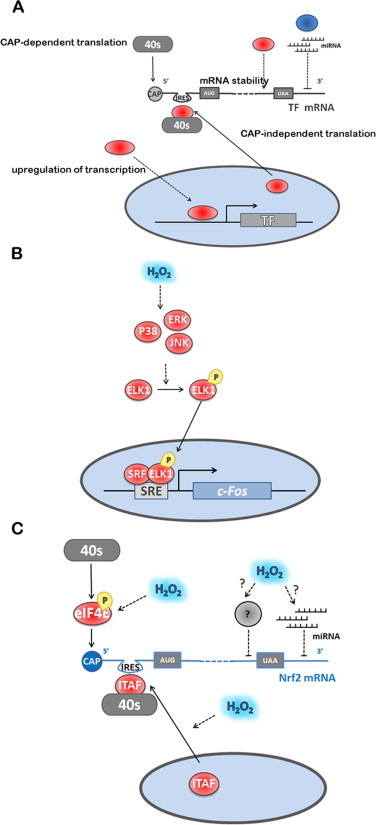
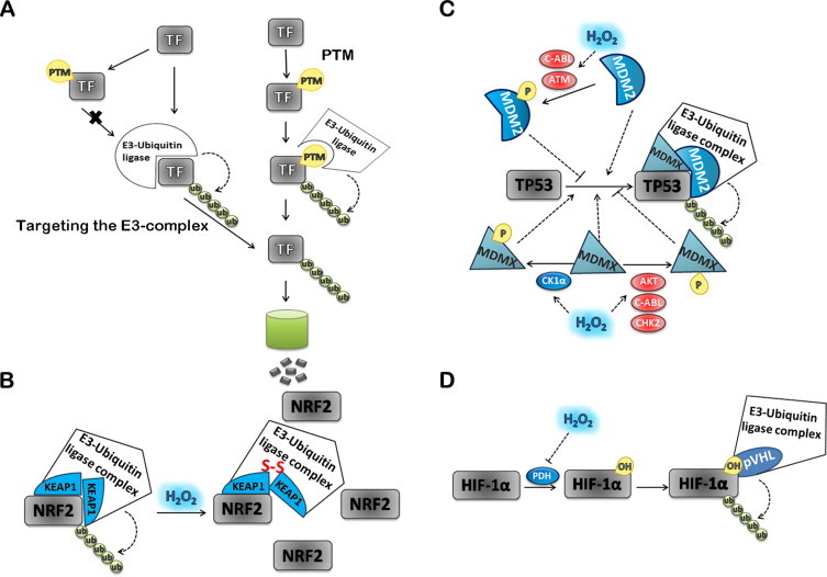
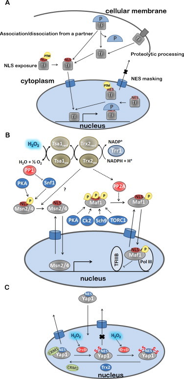
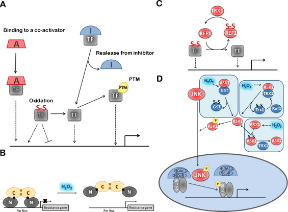
The regulatory mechanisms by which hydrogen peroxide (H2O2) modulates the activity of transcription factors in bacteria (OxyR and PerR), lower eukaryotes (Yap1, Maf1, Hsf1 and Msn2/4) and mammalian cells (AP-1, NRF2, CREB, HSF1, HIF-1, TP53, NF-κB, NOTCH, SP1 and SCREB-1) are reviewed. The complexity of regulatory networks increases throughout the phylogenetic tree, reaching a high level of complexity in mammalians. Multiple H2O2 sensors and pathways are triggered converging in the regulation of transcription factors at several levels: (1) synthesis of the transcription factor by upregulating transcription or increasing both mRNA stability and translation; (ii) stability of the transcription factor by decreasing its association with the ubiquitin E3 ligase complex or by inhibiting this complex; (iii) cytoplasm-nuclear traffic by exposing/masking nuclear localization signals, or by releasing the transcription factor from partners or from membrane anchors; and (iv) DNA binding and nuclear transactivation by modulating transcription factor affinity towards DNA, co-activators or repressors, and by targeting specific regions of chromatin to activate individual genes. We also discuss how H2O2 biological specificity results from diverse thiol protein sensors, with different reactivity of their sulfhydryl groups towards H2O2, being activated by different concentrations and times of exposure to H2O2. The specific regulation of local H2O2 concentrations is also crucial and results from H2O2 localized production and removal controlled by signals. Finally, we formulate equations to extract from typical experiments quantitative data concerning H2O2 reactivity with sensor molecules. Rate constants of 140 M(-1) s(-1) and ≥1.3 × 10(3) M(-1) s(-1) were estimated, respectively, for the reaction of H2O2 with KEAP1 and with an unknown target that mediates NRF2 protein synthesis. In conclusion, the multitude of H2O2 targets and mechanisms provides an opportunity for highly specific effects on gene regulation that depend on the cell type and on signals received from the cellular microenvironment.

Keywords: AD, activation domain; Cytosol-nuclear traffic; DNA binding and transactivation; ER, endoplasmic reticulum; GPx, glutathione peroxidases; Localized H2O2 concentrations; NES, nuclear exporting signal; NLS, nuclear localization signal; PHD, prolyl hydroxylase; Prxs, peroxiredoxins; Rate constants; Redox signaling; TF, transcription factor; Thiol reactivity; Ub, Ubiquitin.


Figures

None
Graphical abstract
Fig. 2
Fig. 2
PTP1B signaling by H2O2 when in the presence of an antioxidant kinetic bottleneck that outcompetes the rate of PTP1B oxidation.The following reactions were included: a rate of H2O2 production of 1.2 × 10⁻² M s−1; H2O2 consumption via glutathione peroxidase (VGPx = kGPx × [GPx] × [H2O2]), via PTP1B (VPTP1B = kPTP1b× [PTP1Brd] × [H2O2]) and via non-enzymatic reaction with GSH (VGSH = kGSH × [GSH] × [H2O2]). kGPx = 6 × 10⁷ M−1 s−1, [GPx] = 2 × 10⁻⁶ M, kPTP1b = 20 M−1 s−1, [PTP1Btot] = 8.3 × 10⁻⁹ M, kGSH = 0.87 M−1 s−1, [GSH] = 5 × 10⁻³ M. With these parameters, the steady state obtained was [H2O2] = 1 × 10⁻⁴ M. [PTP1Btot] = [PTP1Brd] + [PTP1Box], where the subscripts tot, rd and ox, refer to the total amount of PTP1B, to the reduced and to the oxidized forms of PTP1B, respectively.
Fig. 1
Fig. 1
Oxidative modifications of cysteine (A and B) and histidine (C) residues in proteins induced by H2O2. In cells, sulfhydryl (SH) groups of cysteine residues with low pKa may ionize forming thiolates. Thiolates are good nucleophiles and form a sulfenic acid (SOH) upon reaction with H2O2 (reaction 1). Once formed, the SOH can be reduced to a disulfide by a reaction with the SH group of another cysteine residue either in the same (reaction 7) or in a second protein (reaction 5). Alternatively, a SOH can react with the low molecular weight thiol glutathione (GSH) (reaction 4) to form a mixed disulfide in a reaction known as S-glutathionylation or S-thiolation. In an event where a neighboring cysteine residue or GSH is absent, the amide nitrogen of a neighboring amino acid residue can attack the SOH to form a sulfenamide (reaction 6). This reaction occurs in PTP1B. The SOH can also react further with H2O2 to generate more oxidized forms of sulfur, the sulfinic acid (SO2H) (reaction 2) and sulfonic acid SO3H (reaction 3). Disulfides can be reduced back to thiols using the thioredoxin/thioredoxin reductase and glutaredoxin/GSH/glutathione reductase systems. Sulfinic acids in 2-cys Prxs, but not other proteins, can be reduced to thiols using the enzyme sulfiredoxin [372]. No known enzyme is able to catalyze the reduction of sulfonic acids in proteins. In proteins containing iron metal centers such as PerR, histidine residues can be oxidized by H2O2 in a Fenton-like reaction possibly involving the formation of the hydroxyl radical as an intermediate, to form 2-oxo-histidine.
Fig. 3
Fig. 3
Application of Eq. (6) to estimate rate constants between cellular targets and H2O2. Plot of the fraction of PTP1B activity observed invitro after 10 min (A) and of the reduced form of KEAP1 observed in HeLa cells after 5 min (B) of incubation with the indicated H2O2 concentrations. Experimental data are taken from [33] and [189], respectively for PTP1B and KEAP1. If a simple exponential decay is considered, that is no regeneration of sensor occurs, the slope of these plots is ktarget + H2O2 × t (Eq. (6)) and, therefore, the rate constants between PTP1B and KEAP1 with H2O2 are estimated at 22 M−1 s−1 and 20 M−1 s−1, respectively. If a gradient between extracellular and intracellular H2O2 of 6.8 is considered in HeLa cells [42], the rate constant for H2O2 reaction with KEAP1 is estimated at 140 M−1 s−1.
Fig. 4
Fig. 4
Localized increase of H2O2 levels mediated through inhibition of peroxiredoxins activity by its phosphorylation. Peroxiredoxins (Prxs) can act as highly reactive H2O2 sensors and transduce the signal to a signaling molecule (SM). Alternatively, upon binding of a ligand to a membrane receptor an SRC family kinase can be activated. This SRC kinase activates NADPH oxidase in the plasma membrane, which leads to the production of H2O2, and catalyzes phosphorylation of Prx at a tyrosine residue leading to its inactivation. Prx inactivation leads to a transient accumulation of H2O2 around membranes, where signaling components are concentrated. This will promote the direct oxidation of H2O2 sensors with intermediate and low reactivity.
Fig 5
Fig 5
Regulation of TF expression by H2O2. (A) TF expression is regulated by H2O2 at both transcriptional and translational levels. The translation process and the regulation of mRNA stability are preferential targets of H2O2. H2O2 modulates CAP-dependent and independent translation through the activation of 40S-mRNA complexes. mRNA stability is modulated by RNA-binding proteins and by specific miRNA, which are modulated by H2O2. (B) c-FOS transcription is regulated by ELK1, which is phosphorylated by MAP kinases activated by H2O2. (C) The known mechanism for NRF2 is exemplified showing the positive regulation by H2O2 of both CAP-dependent and CAP-independent initiation. The ITAF for NRF2 is La Autoantigen, whose translocation to the cytoplasm is promoted by H2O2. Also shown, are hypothetical mechanisms for H2O2 modulation of microRNAs that negatively control translation by binding to the 3′ UTR region of NRF2 mRNA, and of unknown factors that mediate repressing of NRF2 translation by binding to the 3′ part of the mRNA coding regions. Factors colored blue are inhibitors of TF-dependent gene expression; factors colored red are activators of TF-dependent gene expression. Dashed lines indicate activation/inhibition.
Fig. 6
Fig. 6
Regulation of TFs activity by degradation modulated by H2O2. TF degradation via poliubiquitin(Ub)-proteasome pathway is an important regulatory mechanism in different signaling pathways (A). Proteasome, a multicatalytic protease oligomeric complex, degrades proteins that have been poly-ubiquitinated. Ubiquitin (Ub)-protein ligase (E3) enzymes transfer the activated Ub from an Ub-conjugating protein E2, first to a lysine residue of the substrate protein and the next Ub to lysine residues present in the last added ubiquitin, originating an Ub chain. NRF2 ubiquitination and degradation involves KEAP1 as the substrate adaptor subunit in the E3 holoenzyme (B). In the presence of H2O2 critical cysteine residues in KEAP1 are oxidized, changing KEAP1 conformation. This conformational change affects the interaction between NRF2 and KEAP1 inhibiting NRF2 ubiquitination and degradation. In normal cells TP53 is maintained at low levels by interaction with the negative regulator MDM2 a p53 highly specific ubiquitin ligase. H2O2 mainly regulates MDM2 activity by activating the ATM and c-ABL kinases involved in MDM2 phosphorylation. MDM2 phosphorylation inhibits its ubiquitination activity and stabilizes TP53 (C). The MDM2 ubiquitin ligase substrate preference for TP53 is enhanced by MDMX. Phosphorylation of MDMX in different residues by c-ABL, AKT and CHK2 kinases inhibit TP53 degradation, while those catalyzed by the CK1α kinase stimulates TP53 degradation. All the referred kinases activities are regulated by H2O2. The association between the TF and the ubiquitination machinery can also be modulated by PTMs of the transcription factor (D). HIF-1α is tagged with ubiquitin for degradation after being hydroxylated by PHD in the presence of O2 (D). H2O2 inhibits PHD that leads to HIF-1α stabilization. Factors colored blue are inhibitors of TF-dependent gene expression; factors colored red are activators of TF-dependent gene expression. Dashed lines indicate activation/inhibition.
Fig. 7
Fig. 7
Regulation of cytoplasm-nuclear trafficking of TF by H2O2. (A) Nuclear localization of TF is essential for gene expression activation and H2O2 plays a key role in TF cellular trafficking. H2O2 modulates NLS exposure by removing PTM or by promoting partner dissociation (P). In certain cases, the association to adaptor proteins might promote NLS exposure. Inversely, NES masking is another mechanism to retain TF in the nucleus that is mediated by H2O2. Conformational changes induced by PTM together with the formation of protein complexes make NES inaccessible inducing activation of transcription. Other TF are associated to cellular membranes in their inactive state. The activation of these TF requires proteolytic cleavage and release to the cytoplasm where it is transported to the nucleus. (B) Msn2/4 and Maf1 translocation to the nucleus is activated after dephosphorylation, which uncovers NLS. This process is activated by H2O2 indirectly through Trx system. Msn2/4 and Maf1 dephosphorylation is dependent upon Trx2 by an unknown mechanism and by PP2A activation, respectively. (C) Yap1-dependent gene activation depends on its retention in the nucleus by CRM1 dissociation that occurs in presence of H2O2. The oxidation of four Cys residues in Yap1 is responsible for the conformational alterations that prevent NES recognition by CRM1. As for Msn2/4 and Maf1, Yap1 does not react directly with H2O2, and its oxidation is mediated by a GPx, Orp1. Trx2 reduces Yap1 inducing its translocation to the cytoplasm, inactivating gene transcription. Factors colored blue are inhibitors of TF-dependent gene expression; factors colored red are activators of TF-dependent gene expression. Dashed lines indicate activation/inhibition.
Fig. 8
Fig. 8
Regulation of DNA binding and transactivation of TF by H2O2. (A) TF affinity to DNA or to transcription activators is regulated by H2O2 through different mechanisms involving either conformational alterations induced by PTM or association with activators/inhibitors. In addition, H2O2 is able to modulate TF activation by direct TF oxidation inside or outside DNA binding domain. (B) PerR DNA affinity is lost by a conformational alteration induced by a Fenton reaction of H2O2 with Fe2+ forming 2-oxo-histidine. Both Fe2+and Mn2+ (black spheres) can bind to the regulatory site but when Mn2+ is bound 2-oxo-histidine is not formed. Structural ZnII ions (yellow spheres). (C) Ref-1 induces the DNA binding activity of several TFs by reducing cysteine residues of those TFs. Ref-1 is regenerated by thioredoxin that reduces Cys65. (D) H2O2 promotes c-Jun phosphorylation releasing its repressor complex, which contains HDAC3. This phosphorylation is dependent of the activity of JNK, which is activated by ASK1. ASK1 activation is regulated by H2O2 through three alternative mechanisms: GST oxidation releasing ASK1, Trx oxidation releasing ASK1 or ASK1 oxidation by PRX1. Factors colored blue are inhibitors of TF-dependent gene expression; factors colored red are activators of TF-dependent gene expression. Dashed lines indicate activation/inhibition.

Similar articles

Cited by

References

    1. Chance B. The state of catalase in the Respiring bacterial cell. Science (New York, N.Y.) 1952;116:202–203. doi: 10.1126/science.116.3008.202. 17792314 - DOI - PubMed
    1. Czech MP, Lawrence JC, Lynn WS. Evidence for the involvement of sulfhydryl oxidation in the regulation of fat cell hexose transport by insulin. Proceedings of the National Academy of Sciences of the United States of America. 1974;71:4173–4177. doi: 10.1073/pnas.71.10.4173. 4372610 - DOI - PMC - PubMed
    1. White AA, Crawford KM, Patt CS, Lad PJ. Activation of soluble guanylate cyclase from rat lung by incubation or by hydrogen peroxide. Journal of Biological Chemistry. 1976;251:7304–7312. 12160 - PubMed
    1. Burke TM, Wolin MS. Hydrogen peroxide elicits pulmonary arterial relaxation and guanylate cyclase activation. American Journal of Physiology. 1987;252:H721–H732. 2882694 - PubMed
    1. Heffetz D, Zick Y. H2O2 potentiates phosphorylation of novel putative substrates for the insulin receptor kinase in intact Fao cells. Journal of Biological Chemistry. 1989;264:10126–10132. 2542323 - PubMed

Publication types

MeSH terms