Review
. 2014 Nov;322(7):500-16.
doi: 10.1002/jez.b.22564. Epub 2014 Mar 14.

Tetrahymena thermophila: a divergent perspective on membrane traffic

Affiliations
Review

Tetrahymena thermophila: a divergent perspective on membrane traffic

Joseph S Briguglio et al. J Exp Zool B Mol Dev Evol. 2014 Nov.

Abstract

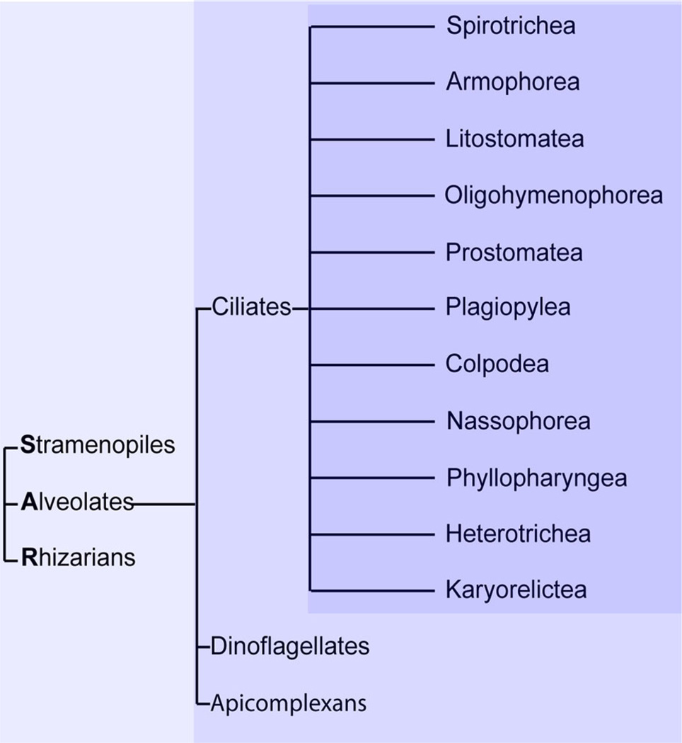
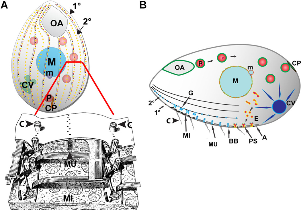
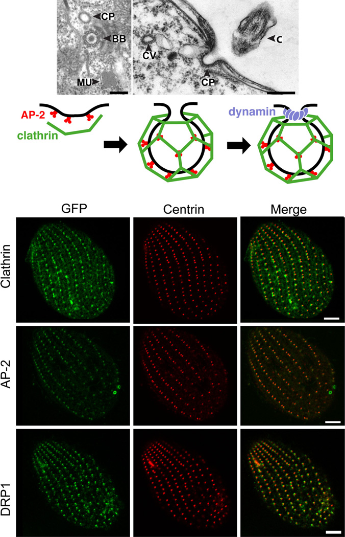
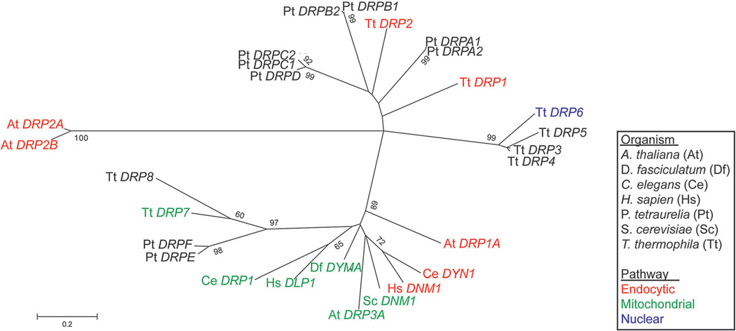
Tetrahymena thermophila, a member of the Ciliates, represents a class of organisms distantly related from commonly used model organisms in cell biology, and thus offers an opportunity to explore potentially novel mechanisms and their evolution. Ciliates, like all eukaryotes, possess a complex network of organelles that facilitate both macromolecular uptake and secretion. The underlying endocytic and exocytic pathways are key mediators of a cell's interaction with its environment, and may therefore show niche-specific adaptations. Our laboratory has taken a variety of approaches to identify key molecular determinants for membrane trafficking pathways in Tetrahymena. Studies of Rab GTPases, dynamins, and sortilin-family receptors substantiate the widespread conservation of some features but also uncover surprising roles for lineage-restricted innovation.


Figures

Figure 1
Figure 1
Ciliates comprise 11 sub-branches, and fall within the Stramenopile/Alveolate/Rhizaria (SAR) lineage. These relationships are illustrated as a cladogram, showing classification but not evolutionary distances. Ciliates, along with Dinoflagellates and Apicomplexans, are Alveolate members of the SAR group (Stramenopiles–Alveolates–Rhizaria), one of the five major eukaryotic lineages (Lynn, 2003; Parfrey et al., 2006; Baldauf, 2008). The Ciliates fall into 11 classes (Lynn, 2003).
Figure 2
Figure 2
Morphology and organization of Tetrahymena thermophila. Figure and legend adapted from Figure 1A and B of Bright et al. (2010) and Figure 22 of Allen (‘67). (A) Top: (Bright et al., 2010). Cartoon of a Tetrahymena cell (length ~50 µM). All surface features of Tetrahymena, with the exception of the oral apparatus (OA) (the site of phagocytic ingestion) and cytoproct (CP) (the site of cellular defecation) occur at repeated positions and therefore form linear arrays, organized by cytoskeletal “ribs” that run the length of the cell, known as 1° and 2° meridians. (A) Bottom: (Allen, ’67). A reconstruction to portray structures positioned just below the cell surface, as seen from outside of a cell in which the plasma membrane and alveoli have been stripped away from the front half. The reconstruction more clearly illustrates the positioning of cilia (C) in longitudinal linear 1° meridians, which can be seen running from top to bottom on either side of the image. Although not prominent in the reconstruction, alveolae are found just beneath the plasma membrane. Alveolae are a monolayer of cisternae underlying the plasma membrane, and function at least in part as a compartment for storage of mobilizable calcium. The reconstruction illustrates a mucocyst (MU) docked at a junction between alveolae. The mucocyst sits on a 2° meridian, positioned between cilia-bearing 1° meridians. (B) Prominent structures involved in membrane trafficking in T. thermophila. Phagocytosis begins at the oral apparatus (OA), resulting in formation of phagosomes (P) that, after undergoing multiple fusion and fission events, eventually egest undigested material via exocytic fusion at the cytoproct (CP). Clathrin-mediated endocytosis occurs at parasomal sacs (PS), giving rise to endocytic vesicles (E). These endocytic vesicles coalesce in tubulovesicular endosomes in the cell posterior. Outbound membrane trafficking, including proteins to be secreted, involves the endoplasmic reticulum (not shown) and Golgi(G), which are present as single cisterna or short stacks near the cell periphery, close to mitochondria (MI). Some of the secretory cargo is packaged into mucocysts (MU) which dock and subsequently undergo exocytosis at sites on 1° and 2° meridians. Another prominent organelle is the water-pumping contractile vacuole (CV). Other structures shown: alveolae (A). macronucleus (polyploid somatic nucleus, M). Micronucleus (diploid germline nucleus, m). Ciliary basal bodies (BB) and the cilia (C) that grow from them. Rows of cilia cover the entire cell surface, but only a subset are shown here for clarity. An excellent review of these structures is provided by Frankel (2000).
Figure 3
Figure 3
Proteins involved in clathrin-mediated endocytosis in Tetrahymena localize to parasomal sacs. Figure and legend adapted from Figures 1B, 2A, 3D, 4C, and 5B of Elde et al. (2005). (A) Upper panel: In Tetrahymena, coated pits (CP) are found at parasomal sacs near the base of cilia, as shown in tangential (left) and cross (right) sections. BB, ciliary basal body; C, cilium; CV, coated vesicle; MU, mucocyst. Bars = 200 nm. (Lower panel) Schematic diagram of clathrin-mediated vesicle formation in animals. AP-2 (red) serves as an adapter. It can interact with receptors destined for internalization while also recruiting clathrin (green) to the plasma membrane. Clathrin assembly at those sites enables membrane invagination. Dynamin (blue) assembles at the neck of a nascent vesicle to promote membrane fission. (B) Left column: Cells expressing GFP-fusions of clathrin (top), AP-2 (middle), and Drp1p (bottom). These fusion proteins localize to linear arrays corresponding to parasomal sacs. The distribution of parasomal sacs was inferred by immunofluorescence using an antibody against centrin, which is used here as a marker for basal bodies (middle column). Centrin localizes to the basal bodies that lie immediately posterior to parasomal sacs. Bar = 5 µm. The merge between columns 1 and 2 is shown in the 3rd column.
Figure 4
Figure 4
Endocytic dynamins appear on multiple unrelated branches. An updated maximum likelihood tree of eukaryotic dynamin-related proteins (DRPs) from Elde et al. (2005). Whereas mitochondria-associated dynamins appear to have a single origin, endocytic dynamins are found on multiple, distantly related branches. The tree was constructed from an alignment of the dynamin N domain sequences from which gaps were deleted. The bootstrap consensus tree was inferred from 1,000 replicates; only values >55% are shown.
Figure 5
Figure 5
Expression profiling identifies potential mucocyst biogenesis genes. Figure and legend adapted from Figures 2A, 3A, and 5C of Briguglio et al. (2013). (A) Electron micrograph of a mucocyst (MU) docked at the plasma membrane illustrating the characteristic elongated shape and electron dense core. Other structures shown include plasma membrane (PM), Alveolus (A), and mitochondrion (MI). Bar = 200 nM. (B) Simultaneous visualization of mucocysts, by indirect immunofluorescence (IF), using monoclonal antibodies against a GRL family protein (Grl3p) and a GRT family protein (Grt1p). In a cross section through the cell (see cartoon to right for reference), Grl3p (red) is found throughout the mucocyst while Grt1p is concentrated at the tip which docks at the plasma membrane. (C) The expression profiles derived from the Tetrahymena Functional Genomics Database illustrate the three peak expression profile that is characteristic of mucocyst genes. The expression profiles of the four Tetrahymena sortilins (SOR1-4, bottom) are similar to those of genes (GRL1, GRL3, GRT1, and IGR1, top) encoding mucocyst cargo proteins. Each expression profile is normalized to that gene’s maximum expression level. Points on the x-axis correspond to successive time-points and represent growing, starved, and mating cultures, including three different culture densities (low (Ll), medium (Lm), and high (Lh)), 7 samples taken during 24 hr of starvation, and 10 samples subsequently taken during 18 hr following conjugation (Xiong et al., 2013).
Figure 6
Figure 6
The sortilin family of VPS10 domain-containing receptors has expanded in Tetrahymena. Figure 2B and legend directly from Briguglio et al. (2013). The maximum likelihood tree illustrates a phylogeny of VPS10 domain-containing receptors (sortilins) in Alveolates, the taxonomic group consisting of Ciliates, Apicomplexans, and Dinoflagellates. Two of the T. thermophila sortilins, marked by black circles, cluster with the sortilins from other Alveolates. In contrast, T. thermophila SOR2 and SOR4, marked by maroon diamonds, belong to an expansion of sortilins restricted to Ciliates. Babesia microti (Bm), Cryptosporidium hominis (Ch), Cryptosporidium muris (Cm), Ichthyophthirius multifiliis (Im), Neospora caninum (Nc), Paramecium tetraurelia (Pt), Perkinsus marinus (Pm), Plasmodium berghei (Pb), Plasmodium cynomolgi (Pc), Plasmodium falciparum (Pf), Plasmodium knowlesi (Pk), Plasmodium vivax(Pv), Plasmodium yoelii yoelii(Py), Tetrahymena thermophila (Tt), Theileria annulata (Ta), Theileria orientalis (To), Theileria parva (Tp), and Toxoplasma gondii (Tg).
Figure 7
Figure 7
Disruption of SOR4 results in the loss of specific mucocyst cargo proteins and aberrant mucocyst morphology. Figure and legend adapted from Figures 3B and 5B of Briguglio et al. (2013). (A) Indirect immunofluorescent visualization of the core protein Grl3p in a cross section of Δsor4 cells reveals that these mutant cells (right) still produce mucocysts, which can be seen at the periphery of the cell, but the mucocysts exhibit an abnormally round morphology compared to the wildtype (left, also compare insets). Bar = 5 µm (B) Indirect immunofluorescent visualization of the tip protein Grt1p in WT cells (left) shows that Grt1p accumulates in the array of docked mucocysts. Here, an optical section at the cell surface is shown, illustrated by the red plane in the cartoon. In contrast, there is only background staining of Grt1p in Δsor4 cells (right), consistent with a failure to sort Grt1p to mucocysts in the absence of Sor4p. Bar = 5 µm

Similar articles

Cited by

References

    1. Adoutte A, De Loubresse NG, Beisson J. Proteolytic cleavage and maturation of the crystalline secretion products of Paramecium. J Mol Biol. 1984;180:1065–1081. - PubMed
    1. Allen RD. Fine structure, reconstruction and possible functions of components of the cortex of Tetrahymena pyriformis . J Protozool. 1967;14:553–565. - PubMed
    1. Allen RD, Wolf RW. Membrane recycling at the cytoproct of Tetrahymena. J Cell Sci. 1979;35:217–227. - PubMed
    1. Arvan P, Castle D. Sorting and storage during secretory granule biogenesis: looking backward and looking forward. Biochem J. 1998;332:593–610. - PMC - PubMed
    1. Asensio CS, Sirkis DW, Edwards RH. RNAi screen identifies a role for adaptor protein AP-3 in sorting to the regulated secretory pathway. J Cell Biol. 2010;191:1173–1187. - PMC - PubMed

Publication types

MeSH terms

LinkOut - more resources