. 2014 Mar 13;156(6):1207-1222.
doi: 10.1016/j.cell.2014.01.063.

Prion-like polymerization underlies signal transduction in antiviral immune defense and inflammasome activation

Affiliations

Prion-like polymerization underlies signal transduction in antiviral immune defense and inflammasome activation

Xin Cai et al. Cell. .

Abstract

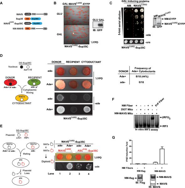
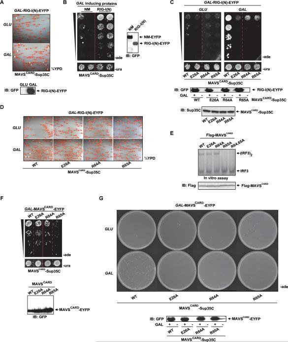
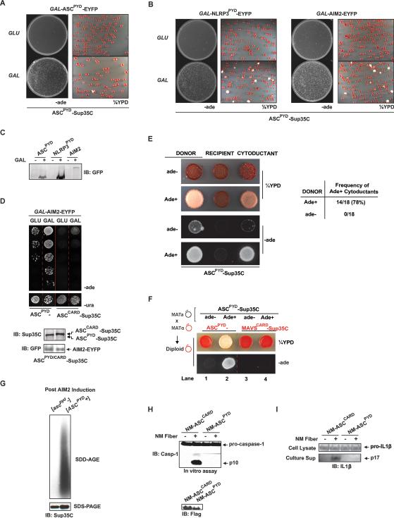
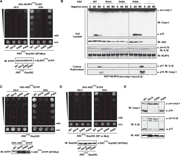
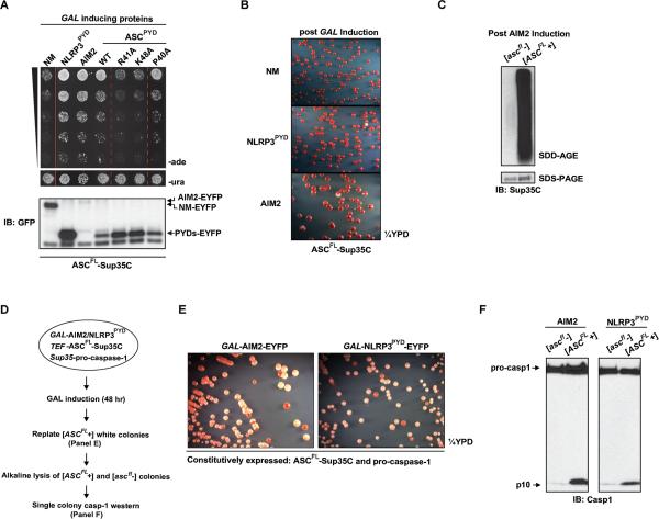
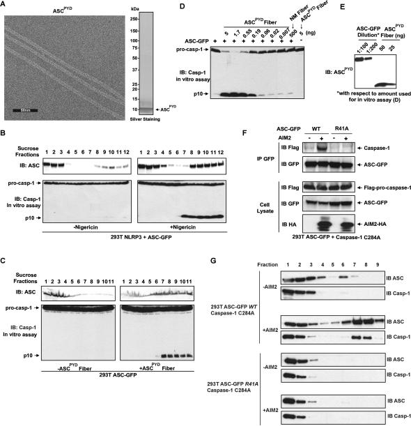
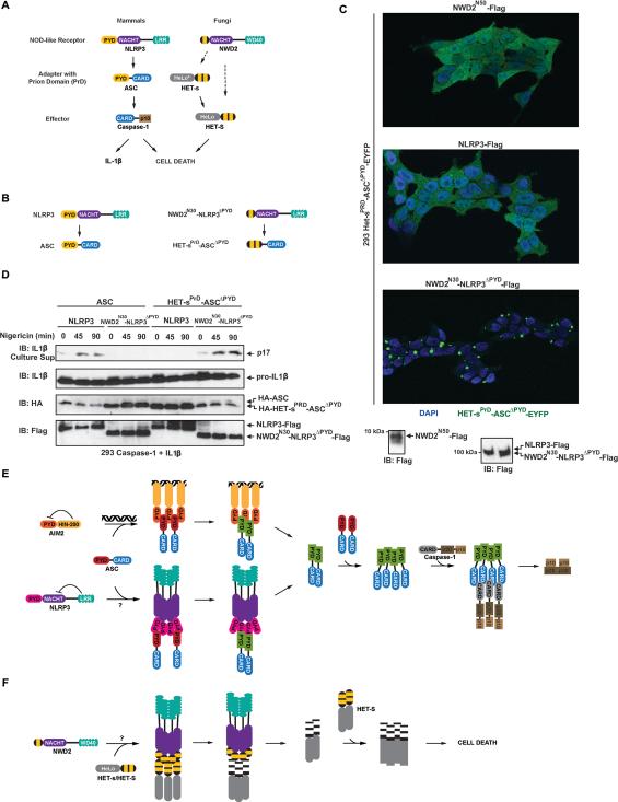
Pathogens and cellular danger signals activate sensors such as RIG-I and NLRP3 to produce robust immune and inflammatory responses through respective adaptor proteins MAVS and ASC, which harbor essential N-terminal CARD and PYRIN domains, respectively. Here, we show that CARD and PYRIN function as bona fide prions in yeast and that their prion forms are inducible by their respective upstream activators. Likewise, a yeast prion domain can functionally replace CARD and PYRIN in mammalian cell signaling. Mutations in MAVS and ASC that disrupt their prion activities in yeast also abrogate their ability to signal in mammalian cells. Furthermore, fibers of recombinant PYRIN can convert ASC into functional polymers capable of activating caspase-1. Remarkably, a conserved fungal NOD-like receptor and prion pair can functionally reconstitute signaling of NLRP3 and ASC PYRINs in mammalian cells. These results indicate that prion-like polymerization is a conserved signal transduction mechanism in innate immunity and inflammation.


Figures

Figure 1
Figure 1. MAVSCARD and Sup35NM Functionally Replace Each Other in Yeast and Mammalian Cells
(A) Cartoon depictions of MAVS, Sup35, MAVSCARD-Sup35C and NM-MAVS proteins. (B) Cells from a single yeast colony harboring constitutively expressed MAVSCARD-Sup35C and galactose inducible (GAL-)MAVSCARD-EYFP (Ura+) were grown for 48 hr in media containing either galactose (SG-ura) or glucose (SD-ura), followed by plating onto ¼YPD at a density of ~500 cells/plate. In this and subsequent panels, induction of EYFP-tagged protein was monitored by western blotting with a GFP antibody. (C) Single yeast colonies harboring constitutively expressed MAVSCARD-Sup35C and GAL-NM-EYFP or GAL-MAVSCARD-EYFP (Ura+) were grown in SD-ura for 24 hr to equal density followed by growth in SG-ura for 48 hr. Two colonies of each were plated onto non prion-selective media (SD-ura) and prion-selective, adenine-deficient media (SD-ade) at five-fold serial dilutions. In this and subsequent figures, one dilution on SD-ura is shown as an indicator of equal plating density among the samples. Dotted red line in this and other figures indicates serial dilutions grown non-adjacently on the same plate. (D) Left: A schematic for the cytoduction experiments. Ade+ or ade- donor strain harboring death domain (DD)-Sup35C fusion (e.g., MAVSCARD-Sup35C) was cytoduced into a ρ recipient strain expressing the same DD-Sup35C fusion and a kar1 mutation (kar1-15) which prevents nuclear fusion. Haploid cytoductants, containing both parental cytoplasms but only the recipient nucleus, were selected. Middle: Representative single clones of Ade+ or ade- donors, ade-recipients, or resultant cytoductants each expressing MAVSCARD-Sup35C were plated onto ¼YPD and SD-ade. Right: A table showing the frequency of Ade+ cytoductants from Ade+ or ade- MAVSCARD-Sup35C donors. (E) Left: A schematic for the mating and plasmid shuffle experiments. Ade+ or ade- haploid yeast of mating type a (MATa) expressing DD-Sup35C fusion from a Leu+ plasmid (black circle) were mated with an ade- MATα strain harboring DD-Sup35C on a Trp+ plasmid (red circle), followed by selection for diploids that have lost the original DD-Sup35C Leu+ plasmid. The resultant leu- Trp+ diploids were assessed for the prion phenotype. Right: An ade- or Ade+ MATa strain of MAVSCARD-Sup35C was mated with an ade- MATα strain harboring either MAVSCARD-Sup35C or ASCPYD-Sup35C following the protocol outlined above. Representative single colonies of leu- Trp+ diploids were plated onto ¼YPD and SD-ade. Lanes are numbered as described in the text. (F) Mitochondrial fractions from parental 293T or those expressing NM-MAVS were incubated with NM or PrP fibers as indicated followed by in vitro IRF3 assay. Dimerization of IRF3 was visualized by autoradiography following native gel electrophoresis. (G) NM-flag or NM-MAVS was transfected into 293T-IFNβ-luciferase reporter cells. Recombinant NM fibers were added to the culture media 24 hr later and allowed to incubate for additional 24 hr, followed by luciferase reporter assay. Error bar represents standard deviation of triplicates. See also Figure S1.
Figure 2
Figure 2. MAVSCARD-Sup35C Prion Assay Faithfully Recapitulates RIG-I Dependent MAVS Activation and Reveals Stepwise Polymerization of MAVSCARD in Yeast
(A) A single yeast colony harboring constitutively expressed MAVSCARD-Sup35C and GAL-RIG-I(N)-EYFP (Ura+) was grown in SD-ura (top) or SG-ura (bottom) for 48 hr followed by plating onto ¼YPD at a density of 500 cells/plate. (B) Single yeast colonies harboring MAVSCARD-Sup35C and NM- or RIG-I(N)-EYFP were grown in SD-ura to equal density followed by growth in SG-ura for 48 hr before plating of two independent colonies at five-fold serial dilutions onto SD-ade and SD-ura. The solid red line in this and other figures indicates serial dilutions grown under the same conditions on separate plates in the same experiment. (C) Similar to (B), except single yeast colonies harbored constitutively expressed WT or mutant MAVSCARD-Sup35C and GAL-RIG-I(N)-EYFP. Expression of MAVSCARD-Sup35C was monitored using a Sup35C antibody. (D) Similar to (C), except that cells were plated onto ¼YPD at a density of 500 cells/plate. (E) Mitochondrial fractions from parental 293T cells were incubated at RT with lysates of 293T cells expressing WT or mutant MAVSCARD, followed by in vitro IRF3 assay. Dimerization of IRF3 was visualized by autoradiography following native gel electrophoresis. (F) Similar to (B), except cells expressed WT or mutant GAL-MAVSCARD-EYFP. (G) Similar to (C), except cells expressed GAL-MAVSCARD-EYFP and were plated at a density of 50,000 cells per plate. See also Figure S2.
Figure 3
Figure 3. ASCPYD Behaves as a Prion in Yeast and can be Functionally Substituted by the NM Prion Domain in Mammalian Cells
(A) A single yeast colony harboring constitutively expressed ASCPYD-Sup35C and GAL-ASCPYD-EYFP (Ura+) was grown in SD-ura or SG-ura for two days followed by plating onto SD-ade and ¼YPD at a density of 100,000 and 500 cells/plate, respectively. (B) Similar to (A), except that NLRP3PYD or AIM2, instead of ASCPYD, was used to induce ASCPYD prion formation. (C) GAL inducible protein expression in (A) and (B) is monitored by immunoblotting with a GFP antibody. (D) Single yeast colonies with constitutively expressed ASCPYD-Sup35C or ASCCARD-Sup35C and GAL-AIM2-EYFP were grown in SD-ura or SG-ura for two days followed by plating of five-fold serial dilutions onto SD-ade and SD-ura. One serial dilution on SD-ura is shown. (E) Left: Following cytoduction as depicted in Figure 1D, representative single clones of Ade+ or ade- donors, ade- recipients, and resultant cytoductants each expressing ASCPYD-Sup35C were plated onto ¼YPD or SD-ade. Right: A table showing the frequency of Ade+ cytoductants from Ade+ or ade- donors harboring ASCPYD-Sup35C. (F) Mating between an ade- or Ade+ MATa strain with an ade- MATα strain each harboring differentially marked DD-Sup35C was carried out as depicted in Figure 1E. Representative resultant diploid leu- Trp+ single colonies were plated onto ¼YPD or SD-ade. (G) Following induction by AIM2 expression, colonies with soluble or prion form of ASCPYD ([ascpyd-] or [ASCPYD+], respectively) were subjected to SDD-AGE analysis for SDS-resistant polymers and SDS-PAGE for expression levels. (H) Cell lysates from NM-ASCPYD or NM-ASCCARD transfected 293T cells were incubated with NM fibers and lysates containing pro-caspase-1 followed by western analysis for caspase-1 activation. (I) NM fibers were added to the culture media of NM-ASCPYD or NM- ASCCARD transfected 293T cells stably expressing pro-caspase-1 and pro-IL1β, followed by immunoblotting analysis of secreted IL1β (p17) and pro-IL1β. See also Figure S3.
Figure 4
Figure 4. ASC Mutants Defective in Prion Formation Are Defective in Inflammasome Signaling
(A) Individual yeast colonies constitutively expressing WT or mutant ASCPYD-Sup35C and GAL-NLRP3PYD-EYFP were grown in SD-ura or SG-ura for two days, followed by plating of five-fold serial dilutions onto SD-ura and SD-ade. (B) Nigericin was added to ASC WT or mutant transfected 293T cells stably expressing NLRP3, pro-caspase-1, and pro-IL-1β, followed by western blot analysis of cell lysates for caspase-1 processing and of culture supernatants for active caspase-1 (p10) and IL-1β (p17) secretion. (C) Similar to (A), except that WT or mutant ASCPYD, instead of NLRP3PYD, was used to induce ASCPYD prion conversion. (D) Individual colonies constitutively expressing WT or mutant ASCPYD-Sup35C and WT GAL-ASCPYD-EYFP were treated as in (A). (E) Lysates of 293T cells expressing WT or mutant ASC were incubated with pro-caspase-1 and pro-IL-1β containing cell extracts, followed by analysis of caspase-1 and IL-1β cleavage by western blotting. Irrelevant lanes were removed for clarity.
Figure 5
Figure 5. Reconstitution of Inflammasome Signaling in Yeast Reveals Caspase-1 Activation Only in Colonies Containing ASC Prions
(A) Single colonies with constitutively expressed ASCFL-Sup35C and GAL inducible NM, NLRP3PYD, AIM2, WT or mutant ASCPYD were grown to similar density in SD-ura, followed by incubation in SG-ura for two days. Five-fold serial dilutions were then plated onto SD-ade and SD-ura. (B) Similar to (A) except that cells were plated to ¼YPD at 500 cells/plate. (C) Following induction of Ade+ by AIM2 expression, colonies with soluble or prion form of ASCFL-Sup35C ([ascfl-] or [ASCFL+], respectively) were subjected to SDD-AGE analysis for SDS-resistant aggregates and SDS-PAGE for expression levels. (D-F) Schematic for the reconstitution of inflammasome signaling in yeast (D). Yeasts expressing AIM2- or NLRP3PYD-EYFP from a GAL inducible promoter (Ura+), ASCFL-Sup35C from the constitutive TEF promoter, and pro-caspase-1 from the constitutive SUP35 promoter were grown in galactose containing media for 2 days. The resultant [ASCFL+] colonies were re-streaked onto ¼YPD (E), from which individual [ascfl-] or [ASCFL+] colonies were lysed and subjected to western analysis for caspase-1 activation (F). See also Figure S4.
Figure 6
Figure 6. Preformed ASCPYD Fibers Convert Inactive ASC into an Active High Molecular Weight Form Capable of Downstream Signaling
(A) Electron microscopy of negatively stained recombinant ASCPYD protein. (B) 293T cells stably expressing NLRP3 and ASCFL-GFP were fractionated on a 20-60% sucrose gradient cushion before and after Nigericin treatment. Individual fractions were western blotted with an ASC specific antibody (top) or incubated with pro-caspase1 containing cell extract, followed by western analysis of caspase-1 activation. (C) Lysate from 293T cells stably expressing ASCFL-GFP were incubated at 30°C for 30 min with or without ASCPYD fibers, followed by the same analysis as in (B). (D) Lysate from 293T cells stably expressing ASCFL-GFP were incubated at 30°C for 60 min with different amounts of ASCPYD fibers or NM fibers as indicated, along with pro-caspase-1 containing cell extract, followed by western blot analysis for caspase-1 activation. (E) Dilutions of lysate of 293T cells stably expressing ASCFL-GFP as in (D) was quantitatively compared to different amounts of ASCPYD fibers by western blotting with an antibody specific to ASCPYD. (F) 293T cells stably expressing flag-caspase-1 C284A and ASC-GFP WT or R41A were transfected with an AIM2 expression plasmid or mock treated. ASC was immunoprecipitated with a GFP antibody and then co-immunoprecipitated flag-caspase-1 was immunoblotted with a flag antibody. (G) Lysate from (F) was fractionated on a 20-60% sucrose gradient cushion followed by immunoblot analysis of each fraction with indicated antibodies. See also Figure S5.
Figure 7
Figure 7. Conserved Fungal Pattern Recognition Receptor and Death Inducing Prion Functionally Replace NLRP3PYD and ASCPYD in Inflammasome Signaling
(A) Cartoon representations of the mammalian NLRP3 inflammasome signaling and fungal NWD2 signaling pathways. Dotted arrow indicates an unconfirmed potential signaling interaction. The predicted HET-sPrD fold of NWD2 N-terminal domain is indicated using a yellow stripe, whereas HET-sPrD harbors two such motifs in tandem. Asterisk indicates inactive HeLo domain. (B) Cartoon representations of fusion proteins between NWD2/HET-s and NLRP3/ASC. (C) Confocal microscopy images of 293T cells stably expressing HET-sPrD-ASCΔPYD-EYFP and transfected NWD2N50, NLRP3, or NWD2N30-NLRP3ΔPYD as indicated on the top of each image. (D) 293T cells stably expressing NLRP3 or NWD2N30-NLRP3ΔPYD along with pro-caspase-1 and pro-IL1β were transfected with either WT ASC or HET-sPrD-ASCΔPYD. Following Nigericin treatment, secretion of mature IL1β and expression of indicated proteins were analyzed by western blotting. (E) Model for ASC-dependent inflammasome signaling. See text. (F) Model for NWD2 mediated HET-S activation. See text. See also Figure S6.

Comment in

Similar articles

Cited by

References

    1. Agostini L, Martinon F, Burns K, McDermott MF, Hawkins PN, Tschopp J. NALP3 forms an IL-1beta-processing inflammasome with increased activity in Muckle-Wells autoinflammatory disorder. Immunity. 2004;20:319–325. - PubMed
    1. Alberti S, Halfmann R, King O, Kapila A, Lindquist S. A systematic survey identifies prions and illuminates sequence features of prionogenic proteins. Cell. 2009;137:146–158. - PMC - PubMed
    1. Broz P, Newton K, Lamkanfi M, Mariathasan S, Dixit VM, Monack DM. Redundant roles for inflammasome receptors NLRP3 and NLRC4 in host defense against Salmonella. The Journal of experimental medicine. 2010;207:1745–1755. - PMC - PubMed
    1. Daskalov A, Paoletti M, Ness F, Saupe SJ. Genomic clustering and homology between HET-S and the NWD2 STAND protein in various fungal genomes. PloS one. 2012;7:e34854. - PMC - PubMed
    1. Ferrao R, Wu H. Helical assembly in the death domain (DD) superfamily. Current opinion in structural biology. 2012;22:241–247. - PMC - PubMed

Publication types

MeSH terms