

Lack of transglutaminase 2 diminished T-cell responses in mice
- PMID: 24628083
- PMCID: PMC4080966
- DOI: 10.1111/imm.12282
Lack of transglutaminase 2 diminished T-cell responses in mice
Abstract
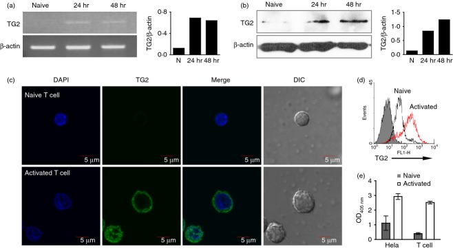
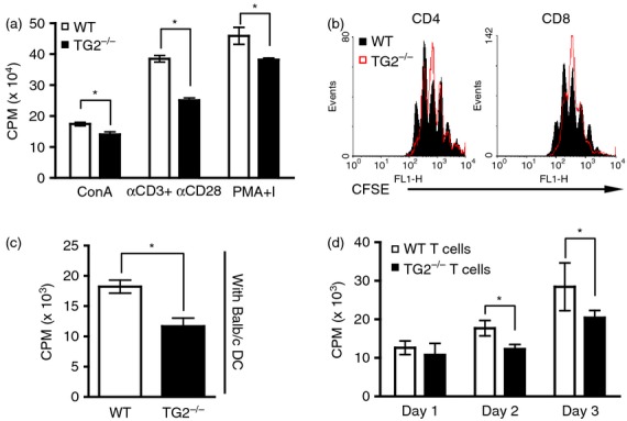
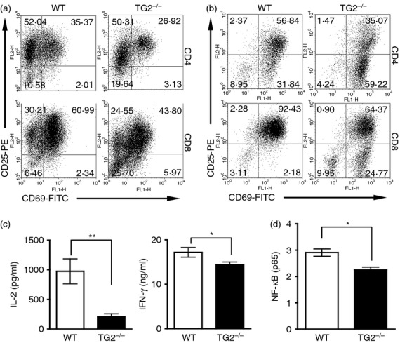
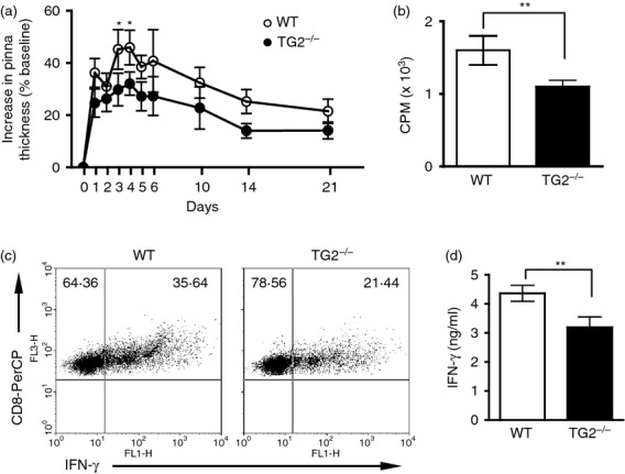
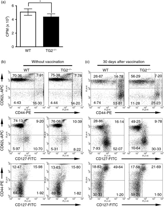
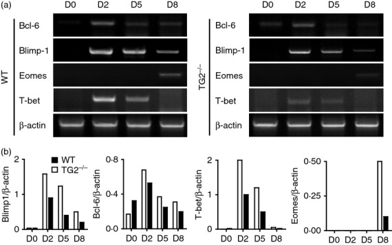
Transglutaminase 2 (TG2) has been reported to play a role in dendritic cell activation and B-cell differentiation after immunization. Its presence and role in T cells, however, has not been explored. In the present study, we determined the expression of TG2 on mouse T cells, and evaluated its role by comparing the behaviours of wild-type and TG2(-/-) T cells after activation. In our results, naive T cells minimally expressed TG2, expression of which was increased after activation. T-cell proliferation, expression of activation markers such as CD69 and CD25, and secretions of interleukin-2 and interferon-γ were suppressed in the absence of TG2, presumably due, in part, to diminished nuclear factor-κB activation. These effects on T cells seemed to be reflected in the in vivo immune response, the contact hypersensitivity reaction elicited by 2,4-dinitro-1-fluorobenzene, with lowered peak responses in the TG2(-/-) mice. When splenic T cells from mice immunized with tumour lysate-loaded wild-type dendritic cells were re-challenged ex vivo with the same antigen, the profile of surface markers including CD44, CD62L, and CD127 strongly indicated lesser generation of memory CD8(+) T cells in TG2(-/-) mice. In the TG2(-/-) CD8(+) T cells, moreover, Eomes expression was markedly decreased. These results indicate possible roles of TG2 in CD8(+) T-cell activation and CD8(+) memory T-cell generation.
Keywords: CD8+ T cells; Eomes; T-cell activation; transglutaminase 2.
© 2014 John Wiley & Sons Ltd.
Figures
Similar articles
-
Transglutaminase 2 on the surface of dendritic cells is proposed to be involved in dendritic cell-T cell interaction.Cell Immunol. 2014 May-Jun;289(1-2):55-62. doi: 10.1016/j.cellimm.2014.03.008. Epub 2014 Mar 31. Cell Immunol. 2014. PMID: 24727157
-
Airway epithelial cells initiate the allergen response through transglutaminase 2 by inducing IL-33 expression and a subsequent Th2 response.Respir Res. 2013 Mar 13;14(1):35. doi: 10.1186/1465-9921-14-35. Respir Res. 2013. PMID: 23496815 Free PMC article.
-
Transglutaminase 2 exacerbates experimental autoimmune encephalomyelitis through positive regulation of encephalitogenic T cell differentiation and inflammation.Clin Immunol. 2012 Nov;145(2):122-32. doi: 10.1016/j.clim.2012.08.009. Epub 2012 Aug 23. Clin Immunol. 2012. PMID: 23001131
-
Some lessons from the tissue transglutaminase knockout mouse.Amino Acids. 2009 Apr;36(4):625-31. doi: 10.1007/s00726-008-0130-x. Epub 2008 Jun 27. Amino Acids. 2009. PMID: 18584284 Review.
-
Transglutaminase 2: an enigmatic enzyme with diverse functions.Trends Biochem Sci. 2002 Oct;27(10):534-9. doi: 10.1016/s0968-0004(02)02182-5. Trends Biochem Sci. 2002. PMID: 12368090 Review.
Cited by
-
Positive Feedback Regulation between Transglutaminase 2 and Toll-Like Receptor 4 Signaling in Hepatic Stellate Cells Correlates with Liver Fibrosis Post Schistosoma japonicum Infection.Front Immunol. 2017 Dec 13;8:1808. doi: 10.3389/fimmu.2017.01808. eCollection 2017. Front Immunol. 2017. PMID: 29321784 Free PMC article.
-
Essential Role for CD30-Transglutaminase 2 Axis in Memory Th1 and Th17 Cell Generation.Front Immunol. 2020 Jul 21;11:1536. doi: 10.3389/fimmu.2020.01536. eCollection 2020. Front Immunol. 2020. PMID: 32793209 Free PMC article.
-
Role of Transglutaminase 2 in Cell Death, Survival, and Fibrosis.Cells. 2021 Jul 20;10(7):1842. doi: 10.3390/cells10071842. Cells. 2021. PMID: 34360011 Free PMC article. Review.
-
A Signature-Based Classification of Gastric Cancer That Stratifies Tumor Immunity and Predicts Responses to PD-1 Inhibitors.Front Immunol. 2021 Jun 11;12:693314. doi: 10.3389/fimmu.2021.693314. eCollection 2021. Front Immunol. 2021. PMID: 34177954 Free PMC article.
-
Computational properties of mitochondria in T cell activation and fate.Open Biol. 2016 Nov;6(11):160192. doi: 10.1098/rsob.160192. Open Biol. 2016. PMID: 27852805 Free PMC article. Review.
References
-
- Maiuri L, Luciani A, Giardino I, et al. Tissue transglutaminase activation modulates inflammation in cystic fibrosis via PPARγ down-regulation. J Immunol. 2008;180:7697–705. - PubMed
-
- Oh K, Park HB, Seo MW, Byoun OJ, Lee DS. Transglutaminase 2 exacerbates experimental autoimmune encephalomyelitis through positive regulation of encephalitogenic T cell differentiation and inflammation. Clin Immunol. 2012;145:122–32. - PubMed
-
- Falasca L, Farracem MG, Rinaldi A, Tuosto L, Melino G, Piacentini M. Transglutaminase type II is involved in the pathogenesis of endotoxic shock. J Immunol. 2008;180:2616–24. - PubMed
Publication types
MeSH terms
Substances
LinkOut - more resources
Full Text Sources
Other Literature Sources
Molecular Biology Databases
Research Materials
Miscellaneous
