Complex formation between S100B protein and the p90 ribosomal S6 kinase (RSK) in malignant melanoma is calcium-dependent and inhibits extracellular signal-regulated kinase (ERK)-mediated phosphorylation of RSK
- PMID: 24627490
- PMCID: PMC4007476
- DOI: 10.1074/jbc.M114.561613
Complex formation between S100B protein and the p90 ribosomal S6 kinase (RSK) in malignant melanoma is calcium-dependent and inhibits extracellular signal-regulated kinase (ERK)-mediated phosphorylation of RSK
Abstract
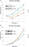
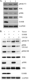
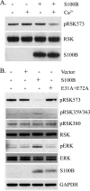
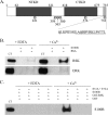
S100B is a prognostic marker for malignant melanoma. Increasing S100B levels are predictive of advancing disease stage, increased recurrence, and low overall survival in malignant melanoma patients. Using S100B overexpression and shRNA(S100B) knockdown studies in melanoma cell lines, elevated S100B was found to enhance cell viability and modulate MAPK signaling by binding directly to the p90 ribosomal S6 kinase (RSK). S100B-RSK complex formation was shown to be Ca(2+)-dependent and to block ERK-dependent phosphorylation of RSK, at Thr-573, in its C-terminal kinase domain. Additionally, the overexpression of S100B sequesters RSK into the cytosol and prevents it from acting on nuclear targets. Thus, elevated S100B contributes to abnormal ERK/RSK signaling and increased cell survival in malignant melanoma.
Keywords: Calcium; Calcium Binding Proteins; Cancer; ERK; MAP Kinases (MAPKs); Melanoma; RSK; S100 Proteins.
Figures


Similar articles
-
The calcium-binding protein S100B reduces IL6 production in malignant melanoma via inhibition of RSK cellular signaling.PLoS One. 2021 Aug 19;16(8):e0256238. doi: 10.1371/journal.pone.0256238. eCollection 2021. PLoS One. 2021. PMID: 34411141 Free PMC article.
-
Structural Basis of Ribosomal S6 Kinase 1 (RSK1) Inhibition by S100B Protein: MODULATION OF THE EXTRACELLULAR SIGNAL-REGULATED KINASE (ERK) SIGNALING CASCADE IN A CALCIUM-DEPENDENT WAY.J Biol Chem. 2016 Jan 1;291(1):11-27. doi: 10.1074/jbc.M115.684928. Epub 2015 Nov 2. J Biol Chem. 2016. PMID: 26527685 Free PMC article.
-
RSK regulates activated BRAF signalling to mTORC1 and promotes melanoma growth.Oncogene. 2013 Jun 13;32(24):2917-2926. doi: 10.1038/onc.2012.312. Epub 2012 Jul 16. Oncogene. 2013. PMID: 22797077 Free PMC article.
-
The RSK factors of activating the Ras/MAPK signaling cascade.Front Biosci. 2008 May 1;13:4258-75. doi: 10.2741/3003. Front Biosci. 2008. PMID: 18508509 Review.
-
Regulation and function of the RSK family of protein kinases.Biochem J. 2012 Jan 15;441(2):553-69. doi: 10.1042/BJ20110289. Biochem J. 2012. PMID: 22187936 Review.
Cited by
-
Disordered Protein Kinase Regions in Regulation of Kinase Domain Cores.Trends Biochem Sci. 2019 Apr;44(4):300-311. doi: 10.1016/j.tibs.2018.12.002. Epub 2019 Jan 2. Trends Biochem Sci. 2019. PMID: 30611608 Free PMC article. Review.
-
S100 proteins in cancer.Nat Rev Cancer. 2015 Feb;15(2):96-109. doi: 10.1038/nrc3893. Nat Rev Cancer. 2015. PMID: 25614008 Free PMC article. Review.
-
Covalent small molecule inhibitors of Ca(2+)-bound S100B.Biochemistry. 2014 Oct 28;53(42):6628-40. doi: 10.1021/bi5005552. Epub 2014 Oct 14. Biochemistry. 2014. PMID: 25268459 Free PMC article.
-
The calcium-binding protein S100B reduces IL6 production in malignant melanoma via inhibition of RSK cellular signaling.PLoS One. 2021 Aug 19;16(8):e0256238. doi: 10.1371/journal.pone.0256238. eCollection 2021. PLoS One. 2021. PMID: 34411141 Free PMC article.
-
Targeting S100 Calcium-Binding Proteins with Small Molecule Inhibitors.Methods Mol Biol. 2019;1929:291-310. doi: 10.1007/978-1-4939-9030-6_19. Methods Mol Biol. 2019. PMID: 30710281 Free PMC article.
References
-
- Zimmer D. B., Cornwall E. H., Landar A., Song W. (1995) The S100 protein family: history, function, and expression. Brain Res. Bull. 37, 417–429 - PubMed
-
- Donato R. (2001) S100: a multigenic family of calcium-modulated proteins of the EF-hand type with intracellular and extracellular functional roles. Int. J. Biochem. Cell Biol. 33, 637–668 - PubMed
-
- Gaynor R., Herschman H. R., Irie R., Jones P., Morton D., Cochran A. (1981) S100 protein: a marker for human malignant melanomas? Lancet 1, 869–871 - PubMed
-
- Nagasaka A., Umekawa H., Hidaka H., Iwase K., Nakai A., Ariyoshi Y., Ohyama T., Aono T., Nakagawa H., Ohtani S. (1987) Increase in S-100b protein content in thyroid carcinoma. Metabolism 36, 388–391 - PubMed
-
- Yang J. F., Zhang X. Y., Qi F. (2004) Expression of S100 protein in renal cell carcinoma and its relation with p53. Zhong Nan. Da Xue Xue Bao. Yi Xue. Ban 29, 301–304 - PubMed
Publication types
MeSH terms
Substances
Grants and funding
LinkOut - more resources
Full Text Sources
Other Literature Sources
Miscellaneous

