. 2014 Apr 25;289(17):11779-11790.
doi: 10.1074/jbc.M114.552307. Epub 2014 Mar 11.

A shrimp C-type lectin inhibits proliferation of the hemolymph microbiota by maintaining the expression of antimicrobial peptides

Affiliations

A shrimp C-type lectin inhibits proliferation of the hemolymph microbiota by maintaining the expression of antimicrobial peptides

Xian-Wei Wang et al. J Biol Chem. .

Abstract

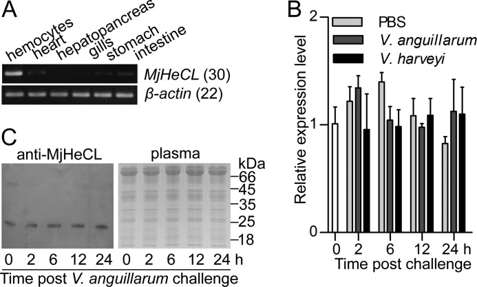
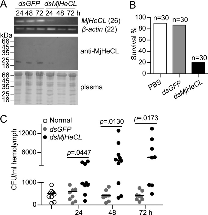
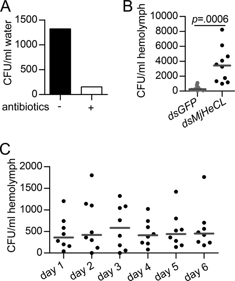
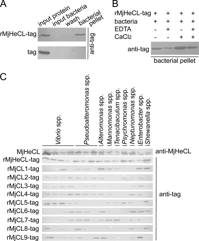
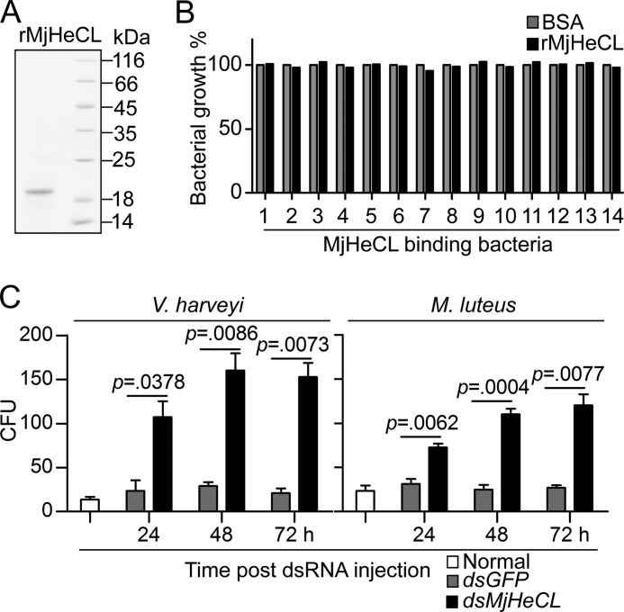
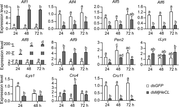
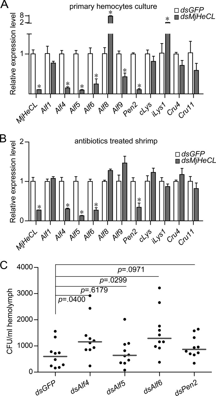
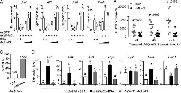
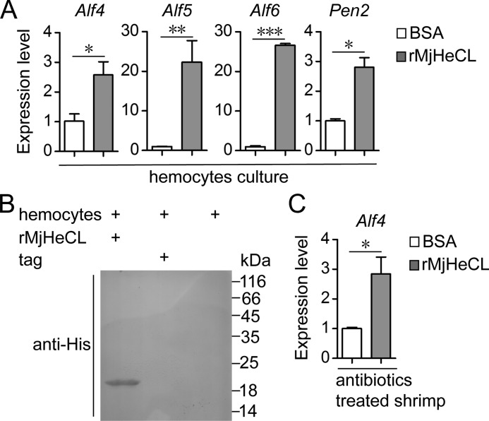
Some aquatic invertebrates such as shrimp contain low albeit stable numbers of bacteria in the circulating hemolymph. The proliferation of this hemolymph microbiota in such a nutrient-rich environment is tightly controlled in healthy animals, but the mechanisms responsible had remained elusive. In the present study, we report a C-type lectin (MjHeCL) from the kuruma shrimp (Marsupenaeus japonicus) that participates in restraining the hemolymph microbiota. Although the expression of MjHeCL did not seem to be modulated by bacterial challenge, the down-regulation of its expression by RNA interference led to proliferation of the hemolymph microbiota, ultimately resulting in shrimp death. This phenotype was rescued by the injection of recombinant MjHeCL, which restored the healthy status of the knockdown shrimp. A mechanistic analysis revealed that MjHeCL inhibited bacterial proliferation by modulating the expression of antimicrobial peptides. The key function of MjHeCL in the shrimp immune homeostasis might be related to its broader recognition spectrum of the hemolymph microbiota components than other lectins. Our study demonstrates the role of MjHeCL in maintaining the healthy status of shrimp and provides new insight into the biological significance of C-type lectins, a diversified and abundant lectin family in invertebrate species.

Keywords: Antimicrobial Peptides; C-type Lectin; Hemolymph Microbiota; Invertebrates; Lectin; Pattern Recognition Receptor; RNA Interference (RNAi); Shrimp.


Figures

FIGURE 1.
FIGURE 1.
Expression of MjHeCL is not significantly affected by bacterial challenge. A, MjHeCL was expressed mainly in hemocytes as analyzed by semiquantitative RT-PCR with β-actin as the reference. B, MjHeCL did not respond to external bacterial challenge. Shrimp were challenged by V. anguillarum or V. harveyi, and the expression of MjHeCL in hemocytes was assessed postchallenge by qRT-PCR. Data represent mean ± SD from three independent repeats. Error bars represent S.D. C, MjHeCL was constitutively secreted into plasma. Shrimp plasma was collected after bacterial challenge, subjected to ultracentrifugation to remove the majority of hemocyanin, concentrated, and subjected to Western blot analyzed by MjHeCL antiserum.
FIGURE 2.
FIGURE 2.
MjHeCL inhibits the proliferation of endogenous bacteria. A, RNAi efficiency of MjHeCL. Shrimp were injected with 10 μg of dsRNA, and the expression of MjHeCL was analyzed at 24, 48, and 72 h postinjection by RT-PCR (upper panel) and Western blot (lower panel). GFP dsRNA (dsGFP) was used as a control. B, knockdown of MjHeCL led to shrimp death. Thirty shrimp were injected with 10 μg of MjHeCL dsRNA (dsMjHeCL) each, and shrimp death was recorded 72 h postinjection. PBS and GFP dsRNA were used as controls. C, the bacterial counts in MjHeCL knockdown shrimp were higher than in the control animals. Shrimp were injected with 10 μg of MjHeCL dsRNA, and the hemolymph was drawn out at 24, 48, and 72 h postinjection and plated onto agar plates, and the bacterial counts were determined. The horizontal bars represent the median. The p value was calculated by the t test for paired samples, and significant differences was accepted when p was <0.05.
FIGURE 3.
FIGURE 3.
The source of proliferating bacteria was the endogenous microbiota. A, antibiotics were used to eliminate the bacteria in the aquarium water. Ampicillin and kanamycin were added to the water to a final concentration of 25 mg/liter. B, endogenous bacteria still proliferated when MjHeCL knockdown shrimp were cultured in the antibiotic-treated water. Shrimp were injected with 10 μg of MjHeCL dsRNA (dsMjHeCL), and the bacterial counts in hemolymph at 24 h postinjection were determined. GFP dsRNA (dsGFP) was used as the control. The horizontal bars represent the median. The p value was calculated by the t test for paired samples. C, healthy shrimp host bacteria in the hemolymph. The bacterial counts in the hemolymph from eight shrimp maintained under standard conditions were determined every day for 6 continuous days. The horizontal bars represent the median.
FIGURE 4.
FIGURE 4.
MjHeCL binds to all strains isolated from the hemolymph microbiota. A, recombinant MjHeCL binds to a Vibrio spp. isolated from hemolymph. Overnight cultured bacteria were collected, resuspended at about 108 CFU/ml, and incubated with 4 μg of recombinant proteins (from bacteria) for 1 h at 25 °C. The bacteria were then washed and analyzed by Western blot using antibodies against the tag of pET32a(+). B, calcium requirement of MjHeCL for binding to bacteria (Vibrio spp.). Either EDTA (5 mm), CaCl2 (5 mm), or both were added to the bacterium-rMjHeCL mixture, and the binding was evaluated by Western blot. C, MjHeCL shows a broader binding spectrum than other shrimp CTLs. Binding of shrimp recombinant CTLs and native MjHeCL was determined as described above. Hemocyte lysate supernatant (1 ml) was used as the source of native MjHeCL, and binding was detected with antibody against MjHeCL.
FIGURE 5.
FIGURE 5.
The antimicrobial activity of plasma was suppressed in the MjHeCL knockdown shrimp. A, expression and purification of recombinant MjHeCL. The recombinant MjHeCL was expressed in P. pastoris GS115 and purified by affinity chromatography. B, the recombinant MjHeCL did not show antimicrobial activity. Recombinant protein (2 μg) was incubated with the bacterial suspension (∼104 CFU) for 2 h, the mixture was added to 200 μl of fresh medium, and the bacterial growth was recorded 24 h later. The strains used here are Vibrio spp. (1–4), Pseudoalteromonas spp. (5–7), Alteromonas spp. (8), Marinomonas spp. (9), Tenacibaculum spp. (10), Psychromonas spp. (11), Neptunomonas spp. (12), Enterobacter spp. (13), and Shewanella spp. (14). C, the plasma antimicrobial activity was suppressed in the MjHeCL knockdown shrimp. Equal volumes (10 μl) of plasma and bacterial suspension were incubated at 25 °C for 1 h. The mixture was then plated onto agar plates to determine the surviving bacteria. Data show the mean ± S.D. from three independent repeats. Error bars represent S.D. The p value was calculated by t test for paired samples, and significant differences were accepted when p was <0.05. dsMjHeCL, MjHeCL dsRNA; dsGFP, GFP dsRNA.
FIGURE 6.
FIGURE 6.
Knockdown of MjHeCL induced changes in expression of AMPs in hemocytes. Shrimp were injected with 10 μg of MjHeCL dsRNA (dsMjHeCL), and the expression of AMPs was analyzed by qRT-PCR at 24, 48, and 72 h postinjection. GFP dsRNA (dsGFP) was injected as a control. Data represent mean ± SD from three independent repeats. Error bars represent S.D. Data were subjected to one-way analysis of variance from triplicate assays. Significant differences (p < 0.05) are represented by different letters.
FIGURE 7.
FIGURE 7.
MjHeCL maintains the expression of Alf4, Alf5, Alf6, and Pen2, which are responsible to inhibit proliferation of endogenous bacteria. A, in vitro knockdown of MjHeCL in hemocytes suppressed the expression of Alf4, Alf5, Alf6, and Pen2. Shrimp hemocytes were precultured in 6-well plates. Two micrograms of dsRNA were transfected into hemocytes with 5 μl of Lipofectamine 2000 according to the manufacturer's instructions. The medium was replaced 10 h later. The expression of AMPs was analyzed after 24 h. GFP dsRNA (dsGFP) was used as the control. B, knockdown of MjHeCL in antibiotic-treated shrimp suppressed the expression of Alf4, Alf5, Alf6, and Pen2. Shrimp were preinjected with ampicillin and kanamycin (25 μg each), and 24 h later, 10 μg of dsRNA were injected into each shrimp. The expression of AMPs was analyzed by qRT-PCR 24 h later. For A and B, data represent mean ± SD from three independent repeats. Error bars represent S.D. Asterisks represent significant differences (calculated by the t test for paired samples from three repeats, and significant differences were accepted when p was <0.05). C, knockdown of Alf4, Alf5, Alf6, and Pen2 triggered proliferation of endogenous bacteria. Shrimp were injected with 10 μg of Alf4 (dsAlf4), Alf5 (dsAlf5), Alf6 (dsAlf6), and Pen2 (dsPen2) dsRNA, respectively. The bacterial counts in the hemolymph were determined 24 h later. The horizontal bars represent the median. GFP dsRNA was used as a control. The p value was calculated by the t test for paired samples against controls, and significant differences were accepted when p was <0.05.
FIGURE 8.
FIGURE 8.
Injection of recombinant MjHeCL rescued the phenotype observed in the MjHeCL knockdown shrimp. A, injection of recombinant MjHeCL restored the expression of Alf4, Alf5, Alf6, and Pen2. Shrimp were injected with MjHeCL dsRNA (dsMjHeCL) (10 μg) together with increasing amounts of rMjHeCL (0.1, 0.5. 2.5, and 8 μg). The expression of Alf4, Alf5, Alf6, and Pen2 was analyzed by qRT-PCR 24 h later. GFP dsRNA (dsGFP) and BSA were used to control MjHeCL dsRNA and rMjHeCL. Data represent mean ± SD from three independent repeats with error bars representing S.D. The data were subjected to one-way analysis of variance analysis. Different letters represent significant differences (p < 0.05). B, injection of recombinant MjHeCL suppressed the bacterial proliferation caused by the MjHeCL knockdown. Shrimp were injected with a mixture of MjHeCL dsRNA (10 μg) and rMjHeCL (2.5 μg). The bacterial counts in hemolymph were determined 24, 48, and 72 h later. BSA was used as a control. The horizontal bars represent the median. C, injection of rMjHeCL restored shrimp survival. Shrimp were injected with a mixture of MjHeCL dsRNA (10 μg) and rMjHeCL (2.5 μg). The survival rate was recorded at 72 h postinjection. BSA was used as a control. p values were calculated by the t test for paired samples, and significant differences were accepted when p was <0.05. D, injection of recombinant MjHeCL restored the expression of AMPs down-regulated in the MjHeCL knockdown shrimp. Shrimp were injected with a mixture of MjHeCL dsRNA (10 μg) and rMjHeCL (2.5 μg). The expression of AMPs was analyzed by qRT-PCR 24 h later. GFP dsRNA and BSA were used to control MjHeCL dsRNA and rMjHeCL. Data represents mean ± SD from three independent repeats with error bars representing S.D. The data were subjected to one-way analysis of variance analysis. Different letters represent significant differences (p < 0.05).
FIGURE 9.
FIGURE 9.
MjHeCL binds to hemocytes to modulate AMP expression. A, recombinant MjHeCL alone stimulates AMPs expression in vitro. Hemocytes were cultured in 6-well plates, and recombinant MjHeCL or BSA (3 μg) was applied to the wells. Expression of AMPs was detected 6 h later. Data represents mean ± SD from three independent repeats with error bars representing S.D. Asterisks represent significant differences (calculated by the t test for paired samples from three repeats: *, p < 0.05; **, p < 0.01; **, p < 0.001). B, recombinant MjHeCL binds to hemocytes. Recombinant MjHeCL or control tag (3 μg) was added to the wells to incubate with the hemocytes for 3 h. Medium was removed, and cells were washed by PBS five times, lysed, and subjected to SDS-PAGE. Bound exogenous proteins were detected with the anti-His tag antibody. C, recombinant MjHeCL alone (in the absence of bacteria) stimulates AMP expression in vivo. Shrimp were pretreated with antibiotics to remove the hemolymph microbiota. Recombinant MjHeCL or BSA (5 μg) was injected into shrimp, and AMP expression was checked 24 h later. Data represent mean ± S.D. from three independent repeats with error bars representing S.D., and the asterisk represents a significant difference calculated by the t test for paired samples.

Similar articles

Cited by

References

    1. Duron O., Hurst G. D. (2013) Arthropods and inherited bacteria: from counting the symbionts to understanding how symbionts count. BMC Biol. 11, 45. - PMC - PubMed
    1. Kamada N., Seo S. U., Chen G. Y., Núñez G. (2013) Role of the gut microbiota in immunity and inflammatory disease. Nat. Rev. Immunol. 13, 321–335 - PubMed
    1. Maynard C. L., Elson C. O., Hatton R. D., Weaver C. T. (2012) Reciprocal interactions of the intestinal microbiota and immune system. Nature 489, 231–241 - PMC - PubMed
    1. Fagutao F. F., Koyama T., Kaizu A., Saito-Taki T., Kondo H., Aoki T., Hirono I. (2009) Increased bacterial load in shrimp hemolymph in the absence of prophenoloxidase. FEBS J. 276, 5298–5306 - PubMed
    1. Olafsen J. A., Mikkelsen H. V., Giaever H. M., Høvik Hansen G. (1993) Indigenous bacteria in hemolymph and tissues of marine bivalves at low temperatures. Appl. Environ. Microbiol. 59, 1848–1854 - PMC - PubMed

Publication types

LinkOut - more resources