Review
. 2014:5:e27952.
doi: 10.4161/sgtp.27952. Epub 2014 Mar 5.

Role of the Rho GTPase Rac in the activation of the phagocyte NADPH oxidase: outsourcing a key task

Affiliations
Review

Role of the Rho GTPase Rac in the activation of the phagocyte NADPH oxidase: outsourcing a key task

Edgar Pick. Small GTPases. 2014.

Abstract

The superoxide-generating NADPH oxidase of phagocytes consists of the membrane-associated cytochrome b 558 (a heterodimer of Nox2 and p22(phox)) and 4 cytosolic components: p47(phox), p67(phox), p40(phox), and the small GTPase, Rac, in complex with RhoGDI. Superoxide is produced by the NADPH-driven reduction of molecular oxygen, via a redox gradient located in Nox2. Electron flow in Nox2 is initiated by interaction with cytosolic components, which translocate to the membrane, p67(phox) playing the central role. The participation of Rac is expressed in the following sequence: (1) Translocation of the RacGDP-RhoGDI complex to the membrane; (2) Dissociation of RacGDP from RhoGDI; (3) GDP to GTP exchange on Rac, mediated by a guanine nucleotide exchange factor; (4) Binding of RacGTP to p67(phox); (5) Induction of a conformational change in p67(phox), promoting interaction with Nox2. The particular involvement of Rac in NADPH oxidase assembly serves as a paradigm for signaling by Rho GTPases, in general.

Keywords: GEF; NADPH oxidase; Rac1; Rac2; Rho GTPases; RhoGDI; cell-free system; cytochrome b558; p67phox; superoxide.


Figures

None
Figure 1. The interface between Rac and p67TPR. (A) Schematic representation of the hydrogen bond interactions at the interface between Rac and p67phox TPRs. Residues from Rac are labeled in red and residues from p67phox TPRs, in blue. Hydrogen bonds are depicted as dotted lines with the bond distances indicated in Å. The positions of switch I and the β hairpin insertion are indicated in red and blue, respectively. (B) Ribbons representation of the RacGTP (red)/p67phox TPR (blue) complex. The effector loop of Rac is colored in yellow, and amino acids 103–107 and the helical insert region (120–135) are indicated in green. The position of Gly30 at the N terminus of the effector loop is indicated to show the orientation of switch I. The positions of mutations in p67phox occurring in CGD are shown as red spheres (Reprinted with permission from ref. . Copyright 2000, Elsevier).
None
Figure 2. Schematic representation of [p67phox – Rac1] chimeras and a [p67phox – CDC42Hs] chimera. The numbering of chimeras are according to Alloul et al. The characteristic feature of each construct is briefly indicated at the right of the scheme of the chimera, with the color of the font corresponding to the color of respective moiety of the chimera. The presence (+) or absence (-) of NADPH oxidase supporting activity was determined in an amphiphile-activated cell-free system, consisting of phagocyte membrane, chimera in the GTPγS-bound form, and p47phox (modified from ref. 83).
None
Figure 3. Intrachimeric bonds between residues in the switch I of the Rac1 moiety and in the TPR domains of the p67phox moiety and an intact activation domain in the p67phox moiety of chimera [(p67phox(1–212)-Rac1(1–192)) (prototype chimera 3) are essential for enabling the chimera to support NADPH oxidase activity. The numbering of chimeras are according to Alloul et al. Four mutants of the prototype chimera 3 were constructed in which residues in the activation domain and one of the TPR domains of the p67phox moiety and 2 residues in the switch I of the Rac1 moiety were mutated. Graphs A and B describe the ability of mutant chimeras to support NADPH oxidase activation in vitro. (A) NADPH oxidase supporting activity of the mutant chimeras, in non-prenylated form, was assessed in an amphiphile-dependent cell-free assay consisting of phagocyte membrane, chimera in the GTPγS-bound form, and p47phox. (B) The activity of the equivalent prenylated chimeras was measured in an amphiphile-independent cell-free system, consisting of membrane and chimera in the GTPγS-bound form, in the absence of p47phox (modified from ref. 83).
None
Figure 4. Hypothetical models of the “closed” (active) and “open” (inactive) conformations of the prototype chimera 3 and Rac1 moiety mutants. Nucleotide exchange to GMPPNP or mutation Q61L, in the Rac1 moiety, assuring that the chimera is permanently in the GTP-bound form, lead to a closed conformation. A GDP-bound form of the chimera or mutations A27K and G30S in the Rac1 moiety of the chimera, in the GMPPNP-bound form, prevent protein – protein interaction with the p67phox moiety and lead to an open conformation (modified from ref. 83).
None
Figure 5. Structure of mutants used in intramolecular FRET studies on the [p67phox – Rac1] chimera. The prototype chimera [p67phox(1–212)-Rac1(1–192)] was subjected to either one (W56F) or 2 mutations (W56F; W97F) in the Rac1 moiety, resulting in the generation of chimeras with 4 tryptophans in the p67phox moiety and 1 or none in the Rac1 moiety. The W56F mutation was confirmed by the lack of response of the mutant Rac1 to the Rac-specific guanine nucleotide exchange factor (GEF), TrioN. The principle of intramolecular FRET is summarized in the bottom part of the figure. In the upper left panel of the figure is a ribbon diagram of Rac1 in the GMPPNP-bound form. Residues W56 and W97 are displayed in dark and light green, respectively. The GMPPNP is displayed as a stick model with atoms colored by atom type (oxygen, red; carbon, white; nitrogen, blue; phosphorous, yellow). The orange sphere represents the Mg2+. The binding surface of Rac1 with p67phox (based on ref. 72) is colored in pink. The upper right side panel illustrates an overlay of characteristic emission spectra (305–485 nm) of the [p67phox – Rac1] chimera containing W56F and W97F mutations, in the mant-GDP and mant-GMPPNP-bound forms, excited at 295 nm. The GMPPNP-bound form exhibits enhanced FRET, in comparison to the GDP-bound form of the same chimera. This is expressed in an increase in mant-dependent fluorescence emission, at 440 nm, and a reduction in tryptophan-dependent fluorescence emission, at 340 nm (modified from ref. 83).
None
Figure 6. A minimal polybasic stretch at the C-terminus of the [p67phox – Rac1] chimeras is essential for the support of NADPH oxidase activity. Four mutants of the prototype chimera 3 were constructed in which 1, 3, 4, or 6 basic residues were replaced by glutamines. The numbering of chimeras are according to Alloul et al. (A) NADPH oxidase supporting activity of the mutant chimeras, in nonprenylated form, was assessed in an amphiphile-dependent cell-free assay consisting of phagocyte membrane, chimera in the GTPγS-bound form, and p47phox. (B) The activity of the equivalent prenylated chimeras was measured in an amphiphile-independent cell-free system, consisting of membrane and chimera in the GTPγS-bound form, in the absence of p47phox (modified from ref. 83).
None
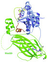
Figure 7. Ribbon representation of the Rac1−RhoGDI complex. Rac1 is depicted in blue and RhoGDI in green. The switch I and II regions in Rac1 are highlighted in yellow and red, respectively. The GDP molecule and geranylgeranyl group are represented in ball and sticks. Mg2+ is shown in gray. Loop (58−66) in GDI (dashed line) is not visible in the crystallographic structure (reprinted with permission from ref. . Copyright 2001, American Chemical Society).
None
Figure 8. Extrapolation of the in vitro mechanism of Rac-RhoGDI dissociation by the cooperative action of PtdIns(3,4,5)P3-containing liposomes, GTP and GEF, to events hypothesized to occur in the course of oxidase activation in the intact phagocyte. The proposed sequence of events is: (1) In the plasma membrane of the resting phagocyte, represented by a phospholipid composition of less than 20% anionic lipids, the Rac-RhoGDI complexes are in the cytosol. (2 and 3) Upon phagocyte stimulation, PtdIns(3,4,5)P3 is generated on the cytosolic aspect of the plasma membrane by PI3K, resulting in a marked increase in negative charge. A small proportion of the Rac-RhoGDI complexes dissociates spontaneously and RacGDP translocates to the PtdIns(3,4,5)P3-enriched plasma membrane. (4) A marked enhancement of the dissociation of Rac-RhoGDI complexes takes place upon the translocation of a Rac-specific GEF to the plasma membrane, by virtue of the affinity of the PH domain of GEF for PtdIns(3,4,5)P3. This leads to GDP to GTP exchange on Rac, also bound to the plasma membrane, preventing reassociation with RhoGDI due to the lower affinity of the latter for RacGTP. (5) Membrane-associated RacGTP interacts with down-stream effectors, exemplified by p67phox. Intrinsic or GAP-enhanced GTPase activity leads to the conversion of RacGTP to RacGDP, which reassociates with RhoGDI and is returned to the cytosol. “Minus” symbols represent the negative charge of the phospholipids on the cytosolic aspect of the plasma membrane; “plus” symbols represent the positive charge of the polybasic C-terminus of Rac, and “ip” stands for isoprenyl (reproduced from ref. 124).
None
Figure 9. Schematic representation of the involvement of a membrane localized NDPK in NADPH oxidase activation. The scheme proposes the existence of a linkage between GEF-induced dissociation of GDP from RacGDP and γ-phosphoryl transfer from ATP to free GDP, by NDPK, resulting in the formation of GTP, which is then bound to Rac, to generate RacGTP. It is also suggested that 3 components participating in GDP dissociation and GDP phosphorylation (prenylated Rac, GEF, and NDPK) are attached to the membrane and are, possibly, co-localized in micro-domains. A hypothetical integration of GEF and NDPK with the subunits of the NADPH oxidase complex is also proposed (modified from ref. 130).

Similar articles

Cited by

References

    1. Nauseef WM. How human neutrophils kill and degrade microbes: an integrated view. Immunol Rev. 2007;219:88–102. doi: 10.1111/j.1600-065X.2007.00550.x. - DOI - PubMed
    1. Nauseef WM. Assembly of the phagocyte NADPH oxidase. Histochem Cell Biol. 2004;122:277–91. doi: 10.1007/s00418-004-0679-8. - DOI - PubMed
    1. Quinn MT, Gauss KA. Structure and regulation of the neutrophil respiratory burst oxidase: comparison with nonphagocyte oxidases. J Leukoc Biol. 2004;76:760–81. doi: 10.1189/jlb.0404216. - DOI - PubMed
    1. Groemping Y, Rittinger K. Activation and assembly of the NADPH oxidase: a structural perspective. Biochem J. 2005;386:401–16. doi: 10.1042/BJ20041835. - DOI - PMC - PubMed
    1. Sumimoto H. Structure, regulation and evolution of Nox-family NADPH oxidases that produce reactive oxygen species. FEBS J. 2008;275:3249–77. doi: 10.1111/j.1742-4658.2008.06488.x. - DOI - PubMed

Publication types

MeSH terms

LinkOut - more resources