. 2014 Feb 17:5:59.
doi: 10.3389/fimmu.2014.00059. eCollection 2014.

Global Inhibition of DC Priming Capacity in the Spleen of Self-Antigen Vaccinated Mice Requires IL-10

Affiliations

Global Inhibition of DC Priming Capacity in the Spleen of Self-Antigen Vaccinated Mice Requires IL-10

Douglas M Marvel et al. Front Immunol. .

Abstract

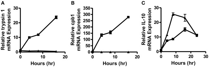
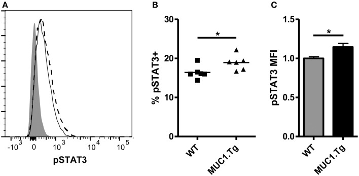
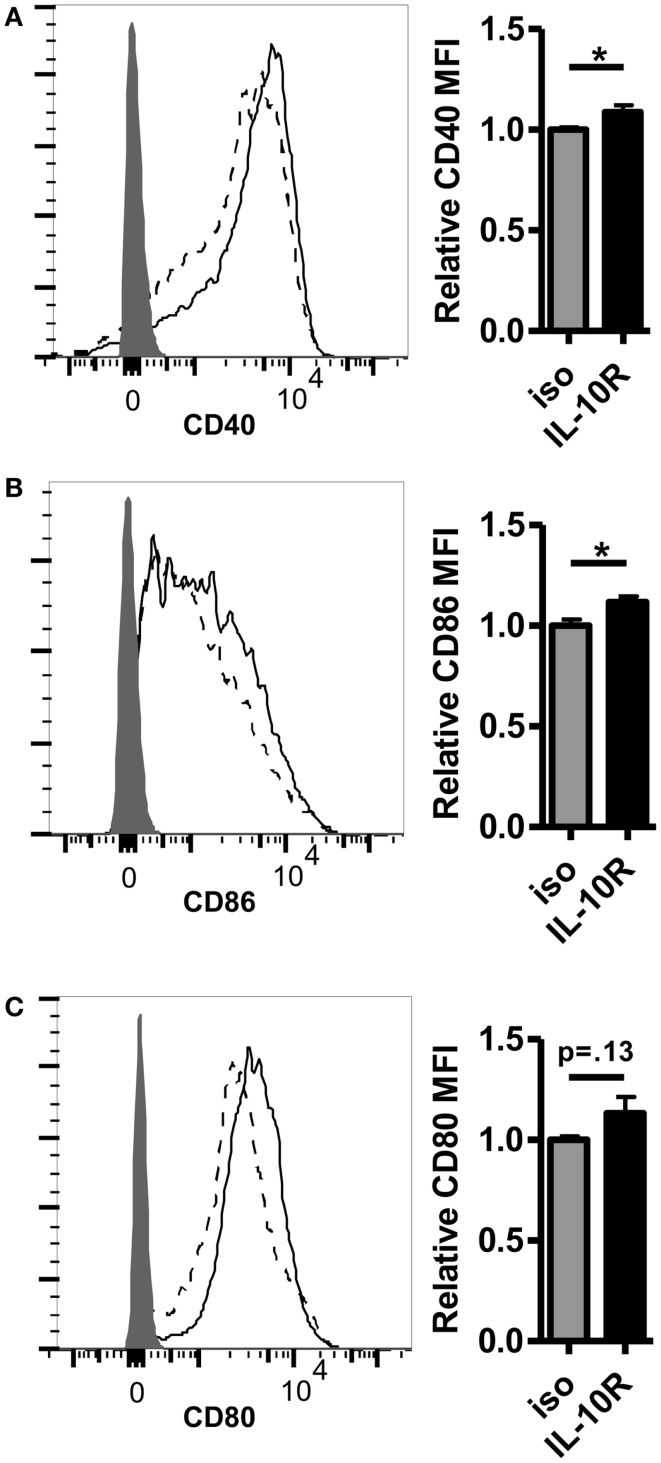
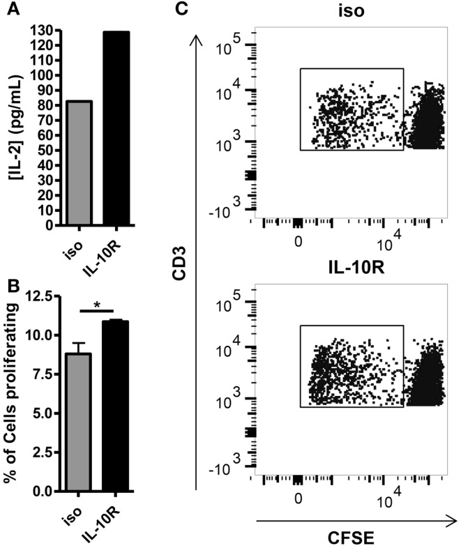
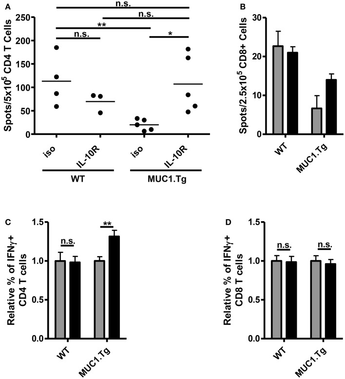
Dendritic cells (DC) in the spleen are highly activated following intravenous vaccination with a foreign-antigen, promoting expansion of effector T cells, but remain phenotypically and functionally immature after vaccination with a self-antigen. Up-regulation or suppression of expression of a cohort of pancreatic enzymes 24-72 h post-vaccination can be used as a biomarker of stimulatory versus tolerogenic DC, respectively. Here we show, using MUC1 transgenic mice and a vaccine based on the MUC1 peptide, which these mice perceive as a self-antigen, that the difference in enzyme expression that predicts whether DC will promote immune response or immune tolerance is seen as early as 4-8 h following vaccination. We also identify early production of IL-10 as a predominant factor that both correlates with this early-time point and controls DC function. Pre-treating mice with an antibody against the IL-10 receptor prior to vaccination results in DC that up-regulate CD40, CD80, and CD86 and promote stronger IFNγ+ T cell responses. This study suggests that transient inhibition of IL-10 prior to vaccination could improve responses to cancer vaccines that utilize self-tumor antigens.

Keywords: IL-10; MUC1; T cell response; cancer vaccine; dendritic cells.


Figures

Figure 1
Figure 1
Splenic DC activation is suppressed as early as 4–8 h post-vaccination with a self-, but not a foreign-antigen and correlated with early IL-10 production in the spleens of these animals. WT (squares) and MUC1.Tg mice (triangles) were vaccinated with MUC1p plus Poly-ICLC via tail vein. Spleens were removed at indicated hours post-vaccination and total splenic mRNA levels of trypsin 1 (A), carboxypeptidase B1 (CPB1) (B), and IL-10 (C) were determined relative to the control gene HPRT. Values shown represent expression relative to the baseline expression in mice of that genotype (WT and MUC1.Tg) at 0 h post-vaccination. Data are representative of three pooled mice were group per time point shown. Data points show mean ± SEM of three technical replicates and are representative of two independent experiments.
Figure 2
Figure 2
Dendritic cell from spleens of mice vaccinated with self-antigen have higher levels of phosphorylated STAT3 after IL-10 treatment than DC from spleens of mice vaccinated with foreign-antigen. WT (solid line) and MUC1.Tg (dashed line) mice were vaccinated with MUC1p via the tail vein. Twenty-four hours post-vaccination splenocytes were removed and treated with 30 ng/mL of IL-10 for 20 min. Following incubation, cells were fixed and phospho-STAT3 expression in CD11c+NK1.1− splenocytes was analyzed via phoshpoflow. (A) A representative flow plot is shown. The shaded histogram represents the fluorescence level when cells are treated with standard surface markers and an isotype-matched control instead of the phosphospecific antibody. pSTAT3 positivity (B) and MFI (C) were analyzed. In (B) symbols correspond to individual animals and are representative of two independent experiments. (C) Values shown have been normalized to the expression level of the control group (WT) in order to allow for pooling of data from separate experiments run on multiple days. Bars are representative of nine mice from two combined experiments and show the mean ± SEM. *Indicates a p-value of <0.05.
Figure 3
Figure 3
Pretreatment with an antibody against the IL-10 receptor increases the level of costimulatory molecule expression on DC in the spleens of self-antigen vaccinated mice. MUC1.Tg mice were pretreated with an antibody against the IL-10 receptor (IL-10R, solid lines) or were given a non-specific isotype control (iso, dashed lines). One to two days later they were vaccinated as in Figure 1 and 24 h post-vaccination, splenocytes were removed and analyzed via flow cytometry. The expression level of CD40 (A), CD86 (B), and CD80 (C) on splenic DC (CD11C+, MHC Class II+) was determined. Shaded histograms represent fluorescence in samples stained with isotype alone. Bar graph values shown have been normalized to the expression level of the control group (iso) in order to allow for pooling of data from separate experiments run on multiple days. (A,C) Data are combined from two independent experiments and representative of six mice. (B) Data are combined from three independent experiments and are representative of 10 mice. Bars represent mean ± SEM. p-Values are as stated unless designated by a *, which indicates a p-value of <0.05.
Figure 4
Figure 4
Blocking of the IL-10 receptor prior to intravenous MUC1 peptide vaccination increases the ability of splenic DC from MUC1.Tg mice to stimulate MUC1 specific CD4 T cells ex vivo. MUC1.tg mice were treated as in Figure 3. Twenty-four hours post MUC1 vaccination, splenocytes from three to four mice per treatment group were pooled and bead isolated DC from these pooled splenocytes were put into culture with CFSE labeled MUC1 specific CD4 T cells (VFT cells) at a ratio 1DC:5VFT. Twenty-four hours after the start of culture, half of the culture media was removed and the concentration of IL-2 was measured by ELISA (A). Cultures were allowed to incubate three more days for a total of four and VFT proliferation was analyzed by CFSE dilution (B,C). (B) Bars represent the mean percentage of CD3+CD4+ T cells that had proliferated at 4 days of three technical replicates ±SEM. (C) A representative flow plot is shown. Data are representative of two to three independent experiments. *Indicates a p-value of <0.05.
Figure 5
Figure 5
Treatment with anti-IL-10R antibody at the time of vaccination increases the number of MUC1p specific, IFNγ+ CD4 T cells without an effect on CD8 T cells. WT and MUC1.Tg mice were pretreated with an antibody against the IL-10 receptor (IL-10R, black bars) or a non-specific isotype control (iso, gray bars). One to two days following antibody treatment, mice were vaccinated with DC loaded with MUC1p. Seven to nine days post-vaccination, spleens were removed and bead isolated CD4 (A,C) and CD8 T cells (B,D) were cultured with MUC1p loaded bone marrow derived DC overnight and analyzed by ELISPOT (A,B), or were cultured for 6–8 h in the presence of brefeldin-A and analyzed by intracellular flow cytometry (C,D). (A) Data are combined from two independent experiments with each spot indicating an individual animal. Data are representative of three independent experiments. (B) Bars indicate the average of three technical replicates pooled from three individual animals per group. Data are representative of two independent experiments. (C,D) Values shown are normalized to the response of mice of that genotype (WT versus MUC1.Tg) given the control treatment (iso). Data are combined from two independent experiments and are representative of five to six mice per group. Bars represent mean ± SEM. *Indicates a p-value of <0.05; **indicates a p-value of <0.005.

Similar articles

Cited by

References

    1. Moore KW, O’garra A, Malefyt RW, Vieira P, Mosmann TR. Interleukin-10. Annu Rev Immunol (1993) 11:165–9010.1146/annurev.iy.11.040193.001121 - DOI - PubMed
    1. Moore KW, De Waal Malefyt R, Coffman RL, O’garra A. Interleukin-10 and the interleukin-10 receptor. Annu Rev Immunol (2001) 19:683–76510.1146/annurev.immunol.19.1.683 - DOI - PubMed
    1. Asadullah K, Sterry W, Volk HD. Interleukin-10 therapy – review of a new approach. Pharmacol Rev (2003) 55:241–6910.1124/pr.55.2.4 - DOI - PubMed
    1. De Smedt T, Van Mechelen M, De Becker G, Urbain J, Leo O, Moser M. Effect of interleukin-10 on dendritic cell maturation and function. Eur J Immunol (1997) 27:1229–3510.1002/eji.1830270526 - DOI - PubMed
    1. Steinbrink K, Wolfl M, Jonuleit H, Knop J, Enk AH. Induction of tolerance by IL-10-treated dendritic cells. J Immunol (1997) 159:4772–80 - PubMed