. 2014 Apr 1;192(7):3121-32.
doi: 10.4049/jimmunol.1302351. Epub 2014 Mar 3.

IκBε is a key regulator of B cell expansion by providing negative feedback on cRel and RelA in a stimulus-specific manner

Affiliations

IκBε is a key regulator of B cell expansion by providing negative feedback on cRel and RelA in a stimulus-specific manner

Bryce N Alves et al. J Immunol. .

Abstract

The transcription factor NF-κB is a regulator of inflammatory and adaptive immune responses, yet only IκBα was shown to limit NF-κB activation and inflammatory responses. We investigated another negative feedback regulator, IκBε, in the regulation of B cell proliferation and survival. Loss of IκBε resulted in increased B cell proliferation and survival in response to both antigenic and innate stimulation. NF-κB activity was elevated during late-phase activation, but the dimer composition was stimulus specific. In response to IgM, cRel dimers were elevated in IκBε-deficient cells, yet in response to LPS, RelA dimers also were elevated. The corresponding dimer-specific sequences were found in the promoters of hyperactivated genes. Using a mathematical model of the NF-κB-signaling system in B cells, we demonstrated that kinetic considerations of IκB kinase-signaling input and IκBε's interactions with RelA- and cRel-specific dimers could account for this stimulus specificity. cRel is known to be the key regulator of B cell expansion. We found that the RelA-specific phenotype in LPS-stimulated cells was physiologically relevant: unbiased transcriptome profiling revealed that the inflammatory cytokine IL-6 was hyperactivated in IκBε(-/-) B cells. When IL-6R was blocked, LPS-responsive IκBε(-/-) B cell proliferation was reduced to near wild-type levels. Our results provide novel evidence for a critical role for immune-response functions of IκBε in B cells; it regulates proliferative capacity via at least two mechanisms involving cRel- and RelA-containing NF-κB dimers. This study illustrates the importance of kinetic considerations in understanding the functional specificity of negative-feedback regulators.


Figures
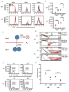
Figure 1
Figure 1
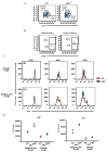
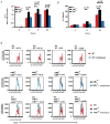
IκBε−/− B cells have increased proliferation and survival in response to both antigenic and inflammatory signals. B cells were isolated and purified from whole splenocytes of wild type and IκBε−/− using negative selection by CD43 magnetic beads. The separated B cells were stained with 1 nM CFSE and stimulated with either 10 μg/ml IgM (Jackson Labs) or 10 μg/ml LPS (Sigma). At designated time points, B cells were harvested and stained with 5 μg/ml 7AAD (Invitrogen) and analyzed for proliferation and death using flow cytometry. (A) B cells from the IκBε−/− mice displayed increased proliferation in response to both IgM and LPS at each time point. (B) The increased number of proliferating IκBε−/− B cells over that of wild type B cells was measured using Flowjo and the cell numbers were graphed. (C) Diagram depicting the fcyton model. In this model, stimulated cells undergo death over time (Tdie0) or enter division (pF0, fraction entering division and Tdiv0 time to division). (D) The CFSE proliferation profiles of the IκBε−/− and wild type B cells stimulated with either IgM or LPS where analyzed using FlowMax running the Pcyton model to predict the fraction of B cells responding to stimulation (pF0), the average time to division of undivided (Tdiv0) and average time to death of undivided (Tdie0) cells. The fraction of responding B cells (pF0) was greatly increased in both IgM and LPS stimulated IκBε−/− B cells when compared to wild type B cells. (E) 7AAD measurements of B cell death show an increased percentage of B cells in the wild type population undergo apoptotic death when compared to IκBε−/− B cells. (F) The percentage of 7AAD positive B cells from several experiments was measured using Flowjo and the percentages of 7AAD positive cells was graphed. Data shown in (A, B, D, E and F) are representative of at least four independent experiments. **P < 0.01, ***P < 0.005 and ****P <0.001 (unpaired t-test).
Figure 2
Figure 2
Follicular and Marginal zone IκBε−/− show increase proliferation. Follicular (FO) and Marginal zone (MZ) B cells population distributions were analyzed from whole splenocyte populations by flow cytometry using anti-B220, anti-CD21 and anti-CD23. (A) IκBε−/− B cells were found to have a distribution of 67% FO B cells and 21.9% MZ B cells. Wild type B cells were distributed as 80.1% FO B cells and 8.75% MZ B cells. (B) Marginal zone and Follicular B cells were separated from each other using FACS sorting of anti-CD9. Purity of the separated CD9+ and CD9- B cell populations was found to be between 90% and 100%. (C) IκBε−/− FO and MZ B cells showed increased proliferation over wild type FO and MZ B cells. (D) The increased number of proliferating IkBe−/− B cells over that of wild type B cells was measured using Flowjo and the cell numbers were graphed. Data shown in (A, B, C and D) are representative of two independent experiments.
Figure 3
Figure 3
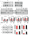
NFκB activity is increased in IκBε−/− B cells. Purified B cells were collected and extracted into cytoplasmic and nuclear fraction at various time points. Nuclear extracts tested for total NFκB activity using EMSAs. (A) IκBε−/− B cells stimulated with either IgM or LPS exhibited increased NFκB activity at 18 and 24 hours following stimulation. (B) The 24 hours nuclear extracts were incubated with antibodies directed towards anti-RelA (αRelA), anti-RelB (αRelB), anti-cRel (αcRel) as well as combination of these antibodies (αRelA/αRelB, αRelA/αcRel, and αRelB/αcRel). Following a 20 minute incubation, 32P-labled probe was added and allowed to incubate for a further 15 minutes. The resulting samples were run on a 5% non-reducing acrylamide gel. (C) The resulting supershifts were quantitated using ImageJ and graphed below each shift. Both IgM and LPS stimulation had increases in the cRel/p50 activity in the IκBε−/− B cell extracts when compared to extracts of wild type B cells stimulated under the same conditions. LPS stimulated IκBε−/− B cell extracts had increased RelA activity. (D) Nuclear western blots for RelA and cRel were run and quantitated to determine if the increased cRel activity observed in the supershifts was the result of increased RelA and cRel levels in the IκBε−/− B cells. Increased cRel protein levels were observed in the nuclear extracts from IκBε−/− B cell when compared to wild type B cell extracts. Similar levels of RelA were found between wild type and IκBε−/− B cell nuclear extracts. Data shown in (A) are representative of two independent experiments. Data shown in (B, C and D) are representative of three independent experiments (n=3, error bars are represented as standard deviations). *P < 0.05, **P < 0.01, ***P < 0.005 and ****P <0.001 (unpaired t-test).
Figure 4
Figure 4
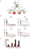
Computation modeling of the IκBε−/− B cells’ NFκB activity identifies IκBε as the dominant regulator of cRel:p50 dimers. Hypothetical model of the IκBα and IκBε control of NFκB within B cells following IgM or LPS stimulation. (A) IgM and LPS stimulation results in two different IKK activity profiles; IgM stimulation has a transient IKK activity, while LPS results in lasting IKK activity. Activation of IKK results in the degradation of IκBs, and can release NFκB dimers into the nucleus. From previous data, we infer that IκBε has a very strong affinity for cRel:p50, while IκBα has a strong affinity for RelA:p50, but not as strong as IκBε-cRel:p50. IκBε has a very low affinity for RelA:p50, while IκBα has a weak affinity for cRel:p50, although the affinity between IκBα-cRel:p50 is stronger than IκBε-cRel:p50. (B) Data from our computation model illustrates the NFκB activity profiles for both wild type and IκBε-deficient B-cells under IgM and LPS stimulus. (C) The model is able to recapitulate the experimental late timepoints, in which IκBε-deficient B-cells have elevated cRel:p50 after both LPS and IgM stimulation. However, RelA:p50 is only elevated after LPS stimulation in IκBε-deficient B-cells.
Figure 5
Figure 5
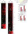
IκBε−/− B cells have an enrichment of NFκB dependent gene expression. Total RNA gene expression was obtained from RNAseq of both wild type and IκBε−/− B cells at 0, 2, 8 and 24 hours stimulated with IgM or LPS. Hyperinduced gene were identified as all genes having a 2-fold induction of at least one time point in wild type B cells and ≥ 2-fold induction at two timepoints in the IκBε−/− B cells were identified using fold change of the FPKM values. (A) The genes identified to be hyperinduced over wild type were displayed as heatmaps for the IgM and LPS stimulation. (B) Motifs for the NFκB dimers were loaded and used in Homer motif discovery software to search the promoter sequences of the identified induced and hyperinduced genes of the IκBε−/− B cells for occurrences of the listed NFκB motifs. The resulting genes containing one of these NFκB motifs were graphed as percentages over the total genes detected to be hyperinduced within the IκBε−/− samples. *P < 0.05 and **P < 0.01.
Figure 6
Figure 6
Release of IL-6 is enhanced in IκBε−/− B cells. B cells from wild type and IκBε−/− mice were isolated and stimulated with LPS. Supernatants and RNA extracts were collected at 0, 4, 8 and 24 hours. (A) Quantitative PCR results of wild type, IκBε−/− and cRel−/− B cells. IL-6 expression is enhanced by the loss of IκBε and is partially dependent on cRel. (B) ELISA assay for IL-6 in wild type, LPS stimulated IκBε−/− B cells have increased cytokine release of IL-6 when compared to wild type IκBε−/− and cRel−/− B cells. The loss of cRel reduced the amount of IL-6 released. (C) Neutralizing antibodies against the cytokines IL-1α, IL-1β and IL-6 or receptor blocking antibodies against the IL-1α, IL-1β and IL-6 receptor were used at a concentration of 2 μg/ml in B cell proliferation assays. Reduced proliferation of IκBε−/− B cells only occurred in the presences of antibodies directed against the individual cytokines or their receptors for the cytokines IL-6. Wild type B cell proliferation is only slightly reduced in the presence of the IL-6 antibody. Data shown in (A and B) are representative of at 3 independent experiments (n=3, error bars are represented as standard deviations). *P < 0.05, **P < 0.01, ***P < 0.005 and ****P < 0.001 (unpaired t-test). Data shown in (C) are representative of 2 independent experiments.

Similar articles

Cited by

References

    1. O’Dea E, Hoffmann A. The regulatory logic of the NF-kappaB signaling system. Cold Spring Harb Perspect Biol. 2010;2:a000216. - PMC - PubMed
    1. Hoffmann A, Levchenko A, Scott ML, Baltimore D. The IkappaB-NF-kappaB signaling module: temporal control and selective gene activation. Science. 2002;298:1241–1245. - PubMed
    1. Shih VF, Kearns JD, Basak S, Savinova OV, Ghosh G, Hoffmann A. Kinetic control of negative feedback regulators of NF-kappaB/RelA determines their pathogen- and cytokine-receptor signaling specificity. Proc Natl Acad Sci U S A. 2009;106:9619–9624. - PMC - PubMed
    1. Doi TS, Takahashi T, Taguchi O, Azuma T, Obata Y. NF-kappa B RelA-deficient lymphocytes: normal development of T cells and B cells, impaired production of IgA and IgG1 and reduced proliferative responses. J Exp Med. 1997;185:953–961. - PMC - PubMed
    1. Gerondakis S, Grumont R, Rourke I, Grossmann M. The regulation and roles of Rel/NF-kappa B transcription factors during lymphocyte activation. Curr Opin Immunol. 1998;10:353–359. - PubMed

Publication types

MeSH terms