. 2014 Feb 19;9(2):e86745.
doi: 10.1371/journal.pone.0086745. eCollection 2014.

The role of IKKβ in Venezuelan equine encephalitis virus infection

Affiliations

The role of IKKβ in Venezuelan equine encephalitis virus infection

Moushimi Amaya et al. PLoS One. .

Abstract

Venezuelan equine encephalitis virus (VEEV) belongs to the genus Alphavirus, family Togaviridae. VEEV infection is characterized by extensive inflammation and studies from other laboratories implicated an involvement of the NF-κB cascade in the in vivo pathology. Initial studies indicated that at early time points of VEEV infection, the NF-κB complex was activated in cells infected with the TC-83 strain of VEEV. One upstream kinase that contributes to the phosphorylation of p65 is the IKKβ component of the IKK complex. Our previous studies with Rift valley fever virus, which exhibited early activation of the NF-κB cascade in infected cells, had indicated that the IKKβ component underwent macromolecular reorganization to form a novel low molecular weight form unique to infected cells. This prompted us to investigate if the IKK complex undergoes a comparable macromolecular reorganization in VEEV infection. Size-fractionated VEEV infected cell extracts indicated a macromolecular reorganization of IKKβ in VEEV infected cells that resulted in formation of lower molecular weight complexes. Well-documented inhibitors of IKKβ function, BAY-11-7082, BAY-11-7085 and IKK2 compound IV, were employed to determine whether IKKβ function was required for the production of infectious progeny virus. A decrease in infectious viral particles and viral RNA copies was observed with inhibitor treatment in the attenuated and virulent strains of VEEV infection. In order to further validate the requirement of IKKβ for VEEV replication, we over-expressed IKKβ in cells and observed an increase in viral titers. In contrast, studies carried out using IKKβ(-/-) cells demonstrated a decrease in VEEV replication. In vivo studies demonstrated that inhibitor treatment of TC-83 infected mice increased their survival. Finally, proteomics studies have revealed that IKKβ may interact with the viral protein nsP3. In conclusion, our studies have revealed that the host IKKβ protein may be critically involved in VEEV replication.


Conflict of interest statement

Competing Interests: The authors' association with Serpin Pharma is strictly academic with the emphasis on NFkB activation in VEEV infections. None of the authors included in the paper (that are not from Serpin Pharma) have any conflicting interests relating to employment, consultancy, patents, products in development or marketed products etc. The authors' association with Serpin Pharma does not alter their adherence to all the PLOS ONE policies on sharing data and materials.

Figures

Figure 1
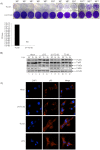
Figure 1. Activation of NF-κB complex in TC-83 infected cells.
A) UV inactivation of the virus was carried out using a Stratalinker UV crosslinker (model 1800). The inactivation was achieved by delivering an energy dose equivalent to 1200 µJoules X 100 per dose five times with a 2 minute interval between dosing. UV-TC-83 and TC-83 were serially diluted and used to infect Vero cells. UV-TC-83 inactivation was confirmed by plaque assay. Plaques were photographed and counted 48 hours post-infection. Plaque counts are represented graphically. B) U87MG cells were either mock infected, treated with LPS (1 µg/mL) or infected with TC-83 or UV-TC-83 (MOI: 1). At 30 minutes, 1 and 2 hours post-infection cells were lysed and protein extracts were resolved by SDS-PAGE and subsequently immunoblotted with antibodies against phosphorylated p65 and phosphorylated IκBα. Total p65, total IκBα and β-actin served as controls. The western blot is representative of 2 independent experiments. C) U87MGs were either mock infected, treated with LPS (1 µg/mL) or UV-TC-83 or TC-83 infected (MOI: 3). One hour post-infection cells were fixed, probed with p65 antibody followed by incubation with Alexa-Fluor 568. The cells were stained with DAPI to observe the nuclei. Images were taken using Nikon Eclipse TE2000-U at 60× magnification and are representative of 2 independent experiments. ND = not detectable.
Figure 2
Figure 2. Macromolecular reorganization of the IKKβ complex in TC-83 infected cells.
U87MG cells were infected with TC-83 or UV-TC-83 (MOI: 5) or the cells were treated with Poly I∶C (10 µg/mL) or Imiquimod (2 µg/mL). Cells were lysed 24 hours post-infection and post treatment and protein extracts were quantified. Two milligrams of total protein was subjected to chromatography using a Superose 6 HR 10/30 size-exclusion chromatography column and an AKTA purifier system. Every 5th fraction was acetone precipitated using 4 volumes of ice-cold 100% acetone and incubated for 15 minutes on ice. Lysates were centrifuged at 4°C for 10 minutes at 12,000 rpm, supernatants were removed, and the pellets were allowed to dry for a few minutes at room temperature. The pellets were resuspended in Laemmli buffer and analyzed by immunoblotting using IKKα (A), IKKβ (B), IKKγ (C) and β-actin (D) antibodies. Every fifth fraction ranging from 2.2 mDa to 100 kDa is represented on the western blot. Smaller IKKβ complexes (highlighted in the red box) in the higher fractions suggests a rearrangement of the IKKβ complex in TC-83 infected cells.
Figure 3
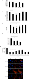
Figure 3. IKKβ inhibitors decrease viral load in TC-83 infected cells pre and post exposure.
A) U87MGs were untreated, DMSO treated or pretreated with IKK inhibitors (1 µM) for 2 hours and 24 hours later cell viability was measured using the Cell-Titer-Glo Luminescent Cell Viability Assay. B) U87MG cells were pretreated with IKK inhibitors (1 µM), BAY-11-7082, BAY-11-7085 and IKK2-IV and non-IKK specific inhibitors (1 µM), O-Phe and DMC for 2 hours. The conditioned media (media containing inhibitor) was removed and viral infections proceeded (MOI: 0.1) for 1 hour. The viral inoculum was removed and replaced with the conditioned media. The cells were incubated for an additional 24 hours. The supernatants were collected from infected and inhibitor treated cells. Infectious viral titers were determined by plaque assay. O-phe and DMC served as positive control inhibitors. C) Supernatants from B) were subjected to q-RT-PCR analysis to determine viral RNA copies using VEEV specific primers. D) U87MGs were infected at MOI: 0.1 for 1 hour and then treated with BAY-11-7082 (1 µM), BAY-11-7085 (1 µM) and IKK2-IV (1 µM) 4 hours post-infection. Supernatants were collected 24 hours post-infection and viral titers were determined by plaque assay. E) U87MGs were pretreated with IKK inhibitors (1 µM) and non-IKK inhibitors (1 µM) for 2 hours and followed by a 1 hour infection. Conditioned media was replaced and cell viability assay was performed 72 hours later using the Cell-Titer-Glo Luminescent Cell Viability Assay. The red line is representative of the base line for luminescence units, where luminescence units above this value are indicative of increased cell viability. F) U87MGs seeded in an 8-well chambered glass slide were either pre-treated with DMSO or BAY-11-7082 (1 µM) for 2 hours and then infected with UV-TC-83 or TC-83 (MOI: 0.1) for 1 hour. The conditioned media was replaced after the infection. One hour post-infection the cells were fixed and probed for p65 with subsequent incubation with Alexa Fluor 568. The cells were stained with DAPI to observe the nuclei. Images were taken using Nikon Eclipse TE2000-U at 60× magnification and are representative of 2 replicate samples within the same experiment. The graphs represent an average of 3 independent experiments. Error bars for the 3 independent experiments were calculated and are represented thusly. **** p≤0.0001, *** p≤0.005, ** p≤0.01 and * p≤0.05.
Figure 4
Figure 4. IKKβ inhibitors decrease TC-83 replication in rat AP7 neuronal cells.
A) Neurons were pre-treated with DMSO or with IKK inhibitors (1 µM) for 2 hours and 24 hours later cell viability was measured using the Cell-Titer-Glo Luminescent Cell Viability Assay. B) Neurons were pretreated with IKK inhibitors (1 µM), BAY-11-7082, BAY-11-7085 and IKK2-IV for 2 hours. Following the pretreatment, the conditioned media (media containing inhibitor) was removed and the cells infected at MOI: 1 for 1 hour. The viral inoculum was removed and the conditioned media replaced. Supernatants were collected 24 hours post-infection, and infectious viral titers were determined by plaque assay. C) Neurons were pretreated with IKK inhibitors (1 µM) for 2 hours and infected with TC-83 for 1 hour. Conditioned media was replaced after removal of the viral inoculum. Cell viability assay was performed 48 hours later using the Cell-Titer-Glo Luminescent Cell Viability Assay. The red line is representative of the base line for luminescence units, such that luminescence units above this are indicative of increased cell viability. The graphs are representative of 2 independent experiments. Error bars for the independent experiments were calculated and are represented thusly. *** p≤0.005, ** p≤0.01 and * p≤0.05.
Figure 5
Figure 5. IKKβ inhibitors are effective in decreasing viral load of wild type VEEV.
A) U87MG cells (A) or neuronal rat AP7 cells (B) were pretreated with 1 µM IKK inhibitors, BAY-11-7082, BAY-11-7085 and IKK2-IV for 2 hours. The cells were infected with the wild type strain of VEEV (TrD) at a MOI: 0.1 (A) or MOI: 1 (B) for 1 hour. The conditioned media (media containing inhibitor) was removed prior to the viral infections and replaced after the viral inoculum was removed. The cells were incubated for an additional 24 hours. The supernatants were collected from all samples and viral titers were determined by plaque assay. The graphs are representative of 2 independent experiments. Error bars (Standard deviations) for 3 replicates within the 2 independent experiments were calculated and are represented thusly. ** p≤0.01.
Figure 6
Figure 6. BAY-11-7082 decreases replication of wild type alphaviruses.
U87MGs were pre-treated with DMSO or BAY-11-7082 (1 µM) for 2 hours. The conditioned media (media containing inhibitors) was removed and the cells infected with VEEV TrD, EEEV GA97 and WEEV (California 1930 strain) for 1 hour at an MOI of 0.1. The conditioned media was replaced and the supernatants collected 24 hours later. Plaque assays were performed to determine viral titers. The graph is the average of n = 3 from a single experiment. Error bars for 3 replicates were calculated and are represented thusly.
Figure 7
Figure 7. Increased survival in TC-83 infected mice when treated with BAY-11-7082.
C3H/HeN mice (n = 7 per group) were infected with TC-83 by the intranasal route (2×10⁷ PFU/mL). Animals were pretreated for one day and post treated once a day with BAY-11-7082 (10 mg/kg) subcutaneously for 10 days. Control mice were TC-83 infected and treated with DMSO. A) Survival was monitored up to a period of 2 weeks. Results from one experiment are represented by a line graph for comparison of survival of the two groups over days of observation. B) Serum samples were collected from 3 animals per group on days 3, 7, and 10. Viral titers were determined by plaque assay. Results are shown in a line graph to indicate differences in viral titers over time. C) Homogenized brains were collected in parallel with serum samples. Plaque assay results of those samples are indicated by a line graph to indicate differences in viral titers over time. Values were obtained by averaging numbers from three brain and serum samples at each time point.
Figure 8
Figure 8. A requirement for IKKβ in TC-83 replication in vitro.
A) U87MG cells were transfected in triplicate with 1 µg of FLAG-IKKβ and 1 µg of pUC19 as a control plasmid for a total of 48 hours. Transfected cells were then infected with TC-83 (MOI: 0.1). Supernatants were collected 24 hours post-infection and plaque assays were performed to determine viral titers. B) Total protein from the cell lysates in A) were subjected to western blot and probed for VEEV capsid, VEEV glycoprotein, IKKβ and β-Actin. FLAG_IKKβ and pUC19 protein bands were quantified using Image J software and normalized to β-Actin. Average fold differences of 2 independent experiments are depicted graphically. C) Mouse embryonic fibroblasts (MEFs) lacking IKKβ (IKKβ−/−) and wild type (WT) MEFs were infected with TC-83 (MOI: 0.1) in triplicate. Supernatants were collected 24 hours post-infection and plaque assays were performed to determine viral titers. D) Western blot analysis of WT MEFs and IKKβ−/− confirmed depletion of IKKβ−/− in the knockout cell line when probed for IKKβ. β-actin served as a loading control. The graphs represent an average of 3 independent experiments. Error bars for the 3 independent experiments were calculated and are represented thusly.
Figure 9
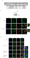
Figure 9. VEEV nsP3 interaction with host IKKβ.
A) IKKβ was immunoprecipitated from VEEV infected U87MGs and Liquid Chromatography Tandem Mass Spectrometry (LC-MS/MS) was performed. Control immunoprecipitations were performed with an IgG antibody. Mass spectrometry analysis revealed that the viral nonstructural protein nsP3 interacted with IKKβ. B) U87MGs were transfected in a 6-well plate with 5 µg of pcDNA3.1 and VEEV_nsP3_HA for 24 and 48 hours. Cell lysates were resolved using SDS-PAGE and subsequently immunoblotted with HA and β-actin served as a loading control. C) U87MGs were transfected in duplicate with (0.2 µg) VEEV_nsP3_HA and pcDNA3.1 (control), cells were fixed after 24 hours and probed with antibodies against the endogenous IKKβ and the HA tag. Cells were subsequently incubated with appropriate secondary Alexa Fluor antibodies and the nuclei stained with DAPI. Co-localization of IKKβ with nsP3 (yellow) was observed as shown by the arrows. The co-localization was confirmed by Z-stack analysis. Panels E–H and J–M serve as examples of transfected cells in a given field of view that show co-localization of IKKβ and VEEV_nsP3_HA at 24 hours post transfection. Panels I and N are magnified images of the outlined cells in red boxes in panels H and M respectively. Co-localization was found to be approximately in 71% of cells (72 cells were counted of which 55% demonstrated expression of nsP3. Of those cells that expressed nsP3, 71% showed co-localization of both IKKβ and VEEV_nsP3_HA proteins). D) U87MGs were transfected in duplicate with (0.2 µg) VEEV_nsP3_HA and pcDNA3.1 (control); cells were treated with BAY-11-7082 (1 µM and 0.1 µM). The cells were fixed 24 hours post transfection and probed with antibodies against the endogenous IKKβ and the HA tag. Cells were subsequently incubated with appropriate secondary Alexa Fluor antibodies and the nuclei stained with DAPI. Co-localization of IKKβ with nsP3 (yellow) was observed as shown by the arrows. The co-localization was confirmed by Z-stack analysis. Panels E–H and I–L serve as examples of transfected cells in a given field of view that show co-localization of IKKβ and VEEV_nsP3_HA at 24 hours post transfection. Panel M is a magnified image of the outlined cells in red boxes in panel L. Panels N–Q and S–V are examples of transfected and treated cells in a given field of view. Panels R and W are magnified images of the outlined cells in red boxes in panels Q and V respectively. Images were taken using Nikon Eclipse TE2000-U at 60× magnification and are representative of 3 independent experiments.
Figure 10
Figure 10. WEEV nsP3 interaction with host IKKβ.
A) U87MGs were transfected in a 6-well plate with 5 µg of pUC19 and WEEV_nsP3_HA for 24 hours. Cell lysates were resolved using SDS-PAGE and subsequently immunoblotted with V5 antibody and β-actin served as a loading control. B) U87MGs were transfected with WEEV_nsP3_V5; cells were fixed after 24 hours and stained with antibodies against the endogenous IKKβ and the V5 tag. Cells were incubated with appropriate secondary Alexa Fluor antibodies and the nuclei stained with DAPI. Co-localization of IKKβ with WEEV_nsP3_V5 (yellow) was observed as shown by the arrows. B) Panels E–H serve as an example of transfected cells in a given field of view that show co-localization of IKKβ and WEEV_nsP3_V5 24 hours post transfection. Panels I-L represent magnified images of other cells showing co-localization of IKKβ and WEEV_nsP3_V5. Panel M is a magnified image of panel L. The co-localization was confirmed by Z-stack analysis. Co-localization was calculated to be approximately in 61% of cells (163 cells were counted of which 44% demonstrated expression of nsP3. Of those cells that expressed nsP3, 61% showed co-localization of both proteins). Images were taken using Nikon Eclipse TE2000-U at 60× magnification and are representative of 2 independent experiments.

Similar articles

Cited by

References

    1. Kim DY, Atasheva S, Frolova EI, Frolov I (2013) Venezuelan equine encephalitis virus nsP2 protein regulates packaging of viral genome into infectious virions. J Virol - PMC - PubMed
    1. Foy NJ, Akhrymuk M, Akhrymuk I, Atasheva S, Bopda-Waffo A, et al. (2013) Hypervariable Domains of nsP3 Proteins of New World and Old World Alphaviruses Mediate Formation of Distinct, Virus-Specific Protein Complexes. J Virol 87: 1997–2010. - PMC - PubMed
    1. Lamb K, Lokesh GL, Sherman M, Watowich S (2010) Structure of a Venezuelan equine encephalitis virus assembly intermediate isolated from infected cells. Virology 406: 261–269. - PMC - PubMed
    1. Aguilar PV, Greene IP, Coffey LL, Medina G, Moncayo AC, et al. (2004) Endemic Venezuelan equine encephalitis in northern Peru. Emerg Infect Dis 10: 880–888. - PMC - PubMed
    1. Steele KE, Twenhafel NA (2010) REVIEW PAPER: pathology of animal models of alphavirus encephalitis. Vet Pathol 47: 790–805. - PubMed

Publication types

MeSH terms

Grants and funding

This work was supported by George Mason University start-up funds to AN, the US Department of Energy grant (DE- SC0001599) to CB and FK, and the generous support of the College of Science to the Center for Applied Proteomics and Molecular Medicine. Publication of this article was funded in part by the George Mason University Libraries Open Access Publishing Fund. The funders had no role in study design, data collection and analysis, decision to publish, or preparation of the manuscript.

LinkOut - more resources