. 2014 Feb 27;10(2):e1003975.
doi: 10.1371/journal.ppat.1003975. eCollection 2014 Feb.

Implication of PMLIV in both intrinsic and innate immunity

Affiliations

Implication of PMLIV in both intrinsic and innate immunity

Faten El Asmi et al. PLoS Pathog. .

Abstract

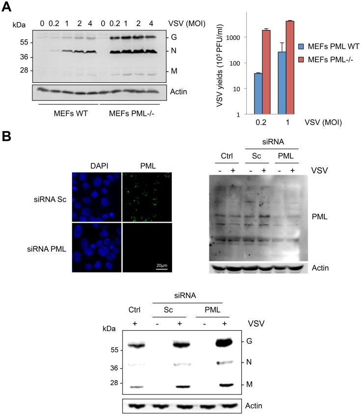
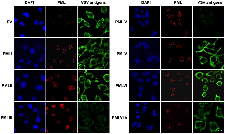
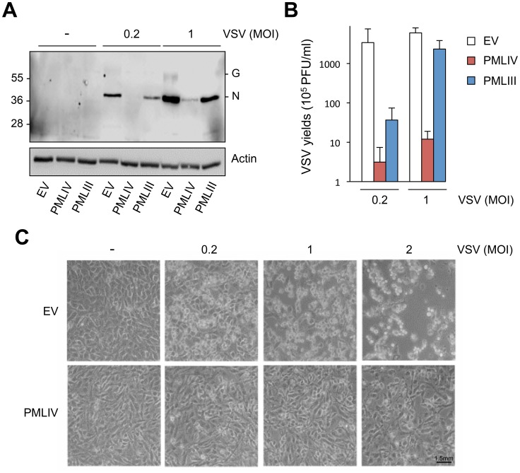
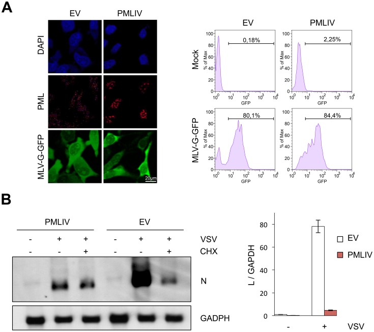
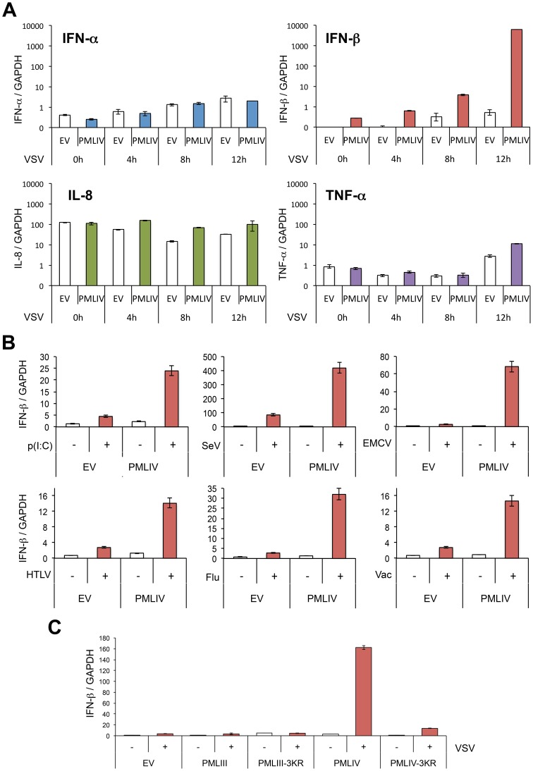
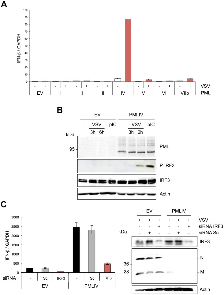
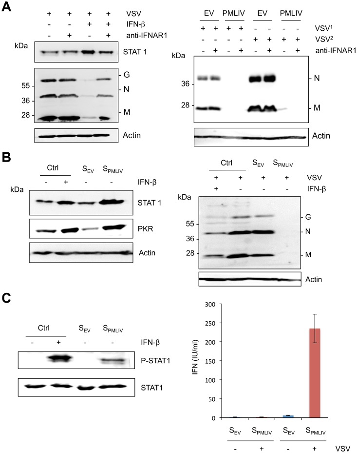
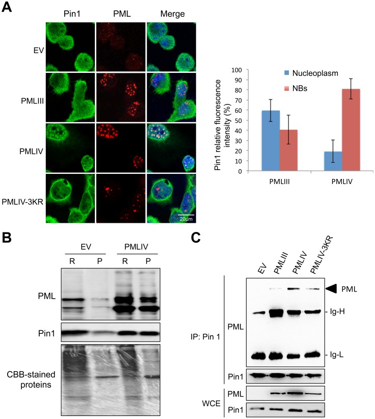
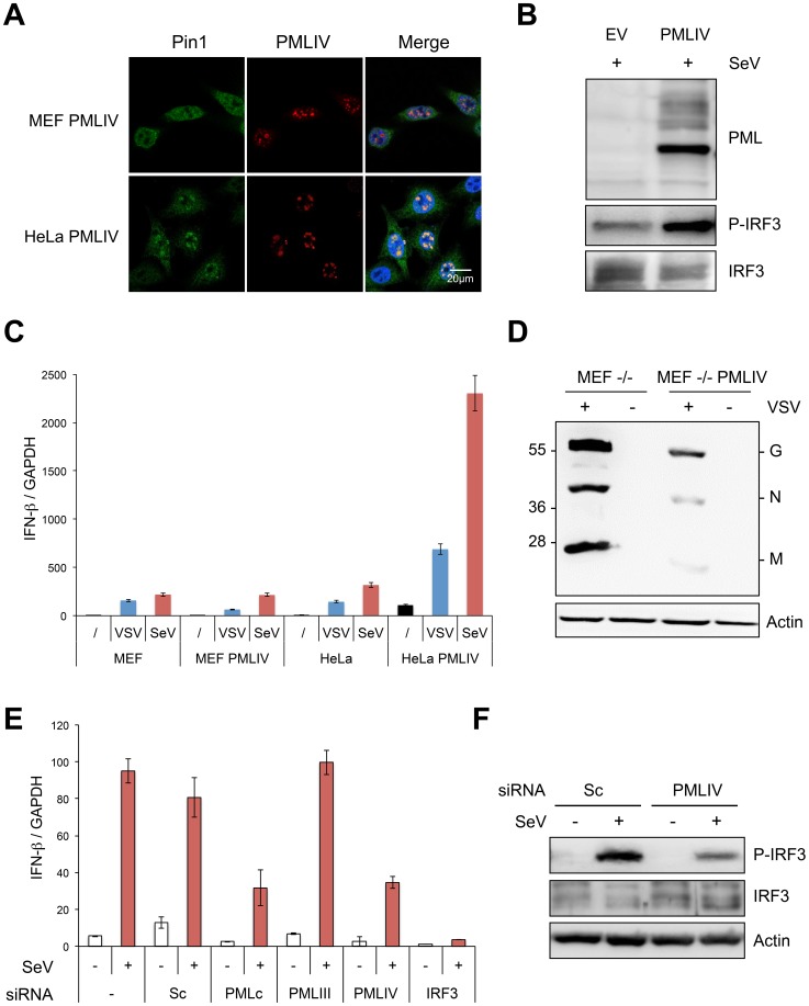
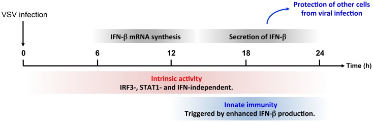
PML/TRIM19, the organizer of nuclear bodies (NBs), has been implicated in the antiviral response to diverse RNA and DNA viruses. Several PML isoforms generated from a single PML gene by alternative splicing, share the same N-terminal region containing the RBCC/tripartite motif but differ in their C-terminal sequences. Recent studies of all the PML isoforms reveal the specific functions of each. The knockout of PML renders mice more sensitive to vesicular stomatitis virus (VSV). Here we report that among PML isoforms (PMLI to PMLVIIb), only PMLIII and PMLIV confer resistance to VSV. Unlike PMLIII, whose anti-VSV activity is IFN-independent, PMLIV can act at two stages: it confers viral resistance directly in an IFN-independent manner and also specifically enhances IFN-β production via a higher activation of IRF3, thus protecting yet uninfected cells from oncoming infection. PMLIV SUMOylation is required for both activities. This demonstrates for the first time that PMLIV is implicated in innate immune response through enhanced IFN-β synthesis. Depletion of IRF3 further demonstrates the dual activity of PMLIV, since it abrogated PMLIV-induced IFN synthesis but not PMLIV-induced inhibition of viral proteins. Mechanistically, PMLIV enhances IFN-β synthesis by regulating the cellular distribution of Pin1 (peptidyl-prolyl cis/trans isomerase), inducing its recruitment to PML NBs where both proteins colocalize. The interaction of SUMOylated PMLIV with endogenous Pin1 and its recruitment within PML NBs prevents the degradation of activated IRF3, and thus potentiates IRF3-dependent production of IFN-β. Whereas the intrinsic antiviral activity of PMLIV is specific to VSV, its effect on IFN-β synthesis is much broader, since it affects a key actor of innate immune pathways. Our results show that, in addition to its intrinsic anti-VSV activity, PMLIV positively regulates IFN-β synthesis in response to different inducers, thus adding PML/TRIM19 to the growing list of TRIM proteins implicated in both intrinsic and innate immunity.


Conflict of interest statement

The authors have declared that no competing interests exist.

Figures

Figure 1
Figure 1. Cells are more susceptible to VSV infection in the absence of PML.
(A) MEFs WT and MEFs PML-/- were infected with VSV at different MOIs for 8 h. Cell extracts were analyzed by Western blot and revealed by anti-VSV and anti-Actin antibodies (left panel) and the supernatants from cells infected at MOI of 0.2 or 1 were used for the determination of virus yields (right panel). Means and standard deviations of two independent experiments are shown. (B) Human U373MG cells control (Ctrl), transfected with siRNA scramble (Sc) or siRNA specific to PML were infected with VSV at an MOI of 0.2 for 8 h. Immunofluorescence analysis was performed using anti-PML antibody (left panel) and the cell extracts were analyzed by Western blot and revealed by anti-PML (right panel), anti-VSV (bottom panel) and anti-Actin antibodies.
Figure 2
Figure 2. Immunofluorescence analysis on infected cells expressing each PML isoform.
U373MG cells transfected with the empty vector (EV) or U373MG cells stably expressing PMLI, PMLII, PMLIII, PMLIV, PMLV, PMLVI, or PMLVIIb were infected with VSV at an MOI of 0.1 for 12 h. Double immunofluorescence analyses were performed using monoclonal anti-PML (red) and rabbit anti-VSV (green) antibodies.
Figure 3
Figure 3. Compared to PMLIII, PMLIV has a higher anti-VSV activity.
(A) U373MG-EV, U373MG-PMLIII or U373MG-PMLIV cells were not infected (-) or infected with VSV at MOIs of 0.2 or 1 for 8 h. Cell extracts were analyzed by Western blotting and revealed by anti-VSV and anti-Actin antibodies. (B) Supernatants from infected U373MG-EV, U373MG-PMLIII or U373MG-PMLIV cells were used for the determination of the virus yields. Mean values and standard deviations of three independent experiments are shown. (C) PMLIV protected cells from VSV-induced cell lysis. U373MG-EV and U373MG-PMLIV cells were left uninfected (-) or infected with VSV at MOIs of 0.2, 1 or 2 for 12 h. The phase contrast picture was acquired using a Nikon Eclipse TS100 microscope and Coolpix MDC lens camera.
Figure 4
Figure 4. PMLIV does not alter VSV entry and inhibits viral transcription.
(A) U373MG-EV and U373MG-PMLIV cells were infected with MLV-GFP/G for 48 h. Double staining of PML and GFP was presented (left panel) and the transduction efficiency, expressed as the percentage of GFP-positive cells, was measured 48 h post-infection using a fluorescence-activated cell sorter (right panel). (B) PMLIV inhibited viral mRNA transcription. U373MG-EV and U373MG-PMLIV cells were infected by VSV at an MOI of 3 in the absence (−CHX) or the presence (+CHX) of cycloheximide. After 4 h of infection, samples were analyzed for the presence of VSV-N and GAPDH mRNAs (left panel). Total RNA were used for VSV-L mRNA quantification by RT-qPCR (right panel). The values obtained for uninfected cells expressing the empty vector (EV) were arbitrarily set to 1. Means and standard deviations of three independent experiments are shown.
Figure 5
Figure 5. PMLIV positively regulates IFN-β synthesis.
(A) PMLIV enhances IFN-β synthesis without affecting that of IFN-α, IL8 or TNF-α upon VSV infection. U373MG-EV and U373MG-PMLIV cells were infected with VSV at an MOI of 0.2 for different lengths of time. Total RNA was extracted and mRNAs encoding IFN-α, IFN-β, IL8, TNF-α, and GAPDH were quantified by RT-qPCR. (B) PMLIV positively regulates IFN-β synthesis in cells infected with SeV or transfected with poly(I:C). U373MG-EV and U373MG-PMLIV cells were infected with SeV (40 HAU/ml) or EMCV (MOI of 0.5), or transfected with poly(I:C) (1 µg/ml). HeLa cells transduced with PMLIV-expressing or with noncoding parental (EV) lentiviral vectors were infected with HTLV-1 (HTLV, 600 ng/ml of p19-equivalent), influenza virus (Flu, 40 HAU/ml) or vaccinia virus (Vac, MOI of 2). After 8 h, mRNAs were extracted and IFN-βtranscripts quantified by RT-qPCR. (C) SUMOylation of PMLIV was required for IFN-β induction. U373MG-EV, U373MG-PMLIV, U373MG-PMLIV-3KR, U373MG-PMLIII and U373MG-PMLIII-3KR cells were infected with VSV for 12 h at an MOI of 0.2. Total RNA was extracted and mRNAs encoding IFN-β and GAPDH were quantified by RT-qPCR. Mean values and standard deviations of two independent experiments are shown.
Figure 6
Figure 6. PMLIV is the only PML isoform able to boost IFN-β synthesis through IRF3 activation.
(A) Quantification of IFN-β mRNA in infected cells expressing different PML isoforms. U373MG-EV cells or cells stably expressing PMLI, PMLII, PMLIII, PMLIV, PMLV, PMLVI or PMLVIIb were infected with VSV at an MOI of 0.2 for 12 h. Total RNA was extracted and mRNA encoding IFN-β and GAPDH quantified by RT-qPCR. Mean values and standard deviations of two independent experiments are shown. (B) IRF-3 phosphorylation in VSV-infected or poly(I:C)-transfected cells expressing PMLIV. U373MG-EV or U373MG-PMLIV cells were left untreated (-), infected with VSV at an MOI of 1 for 3 or 6 h or transfected with poly(I:C). Cell extracts were analyzed by Western blotting using antibodies directed against PML, P-IRF3, IRF3 or Actin. (C) Effect of IRF3 knockdown on VSV inhibition and IFN-β induction. U373MG-EV and U373MG-PMLIV cells, prepared in duplicate, were transfected with scramble (Sc) or IRF3-specific siRNA. Two days later, cells were infected with VSV at an MOI of 1 for 8 h. Total RNA was extracted in one series and mRNA encoding IFN-βand GAPDH was quantified by RT-qPCR (left panel). Means and standard deviations of three independent experiments are shown. The second series was used for the determination by Western blot of IRF3 and VSV protein expression (right panel).
Figure 7
Figure 7. Intrinsic and innate immune properties of PMLIV.
(A) U373MG cells were left untreated or treated for 24 h with 100 units/ml of IFN-β in the absence or the presence of 20 µg/ml of purified anti-IFNAR1 mAb before infection with VSV at an MOI of 1 for 8 h (left panel). U373MG and U373MG-PMLIV cells were infected with VSV at an MOI of 0.2 for 8 h (VSV1) or 12 h (VSV2) in the absence or the presence of 20 µg/ml of anti-IFNAR1 mAb (right panel). Cell extracts were analyzed by Western blotting and revealed by antibodies directed against VSV, STAT1 or Actin. (B) Activity of secreted IFN in infected U373MG-PMLIV cells. Culture supernatants (from U373MG-EV cells [SEV] and U373MG-PMLIV cells [SPMLIV]) were treated with acid buffer to inactivate virus, and the pH was then neutralized. One series of HeLa cells were left untreated as a control (Ctrl) or treated for 24 h with 100 units/ml of IFN-β, SEV or SPMLIV, then their extracts were analyzed by Western blot for PKR and STAT1 (left panel). The second series of HeLa cells were infected with VSV at an MOI of 0.2 for 8 h and the cell extracts were analyzed by Western blotting using anti-VSV and anti-Actin antibodies (right panel). (C) Secreted IFN activates STAT1 phosphorylation (left panel). HeLa cells were left untreated (Ctrl) or treated for 30 min with 1000 units/ml of IFN-β, SEV or SPMLIV, then their extracts were analyzed by Western blot for P-STAT1 and STAT1. Quantification of produced type I IFN upon VSV infection (right panel). IFN was quantified using the reporter cell line HL116 that carries the luciferase gene under the control of the IFN-inducible 6–16 promoter . HL116 cells were incubated for 8 h with a standard containing titrated human IFN-β, the supernatants from U373MG-EV [SEV] and U373MG-PMLIV [SPMLIV] cells infected with VSV at an MOI of 1 for 20 h or the supernatants from uninfected U373MG-EV and U373MG-PMLIV. Cells were then lysed and luciferase activity measured. IFN levels were expressed as equivalent of IFN-β concentration, in IU/ml. Means and standard deviations of two independent experiments are shown.
Figure 8
Figure 8. PMLIV interacts with Pin1 and recruits it within PMLNBs.
(A) Double immunofluorescence analyses were performed using monoclonal anti-PML (red) and rabbit anti-Pin1 (green) antibodies in U373MG-EV cells or cells stably expressing PMLIII, PMLIV or PMLIV-3KR (left panel). Images obtained in PMLIII- and PMLIV-expressing cells were quantified using Image-J software. The resulting relative values (mean ± s.e.m.) corresponding to Pin1 localization are shown in histograms (n = 30) (right panel). (B) PMLIV induces the recruitment of Pin1 to the nuclear matrix. RIPA soluble (R) and RIPA insoluble fractions (P) from U373MG-EV and U373MG-PMLIV cells were analyzed by Western blotting using anti-PML and anti-Pin1 antibodies. (C) PMLIV and PMLIV-3KR interacted with Pin1. Whole-cell lysates from U373MG-EV cells and cells expressing PMLIV, PMLIV-3KR or PMLIII were immunoprecipitated with anti-Pin1 antibodies. The immunopellets were separated by SDS-PAGE and immunoblotting was performed with anti-PML and anti-Pin1 antibodies. Ten percent input is shown (WCE).
Figure 9
Figure 9. PMLIV is responsible for Pin1 recruitment in PML NBs, enhanced P-IRF3 and IFN-β synthesis upon viral infection.
(A) PMLIV recruits Pin1 within the NBs in HeLa cells but not in MEFs. MEFs or HeLa cells were transduced with HIV-derived lentiviral vectors expressing PMLIV. Two days post-transduction, double immunofluorescence analyses were performed using monoclonal anti-PML (red) and rabbit anti-Pin1 (green) antibodies. (B) HeLa cells transduced with HIV-derived lentiviral vectors expressing EV or PMLIV were infected with SeV (40 HAU/ml) for 8 h and their extracts were analyzed by Western blot for PML, P-IRF3 and IRF3 expression. (C) Wild-type MEF and HeLa cells transduced with PMLIV-expressing or with the noncoding parental lentiviral vector (EV) were infected with VSV (MOI of 0.2) for 12 h or with SeV (40 HAU/ml) for 8 h. After RNA extraction, GAPDH and IFN-β transcripts were quantified by RT-qPCR. Mean values and standard deviations of two independent experiments are shown. (D) MEF PML-/- cells were transduced with pTRIP-PMLIV and infected with VSV at a MOI of 1 for 8 h. Cell extracts were analyzed by Western blot and revealed using anti-VSV and anti-actin antibodies. (E) U373MG cells were transfected with scramble siRNA (Sc), or with a siRNA targeting IRF3, PMLIII, PMLIV or common to all PML isoforms (PMLc), before infection with SeV (40 HAU/ml) for 12 h. After RNA extraction, GAPDH and IFN-β transcripts were quantified by RT-qPCR. Mean values and standard deviations of two independent experiments are shown. (F) Extracts of U373MG cells transfected with siRNA (Sc), or with siRNA specific to PMLIV before infection with SeV, were analyzed by Western blot for P-IRF3, IRF3 and Actin expression.
Figure 10
Figure 10. Intrinsic and innate immune properties of PMLIV during viral infection.
The anti-VSV intrinsic activity of PMLIV is independent of IRF3, STAT1 and IFN and can be observed during the whole duration of the experiments. In parallel, PMLIV enhances IFN-β mRNA synthesis around 12 h post-infection. This produced IFN is secreted between 18 and 24 h and then acts on other cells by activating STAT1, increasing the expression ISG products thus protecting them from uncoming infection.

Similar articles

Cited by

References

    1. Taniguchi T, Ogasawara K, Takaoka A, Tanaka N (2001) IRF family of transcription factors as regulators of host defense. Annu Rev Immunol 19: 623–655. - PubMed
    1. Chelbi-Alix MK, Wietzerbin J (2007) Interferon, a growing cytokine family: 50 years of interferon research. Biochimie 89 6–7: 713–718. - PubMed
    1. Sadler AJ, Williams BR (2008) Interferon-inducible antiviral effectors. Nat Rev Immunol 8: 559–568. - PMC - PubMed
    1. Weissenhorn W, Hinz A, Gaudin Y (2007) Virus membrane fusion. FEBS Lett 581: 2150–2155. - PMC - PubMed
    1. Albertini AA, Ruigrok RW, Blondel D (2011) Rabies virus transcription and replication. Adv Virus Res 79: 1–22. - PubMed

Publication types

MeSH terms

Grants and funding

This work was supported by grants from the Agence Nationale de la Recherche (ANR) and Agence Nationale de Recherches sur le SIDA et les hépatites virales (ANRS). FEA is funded by University Paris XI, France and University of Carthage, Tunisia. MAM and JD are funded by ANR and ANRS, respectively. The funders had no role in study design, data collection and analysis, decision to publish, or preparation of the manuscript.