

Inflammatory cytokines drive CD4+ T-cell cycling and impaired responsiveness to interleukin 7: implications for immune failure in HIV disease
- PMID: 24585897
- PMCID: PMC4172041
- DOI: 10.1093/infdis/jiu125
Inflammatory cytokines drive CD4+ T-cell cycling and impaired responsiveness to interleukin 7: implications for immune failure in HIV disease
Abstract
Background: Systemic inflammation has been linked to a failure to normalize CD4(+) T-cell numbers in treated human immunodeficiency virus (HIV) infection. Although inflammatory cytokines such as interleukin 6 (IL-6) are predictors of disease progression in treated HIV infection, it is not clear how or whether inflammatory mediators contribute to immune restoration failure.
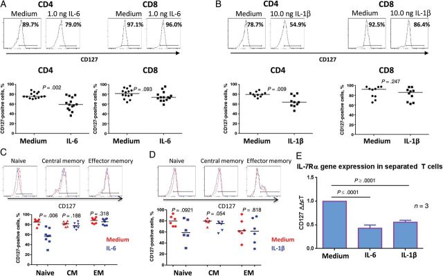
Methods: We examined the in vitro effects of IL-6 and interleukin 1β (IL-1β) on peripheral blood T-cell cycling and CD127 surface expression.
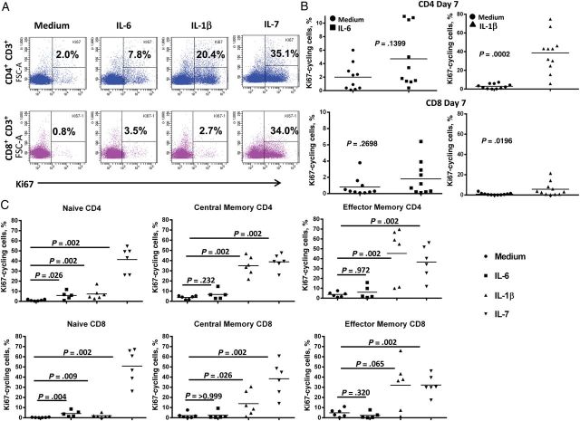
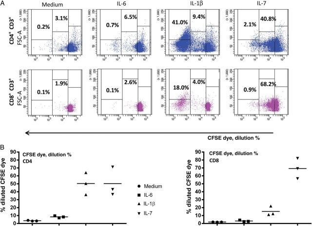
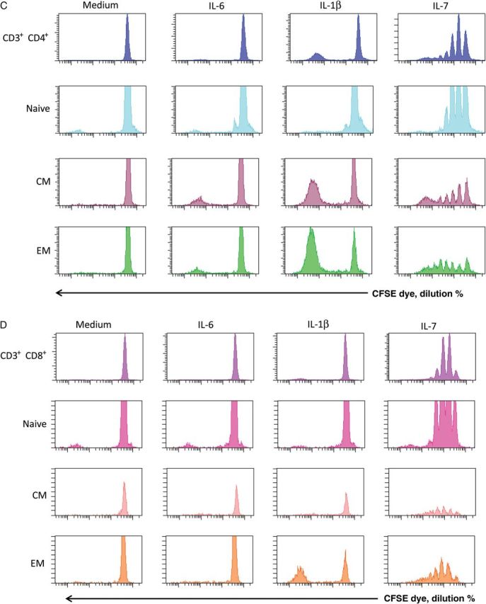
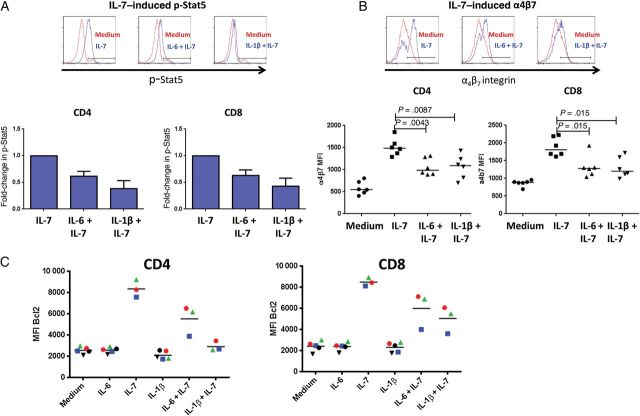
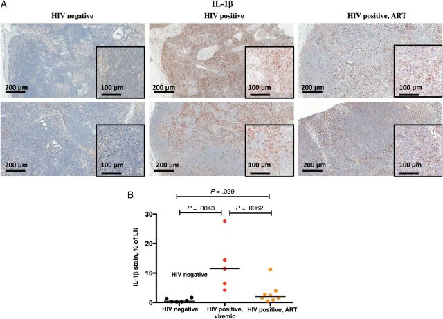
Results: The proinflammatory cytokine IL-1β induces cell cycling and turnover of memory CD4(+) T cells, and IL-6 can induce low-level cycling of naive T cells. Both IL-1β and IL-6 can decrease T-cell surface expression and RNA levels of CD127, the interleukin 7 receptor α chain (IL-7Rα). Preexposure of healthy peripheral blood mononuclear cells (PBMCs) to IL-6 or IL-1β attenuates IL-7-induced Stat5 phosphorylation and induction of the prosurvival factor Bcl-2 and the gut homing integrin α4β7. We found elevated expression of IL-1β in the lymphoid tissues of patients with HIV infection that did not normalize with antiretroviral therapy.
Conclusions: Induction of CD4(+) T-cell turnover and diminished T-cell responsiveness to IL-7 by IL-1β and IL-6 exposure may contribute to the lack of CD4(+) T-cell reconstitution in treated HIV-infected subjects.
Keywords: HIV; immune failure; inflammation; interleukin 1 beta; interleukin 6; interleukin 7.
© The Author 2014. Published by Oxford University Press on behalf of the Infectious Diseases Society of America. All rights reserved. For Permissions, please e-mail: journals.permissions@oup.com.
Figures
Similar articles
-
Inflammation Perturbs the IL-7 Axis, Promoting Senescence and Exhaustion that Broadly Characterize Immune Failure in Treated HIV Infection.J Acquir Immune Defic Syndr. 2016 Apr 15;71(5):483-92. doi: 10.1097/QAI.0000000000000913. J Acquir Immune Defic Syndr. 2016. PMID: 26627102 Free PMC article.
-
Biological determinants of immune reconstitution in HIV-infected patients receiving antiretroviral therapy: the role of interleukin 7 and interleukin 7 receptor α and microbial translocation.J Infect Dis. 2010 Oct 15;202(8):1254-64. doi: 10.1086/656369. J Infect Dis. 2010. PMID: 20812848
-
The correlation between levels of IL-7Ralpha expression and responsiveness to IL-7 is lost in CD4 lymphocytes from HIV-infected patients.AIDS. 2007 Jan 2;21(1):101-3. doi: 10.1097/QAD.0b013e3280115b6a. AIDS. 2007. PMID: 17148974
-
Blocking Formation of the Stable HIV Reservoir: A New Perspective for HIV-1 Cure.Front Immunol. 2019 Aug 22;10:1966. doi: 10.3389/fimmu.2019.01966. eCollection 2019. Front Immunol. 2019. PMID: 31507594 Free PMC article. Review.
-
Interleukin-7 (IL-7): immune function, involvement in the pathogenesis of HIV infection and therapeutic potential.Eur Cytokine Netw. 2004 Oct-Dec;15(4):279-89. Eur Cytokine Netw. 2004. PMID: 15627636 Review.
Cited by
-
Elevation and persistence of CD8 T-cells in HIV infection: the Achilles heel in the ART era.J Int AIDS Soc. 2016 Mar 3;19(1):20697. doi: 10.7448/IAS.19.1.20697. eCollection 2016. J Int AIDS Soc. 2016. PMID: 26945343 Free PMC article. Review.
-
Replicative fitness of transmitted HIV-1 drives acute immune activation, proviral load in memory CD4+ T cells, and disease progression.Proc Natl Acad Sci U S A. 2015 Mar 24;112(12):E1480-9. doi: 10.1073/pnas.1421607112. Epub 2015 Feb 17. Proc Natl Acad Sci U S A. 2015. PMID: 25730868 Free PMC article.
-
Mitochondrial Functions Are Compromised in CD4 T Cells From ART-Controlled PLHIV.Front Immunol. 2021 May 4;12:658420. doi: 10.3389/fimmu.2021.658420. eCollection 2021. Front Immunol. 2021. PMID: 34017335 Free PMC article.
-
Single-cell sequencing resolves the landscape of immune cells and regulatory mechanisms in HIV-infected immune non-responders.Cell Death Dis. 2022 Oct 4;13(10):849. doi: 10.1038/s41419-022-05225-6. Cell Death Dis. 2022. PMID: 36195585 Free PMC article.
-
HIV infection modulates IL-1β response to LPS stimulation through a TLR4-NLRP3 pathway in human liver macrophages.J Leukoc Biol. 2019 Apr;105(4):783-795. doi: 10.1002/JLB.4A1018-381R. Epub 2019 Feb 18. J Leukoc Biol. 2019. PMID: 30776150 Free PMC article.
References
-
- Tenorio A, Zheng E, Bosch R, et al. Soluble markers of inflammation and coagulation, but not T cell activation, predict non-AIDS-defining events during suppressive ART [abstract 790]. CROI 3–6 March 2013: 20th Conference on Retroviruses and Opportunistic Infections; Atlanta, Georgia.
-
- Hunt P, Sinclair E, Rodriguez B, et al. Gut epithelial barrier dysfunction, inflammation, and coagulation predict higher mortality during treated HIV/AIDS [abstract 278]. CROI 5–8 March 2012: 19th Conference on Retroviruses and Opportunistic Infections; Seattle, Washington.
-
- Hunt PW, Martin JN, Sinclair E, et al. T cell activation is associated with lower CD4+ T cell gains in human immunodeficiency virus-infected patients with sustained viral suppression during antiretroviral therapy. J Infect Dis. 2003;187:1534–43. - PubMed
Publication types
MeSH terms
Substances
Grants and funding
LinkOut - more resources
Full Text Sources
Other Literature Sources
Medical
Research Materials
Miscellaneous
