. 2014 Feb 27;156(5):893-906.
doi: 10.1016/j.cell.2013.12.043.

Hippo signaling regulates microprocessor and links cell-density-dependent miRNA biogenesis to cancer

Affiliations

Hippo signaling regulates microprocessor and links cell-density-dependent miRNA biogenesis to cancer

Masaki Mori et al. Cell. .

Abstract

Global downregulation of microRNAs (miRNAs) is commonly observed in human cancers and can have a causative role in tumorigenesis. The mechanisms responsible for this phenomenon remain poorly understood. Here, we show that YAP, the downstream target of the tumor-suppressive Hippo-signaling pathway regulates miRNA biogenesis in a cell-density-dependent manner. At low cell density, nuclear YAP binds and sequesters p72 (DDX17), a regulatory component of the miRNA-processing machinery. At high cell density, Hippo-mediated cytoplasmic retention of YAP facilitates p72 association with Microprocessor and binding to a specific sequence motif in pri-miRNAs. Inactivation of the Hippo pathway or expression of constitutively active YAP causes widespread miRNA suppression in cells and tumors and a corresponding posttranscriptional induction of MYC expression. Thus, the Hippo pathway links contact-inhibition regulation to miRNA biogenesis and may be responsible for the widespread miRNA repression observed in cancer.


Figures

Figure 1
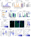
Figure 1. YAP regulates Microprocessor activity in a cell density-dependent manner
(A) qRT-PCR analysis of miRNA expression in HaCaT cells. Data were normalized to U6. *p < 0.05, Student’s t test. (B) Relative expression of pri-miRNAs and DROSHA and DGCR8 at low and high densities. qRT-PCR data normalized to GAPDH. (C) Western blot analysis. (D) Schematic representation of the Microprocessor (MP) reporter. The Δstem-loop mutant lacks the pre-miRNA stem-loop crucial for the recognition by Microprocessor. (E) Microprocessor reporter assays. (F) Expression levels of pri-, pre- and mature miR-125b after indicated siRNA-mediated knockdown, normalized to GAPDH for the pri-miRNA and to U6 for the pre- and mature miRNA. (G) Microprocessor reporter activity at different cell densities. *p < 0.05 versus empty, Student’s t test. (H) Immunocytochemistry analysis of YAP localization. YAP nuclear translocation was induced by knockdown of NF2 and LATS2. Scale bar, 30μm. (I-M) Microprocessor reporter assays *p < 0.05, **p < 0.01, Student’s t test. Data are represented as mean +/− SEM. See also Figure S1.
Figure 2
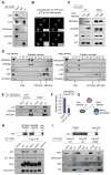
Figure 2. YAP sequesters p72 from Microprocessor complex in a cell density-dependent manner
(A) Co-immunoprecipitation assays (Co-IPs) with endogenous YAP in HaCaT cells. (B) Immunocytochemistry of YAP and p72 in HaCaT cells at low density. Nuclei were stained with DAPI. Scale bar, 30μm. (C) Co-IP with HA-p72 in HaCaT cells at low and high density. (D) Western blot analysis of Superose 6 gel-filtration fractions. Whole cell lysates from HaCaT cells cultured at low and high densities were fractionated. β-tubulin served as a control. (E) Co-IP with Flag-DROSHA in HaCaT cells transfected with YAP or control EGFP. (F) Densitometry measurement for the amount of p72 bound by DROSHA in the HaCaT cells transfected with YAP or control EGFP (n=3). *p < 0.05, Student’s t test. (G) The scheme of interactions among YAP, p72 and Microprocessor. (H) Co-IP with Flag-YAP and YAP mutants. Mutations in YAP are represented in the upper panel. WT, wild-type. (I) Co-IP with HA-p72 and p72 mutants. Mutations in p72 are represented in the upper panel. See also Figure S2.
Figure 3
Figure 3. Hippo pathway and p72 regulate pri-miRNA processing efficiency of Microprocessor
(A) Scheme of the Microprocessor assay. IVT, in vitro-transcribed. (B) Western blot analysis showing the siRNA efficacies and expression of Flag-DROSHA. (C) Western blot of purified protein complexes. (D) Microprocessor assays with in vitro transcribed pri-miR-125b. The numbers for Microprocessor indicate the relative amounts of Flag-IP products used for western blot (C) and Microprocessor assay (D).
Figure 4
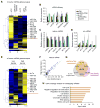
Figure 4. Global impact of Hippo pathway on miRNA biogenesis
(A) Global miRNA expression analysis of HaCaT with indicated knockdown. The miRNAs <0.8 or 1.2< fold compared to the siCtrl were analyzed by hierarchical clustering. (B) The efficacy of siRNAs used in (A). The expression values were normalized to GAPDH. (C) qPCR-quantification of mature miRNAs normalized to U6. (D) Pri-miRNA expression levels measured by qRT-PCR. Data were normalized to GAPDH. (E) miRNA expression analysis in RNA samples from low- and high-density HaCaT cells. miRNAs changed by <0.8 or 1.2< fold were analyzed by hierarchical clustering. (F) Scatter plot of miRNA expression levels (log10) in the low and the high densities (G) Venn diagram showing the overlap of miRNAs repressed by siNF2/LATS2, si p72, or low density. (H) Gene ontology analysis of the overlapping miRNAs in (G). Bonferroni-corrected p-values were indicated. Data are represented as mean +/− SEM. See also Figure S3.
Figure 5
Figure 5. p72 DEAD box RNA helicase binds to a sequence motif in the 3′ flanking segment of pri-miRNA
(A–D) EMSA with recombinant p72 protein and in vitro transcribed pri-miR-21, pri-miR-125b-1 or deletion mutants of pri-miR-21. Stem loop (Δstem-loop), 5′ flanking segment (Δ5′) or 3′ flanking segment (Δ3′) were deleted. (E) Identification of a sequence motif in the miRNAs repressed by both knockdown of NF2/LATS2 and p72. (F) Schematics showing the motif in the 3′ flanking segments of pri-miR-21 and pri-miR-125b-1. (G) Pri-miR-21 schematic indicating the motif mutations introduced and the control mutant. Arrowheads indicate cleavage sites by the Microprocessor. (H) EMSA with recombinant p72 protein and the +55 mutant, the control mutant and the motif mutant of the pri-miR-21. See also Figure S4.
Figure 6
Figure 6. YAP-regulated miRNAs repress MYC expression
(A) qRT-PCR analysis with data normalized to GAPDH. (B) miRNA Northern blot performed with spike-in of luciferase siRNA for normalization. (C) qRT-PCR analysis of mature miRNA levels normalized to U6. *p < 0.05, Student’s t test. (D) Relative pri-miRNA expression measured by qRT-PCR normalized to GAPDH. (E–F) Western blot analysis using indicated antibodies. (G–H) Luciferase assays with a MYC 3′UTR reporter. HaCaT cells were co-transfected with the luciferase and the expression plasmids for YAP or control EGFP. Luciferase activity was normalized to that of pRL-Tk. **p < 0.01, Student’s t test. n.s., not significant. Data are represented as mean +/− SEM. See also Figure S5.
Figure 7
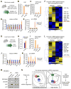
Figure 7. YAP mediates the global repression of miRNA biogenesis in tumors
(A) Mouse model of YAP-induced skin tumorigenesis. (B) Expression of exogenous human YAP S127A and endogenous mouse Yap normalized to Hprt1 in isolated epidermal cells. (C) Expression levels of YAP-target genes normalized to Hprt1. (D) Mature miRNA expression levels normalized to sno142. *p < 0.05, Student’s t test. (E) Expression levels of the pri-miRNAs normalized to Hprt1. (F) Global miRNA analysis. (G) Mouse model of YAP-induced tumorigenesis in the liver. (H) Expression levels of mouse Yap normalized to Hprt1. (I) The expression levels of YAP-target genes normalized to Hprt1. (J) Mature miRNA expression levels normalized to sno142. *p < 0.05, Student’s t test. (K) Relative expression levels of the pri-miRNAs normalized to Hprt1. (L) Global miRNA analysis in the liver tissues. (M) Co-IP with p72 in the normal tissues and tumors from the mouse livers. (N) Proposed model. Data are represented as mean +/− SEM. See also Figure S6.

Comment in

Similar articles

Cited by

References

    1. Auyeung VC, Ulitsky I, McGeary SE, Bartel DP. Beyond secondary structure: primary-sequence determinants license pri-miRNA hairpins for processing. Cell. 2013;152:844–858. - PMC - PubMed
    1. Barry ER, Morikawa T, Butler BL, Shrestha K, De la Rosa R, Yan KS, Fuchs CS, Magness ST, Smits R, Ogino S, et al. Restriction of intestinal stem cell expansion and the regenerative response by YAP. Nature. 2012;493:106–110. - PMC - PubMed
    1. Bartel DP. MicroRNAs: target recognition and regulatory functions. Cell. 2009;136:215–233. - PMC - PubMed
    1. Benhamouche S, Curto M, Saotome I, Gladden AB, Liu CH, Giovannini M, McClatchey AI. Nf2/Merlin controls progenitor homeostasis and tumorigenesis in the liver. Genes Dev. 2010;24:1718–1730. - PMC - PubMed
    1. Camargo FD, Gokhale S, Johnnidis JB, Fu D, Bell GW, Jaenisch R, Brummelkamp TR. YAP1 increases organ size and expands undifferentiated progenitor cells. Curr Biol. 2007;17:2054–2060. - PubMed

Publication types

Associated data