. 2014 May;88(9):5109-21.
doi: 10.1128/JVI.03057-13. Epub 2014 Feb 26.

Dendritic cell-lymphocyte cross talk downregulates host restriction factor SAMHD1 and stimulates HIV-1 replication in dendritic cells

Affiliations

Dendritic cell-lymphocyte cross talk downregulates host restriction factor SAMHD1 and stimulates HIV-1 replication in dendritic cells

Bin Su et al. J Virol. 2014 May.

Abstract

Human immunodeficiency virus type 1 (HIV-1) replication in dendritic cells (DCs) is restricted by SAMHD1. This factor is counteracted by the viral protein Vpx; Vpx is found in HIV-2 and simian immunodeficiency virus (SIV) from sooty mangabeys (SIVsm) or from macaques (SIVmac) but is absent from HIV-1. We previously observed that HIV-1 replication in immature DCs is stimulated by cocultivation with primary T and B lymphocytes, suggesting that HIV-1 restriction in DCs may be overcome under coculture conditions. Here, we aimed to decipher the mechanism of SAMHD1-mediated restriction in DC-lymphocyte coculture. We found that coculture with lymphocytes downregulated SAMHD1 expression and was associated with increased HIV-1 replication in DCs. Moreover, in infected DC-T lymphocyte cocultures, DCs acquired maturation status and secreted type 1 interferon (alpha interferon [IFN-α]). The blockade of DC-lymphocyte cross talk by anti-ICAM-1 antibody markedly inhibited the stimulation of HIV-1 replication and prevented the downregulation of SAMHD1 expression in cocultured DCs. These results demonstrate that, in contrast to purified DCs, cross talk with lymphocytes downregulates SAMHD1 expression in DCs, triggering HIV-1 replication and an antiviral immune response. Therefore, HIV-1 replication and immune sensing by DCs should be investigated in more physiologically relevant models of DC/lymphocyte coculture.

Importance: SAMHD1 restricts HIV-1 replication in dendritic cells (DCs). Here, we demonstrate that, in a coculture model of DCs and lymphocytes mimicking early mucosal HIV-1 infection, stimulation of HIV-1 replication in DCs is associated with downregulation of SAMHD1 expression and activation of innate immune sensing by DCs. We propose that DC-lymphocyte cross talk occurring in vivo modulates host restriction factor SAMHD1, promoting HIV-1 replication in cellular reservoirs and stimulating immune sensing.


Figures

FIG 1
FIG 1
Schematic representation of HIV-1 cell-to-cell transfer assay and the gating strategy for flow cytometry analysis. (A) HIV-1 cell-to-cell transfer assay. Immature MoDCs were pulsed with primary R5 HIV-1 isolates for 2 h and then thoroughly washed to remove unbound free viral particles. HIV-1-loaded MoDCs were incubated with or without autologous PHA-activated or nonactivated CD4 T or B lymphocytes or cells of the MT4 T cell line in 96-well plates. Where indicated, virus-like particles containing Vpx (VLP-Vpx) or exogenous dNTPs were added to infected MoDCs at the same time as lymphocytes as controls to decrease SAMHD1 levels and stimulate HIV-1 replication, respectively. After 48 and 72 h of culture, productive infection in MoDCs was quantified by detection of intracellular viral p24 antigen and intracellular SAMHD1 levels simultaneously by flow cytometry using cell-specific marker staining. (B) Quantification of productive infection and intracellular SAMHD1 levels by flow cytometry. Among all events, forward width and forward area were used to exclude doublet cells; forward angle and side scatter light gating were used to exclude cell debris. The DC population can easily be distinguished from lymphocytes according to size. Within the DC population, Ab directed against human CD209 (DC-SIGN, a DC-specific surface marker) was used to select DC-SIGN+ CD3 MoDCs; in the T cell population, Ab directed against human CD3 was used to select CD3+ CD4 T cells. Dead cells were then excluded with LIVE/DEAD fixable dead cell stain fluorescence kits. Percentages of living DC-SIGN+ MoDCs that are infected and contain intracellular SAMHD1 can be determined simultaneously. Double staining for HIV-1 infection and SAMHD1 expression in living DC-SIGN+ MoDCs is shown. The final analysis was performed with FACS Diva software, which generated a graphical output. FSC, forward scatter; SSC, side scatter.
FIG 2
FIG 2
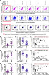
Detection of intracellular HIV-1 p24 antigen and SAMHD1, and the correlation between the two markers in immature MoDCs. (A) Dot plot showing the percentages of HIV-1 intracellular p24 Ag (top), intracellular SAMHD1 expression (middle), and double intracellular p24 Ag and SAMHD1 staining (bottom) within DC-SIGN+ MoDCs at 48 h postinfection (PI) by primary HIV-1BaL in the presence or absence of activated CD4 T lymphocytes or VLP-Vpx. One representative experiment from 12 independent experiments performed with 12 different healthy blood donors is shown. Shown are percentages of infected MoDCs with primary HIV-1BaL (B and E) and SAMHD1 expression in DC-SIGN+ MoDCs (C and F) after 48 h (B and C) or 72 h (E and F) of infection as percentages of cells in the absence or presence of activated CD4 T cells. The percentages of infected MoDCs (H) and SAMHD1 expression (I) among DC-SIGN+ MoDCs were measured 48 and 72 h postinfection in the absence or presence of VLP-Vpx. Data are means ± standard deviations (SD) from at least 12 independent experiments performed with cells from 12 healthy blood donors in duplicate. (Each point represents the result from one donor.) Horizontal bars denote medians, and whiskers denote interquartile ranges. The significance of differences was determined with two-tailed Wilcoxon matched-pairs signed rank test, and P < 0.05 is considered significant. Plotted is the correlation between the percent decrease of SAMHD1 levels (with respect to control infected MoDCs alone) and the percent increase of infection in cocultured DC-SIGN+ MoDCs with activated CD4 T lymphocytes at 48 h (D) and 72 h (G) postinfection and infected DC-SIGN+ MoDCs alone in the presence of VLP-Vpx as a control (J). Repeated independent experiments were performed with cells from at least 12 healthy donors. Correlations were analyzed by calculating Pearson's correlation coefficient, with P < 0.05 considered significant.
FIG 3
FIG 3
Single cycle of HIV-1 infection in cocultured MoDCs. (A) Percentages of HIV-1BaL-infected DC-SIGN+ MoDCs (left panel) were measured at 48 h postinfection in the presence or absence of activated CD4 T lymphocytes treated with HIV-1 protease inhibitor indinavir (IDV) at 1 μM to prevent infection of new virus produced by MoDCs or T lymphocytes or treated with HIV-1 reverse transcriptase inhibitor azidothymidine (AZT) at 5 μM to prevent HIV-1 replication as negative controls. Data are expressed as means ± SD from 10 independent experiments performed with 10 healthy blood donors. Under the same conditions, we measured intracellular SAMHD1 levels in DC-SIGN+ MoDCs (right panel). Data are expressed as means ± SD from four healthy blood donors. (B) Quantification of the amounts of viral p24 released into the supernatant when HIV-1-infected immature MoDCs were cultured with or without activated CD4 T cells in the presence or absence of negative controls treated with AZT. The means ± SD from eight different donors after 48 h of infection are shown. The significance of differences was determined with two-tailed Wilcoxon matched-pairs signed rank test. P values of <0.05 were considered statistically significant.
FIG 4
FIG 4
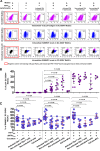
Measurement of HIV-1 infection and SAMHD1 expression in immature MoDCs cocultivated with various primary lymphocyte subtypes and MT4. (A) Dot plot showing the percentages of HIV-1 infection (top), intracellular SAMHD1 expression (middle), and double intracellular p24 Ag and SAMHD1 staining (bottom) among DC-SIGN+ MoDCs in the absence or presence of virus, PHA-activated CD4 T lymphocytes, or nonactivated CD4 T and B lymphocytes at 48 h postinfection. One representative experiment from seven independent experiments performed with seven different healthy blood donors is shown. HIV-1 infection (B) and SAMHD1 expression (C) were measured as percentages of DC-SIGN+ MoDCs at 48 h postinfection in the absence or presence of virus, autologous primary lymphocytes, T cell line MT4, or exogenous dNTPs. The data are the means ± SD from at least seven independent experiments performed with DCs from seven healthy blood donors in duplicate (except for coculture with MT4, where n = 4). Horizontal bars denote the medians, and error bars denote the interquartile ranges. The significance of differences was determined with two-tailed Wilcoxon matched-pairs signed rank test, with P < 0.05 considered significant.
FIG 5
FIG 5
Measurement of HIV-1 infection and SAMHD1 expression in immature MoDCs infected with primary HIV-1 isolates HIV-1QH0 and HIV-1SF162 when cocultured with autologous primary lymphocyte populations. Dot plots show the percentages of HIV-1 infection and SAMHD1 levels in DC-SIGN+ MoDCs in the absence or presence of PHA-activated CD4 T lymphocytes or nonactivated CD4 T or B lymphocytes at 48 h postinfection, infected with primary clinical isolate HIV-1QH0 (A and B) or HIV-1SF162 (C and D). The data are the means ± SD from one representative experiment.
FIG 6
FIG 6
Blockade of dendritic cell-lymphocyte cross talk by anti-ICAM-1. Anti-ICAM-1 antibody (1H4; 20 μg/ml) was added to HIV-1-infected or uninfected MoDCs prior to coculture with autologous lymphocytes. At 48 h postinfection, intracellular HIV-1 infection (A) and SAMHD1 expression (B) were measured and expressed as a percentage of the p24+ or SAMHD1+ DC-SIGN+ MoDCs compared to control cells in the absence of coculture. The percent decrease of HIV-1 infection was calculated and is shown in red. The data are means ± SD from two independent healthy blood donors.
FIG 7
FIG 7
Coculture with CD4 T lymphocytes induces innate sensing of HIV-1-infected MoDCs. (A) The kinetics of MoDC maturation were assessed at various time points. The percentage of CD83+ DC-SIGN+ MoDCs was determined in the absence or presence of virus and/or activated CD4 T lymphocytes. Each point represents the result from one donor. (B) Detection of the production of IFN-α by MoDCs in the supernatants of uninfected or infected MoDCs cultured alone or cocultured with activated CD4 T lymphocytes, VLP-Vpx, or dNTPs or with combinations of these factors collected at 48 h (black diamonds) and 72 h (pink circles) postinfection (P.I.). At least 12 independent experiments were performed with DCs from 12 healthy blood donors. (C) IFN-α was assessed under same conditions as in panel B, cocultured with nonactivated CD4 T or B lymphocytes. Data represent the results from seven independent experiments performed with DCs from seven healthy blood donors. Each point represents the result for one donor, and horizontal red bars denote the medians in the plots. (D) IFN-α was measured under same conditions as in panel B and cocultured with PHA-activated CD4 T lymphocytes or VLP-Vpx-transduced MoDCs in the absence or presence of HIV-1 protease inhibitor IDV at 1 μM to prevent the final assembly and maturation of newly synthesized virions. Data are expressed as means ± SD from four healthy blood donors. Groups were compared by one-way ANOVA (Kruskal-Wallis test) and two-tailed pairwise comparison based on Wilcoxon matched-pairs signed rank test, with P < 0.05 considered significant.

Similar articles

Cited by

References

    1. Granelli-Piperno A, Delgado E, Finkel V, Paxton W, Steinman RM. 1998. Immature dendritic cells selectively replicate macrophagetropic (M-tropic) human immunodeficiency virus type 1, while mature cells efficiently transmit both M- and T-tropic virus to T cells. J. Virol. 72:2733–2737 - PMC - PubMed
    1. Goujon C, Jarrosson-Wuilleme L, Bernaud J, Rigal D, Darlix JL, Cimarelli A. 2006. With a little help from a friend: increasing HIV transduction of monocyte-derived dendritic cells with virion-like particles of SIV(MAC). Gene Ther. 13:991–994. 10.1038/sj.gt.3302753 - DOI - PubMed
    1. Laguette N, Sobhian B, Casartelli N, Ringeard M, Chable-Bessia C, Segeral E, Yatim A, Emiliani S, Schwartz O, Benkirane M. 2011. SAMHD1 is the dendritic- and myeloid-cell-specific HIV-1 restriction factor counteracted by Vpx. Nature 474:654–657. 10.1038/nature10117 - DOI - PMC - PubMed
    1. Hrecka K, Hao C, Gierszewska M, Swanson SK, Kesik-Brodacka M, Srivastava S, Florens L, Washburn MP, Skowronski J. 2011. Vpx relieves inhibition of HIV-1 infection of macrophages mediated by the SAMHD1 protein. Nature 474:658–661. 10.1038/nature10195 - DOI - PMC - PubMed
    1. Berger A, Sommer AF, Zwarg J, Hamdorf M, Welzel K, Esly N, Panitz S, Reuter A, Ramos I, Jatiani A, Mulder LC, Fernandez-Sesma A, Rutsch F, Simon V, Konig R, Flory E. 2011. SAMHD1-deficient CD14+ cells from individuals with Aicardi-Goutieres syndrome are highly susceptible to HIV-1 infection. PLoS Pathog. 7:e1002425. 10.1371/journal.ppat.1002425 - DOI - PMC - PubMed

Publication types

Substances

LinkOut - more resources