Review
. 2014 Feb 6:7:3.
doi: 10.3389/fnmol.2014.00003. eCollection 2014.

Presynaptic [Ca(2+)] and GCAPs: aspects on the structure and function of photoreceptor ribbon synapses

Affiliations
Review

Presynaptic [Ca(2+)] and GCAPs: aspects on the structure and function of photoreceptor ribbon synapses

Frank Schmitz. Front Mol Neurosci. .

Abstract

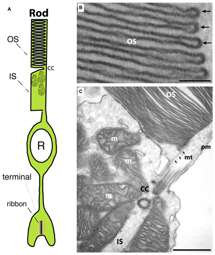
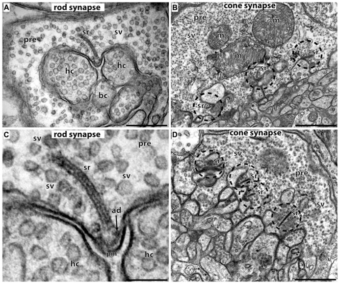
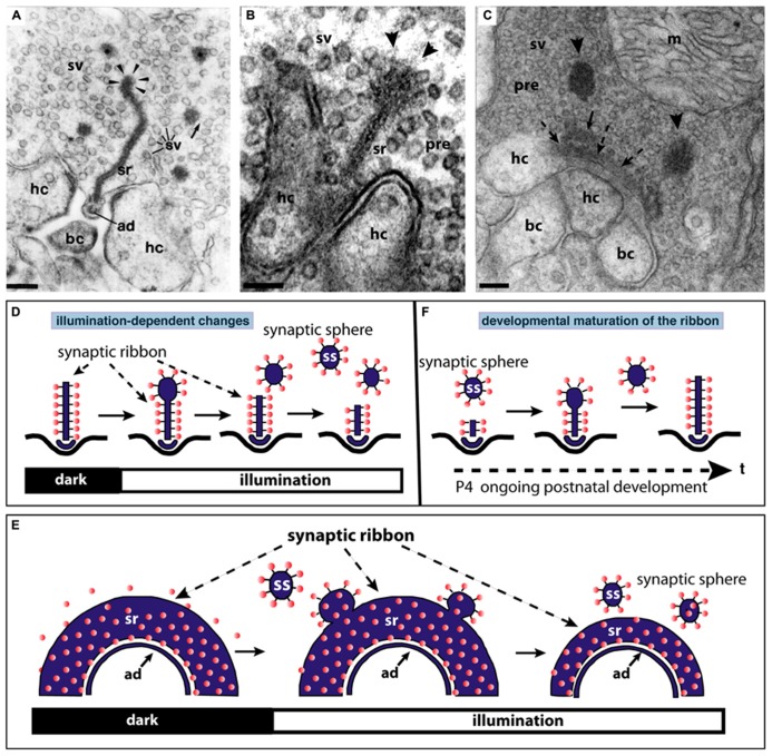
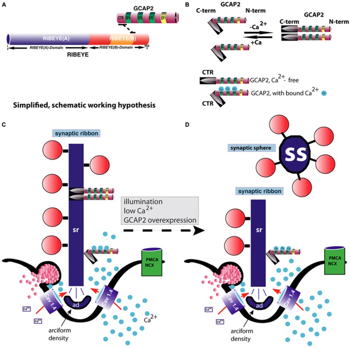
Changes in intracellular calcium ions [Ca(2+)] play important roles in photoreceptor signaling. Consequently, intracellular [Ca(2+)] levels need to be tightly controlled. In the light-sensitive outer segments (OS) of photoreceptors, Ca(2+) regulates the activity of retinal guanylate cyclases thus playing a central role in phototransduction and light-adaptation by restoring light-induced decreases in cGMP. In the synaptic terminals, changes of intracellular Ca(2+) trigger various aspects of neurotransmission. Photoreceptors employ tonically active ribbon synapses that encode light-induced, graded changes of membrane potential into modulation of continuous synaptic vesicle exocytosis. The active zones of ribbon synapses contain large electron-dense structures, synaptic ribbons, that are associated with large numbers of synaptic vesicles. Synaptic coding at ribbon synapses differs from synaptic coding at conventional (phasic) synapses. Recent studies revealed new insights how synaptic ribbons are involved in this process. This review focuses on the regulation of [Ca(2+)] in presynaptic photoreceptor terminals and on the function of a particular Ca(2+)-regulated protein, the neuronal calcium sensor protein GCAP2 (guanylate cyclase-activating protein-2) in the photoreceptor ribbon synapse. GCAP2, an EF-hand-containing protein plays multiple roles in the OS and in the photoreceptor synapse. In the OS, GCAP2 works as a Ca(2+)-sensor within a Ca(2+)-regulated feedback loop that adjusts cGMP levels. In the photoreceptor synapse, GCAP2 binds to RIBEYE, a component of synaptic ribbons, and mediates Ca(2+)-dependent plasticity at that site. Possible mechanisms are discussed.

Keywords: Ca2+; GCAP2; RIBEYE; photoreceptor; ribbon synapse; synaptic ribbon.


Figures

FIGURE 1
FIGURE 1
(A) Photoreceptors show a bipolar morphology. A distal process forms the outer segment (OS) and the inner segment (IS). The OS is the place where phototransduction takes place. The IS represents the metabolic center of the photoreceptor. An inner, vitread process ends in the presynaptic terminal that contains a synaptic ribbon at the active zone. (B) Transmission electron micrograph of an outer segment (OS). Densely packed arrays of membrane disks can be observed. The arrows in (B) point to individual disks. In the OS, phototransduction occurs and GCAP proteins perform a Ca2+-dependent feedback on guanylate cyclases. (C) Transmission electron micrograph at the junctional zone between OS and IS. OS and IS are connected by the connecting cilum (cc) that organizes mt-dependent vesicle transport into the outer segment (Sung and Chuang, 2010). OS, outer segment; IS, inner segment; m, mitochondrion; cc, connecting cilium linking OS and IS; mt, microtubule; pm, plasma membrane. Scale bars: 50 nm (B), 250 nm (C).
FIGURE 2
FIGURE 2
Photoreceptor ribbon synapses. In (A,C) rod photoreceptor ribbon synapses are shown by transmission electron microscopy; in (B,D) cone photoreceptor ribbon synapses. (A,B) The large presynaptic terminals are filled with numerous synaptic vesicles (sv). The active zone is characterized by specialized presynaptic densities, the arciform densities (ad). The synaptic ribbon (sr) is anchored to the arciform density. The synaptic ribbon is associated with large numbers of synaptic vesicles. Opposite to the active zones are the dendritic tips of horizontal cells (hc) and bipolar cells (bc) that contain ionotropic and metabotropic glutamate receptors for signaling. The dendritic tips are located within an invagination of the presynaptic terminal. Cone synapses (B,D) are larger in diameter than rod synapses and contain multiple active zones (dashed circles) and multiple synaptic ribbons (sr). sr, synaptic ribbon; sv, synaptic vesicles; ad, arciform density; pre, presynaptic terminal; m, mitochondrium; hc, dendritic tips of horizontal cells; bc, dendritic tips of bipolar cells; pm, presynaptic plasma membrane. Scale bars: 400 nm (A), 1 μm (B), 150 nm (C), 800 nm (D).
FIGURE 3
FIGURE 3
The synaptic ribbon is a dynamic structure, both in the mature retina (A,B,D,E) as well as in the immature retina during early postnatal retinal development (C,F). Panels (A,B) show disassemblying synaptic ribbons in the mature retina. The mature, bar-shaped synaptic ribbon disassembles via spherical structures, the synaptic spheres (arrowheads in A,B). Disassembly into synaptic spheres occurs at the cytosolic, non-plasma membrane-anchored end of the synaptic ribbon (arrowheads in A,B). At this end, the synaptic ribbon appears enlarged before the spherical synaptic spheres detach from it (A,B). The spherical synaptic ribbons are still associated with synaptic vesicles via thin connections. But they are no longer anchored to the presynaptic plasma membrane. Panel (A) is modified from Schmitz and Drenckhahn (1993) with permission from Springer-Verlag, Berlin, Heidelberg, Germany. (C) Assembly of the mature, bar-shaped synaptic ribbon during postnatal development. The arciform density (ad) is clearly visible at the active zone (dashed arrowheads in C). In the synapse shown in (C), the assembly of the synaptic ribbon just started. A small piece of the ribbon is already assembled (small black arrow in C) and anchored at the arciform density. Most of the ribbon is still present as immature synaptic spheres (arrowheads) located in vicinity to the small anchored ribbon primer (arrow in C). Later in development, the spherical synaptic ribbons (arrowheads) coalesce with the anchored ribbon primer (small black arrow in C) to form the mature, bar-shaped synaptic ribbon. RIBEYE–RIBEYE interactions might mediate this process. Panel (C) is taken from Schmitz et al. (2006) with permission from PNAS (copyright (2006) National Academy of Sciences, USA). Panels (D,E) schematically depict the illumination-induced disassembly of the synaptic ribbon (see also A,B). Panel (F) summarizes the possible sequence of synaptic ribbon assembly that occurs during postnatal development. sr, synaptic ribbon; ss, synaptic sphere; pre, presynaptic, sv, synaptic vesicle; bc, bipolar cell dendritic tip; hc, horizontal cell dendritic tip; m, mitochondrium; P4, postnatal day 4. Arrowheads in (A,B) point to synaptic spheres forming at the distal, non-membrane-anchored end of the synaptic ribbon. Dashed arrows in (C) point to the arciform density, which is the presynaptic density in ribbon synapses. The spheres colored in red in (D–F) represent synaptic vesicles. Panels (D–F) were re-drawn based on figures by Adly et al. (1999) and Spiwoks-Becker et al. (2004). Scale bars: 100 nm (A–C).
FIGURE 4
FIGURE 4
GCAP2-mediated disassembly of the synaptic ribbon. (A) Previous studies have shown that GCAP2 interacts with RIBEYE, the main component of synaptic ribbons (Venkatesan et al., 2010). The drawing in (A) schematically summarizes the interaction between GCAP2 and RIBEYE. The C-terminal region (CTR) of GCAP2 interacts with the hinge region of RIBEYE(B)-domain that connects the NAD(H)-binding sub-domain with the substrate-binding sub-domain of RIBEYE (Venkatesan et al., 2010; Schmitz et al., 2012). This interaction is promoted by NAD(H). (B) GCAP2 performs a Ca2+-dependent conformational change of its C-terminal region (CTR). In the Ca2+-free form, the CTR is exposed, whereas in its Ca2+-bound form it is less exposed (Peshenko et al., 2004). In addition, GCAP2 dimerizes at low Ca2+ (Olshevskaya et al., 1999). The CTR is responsible for the binding to RIBEYE (Venkatesan et al., 2010). (C,D) Binding of GCAP2 to RIBEYE of synaptic ribbons might lead to severing of the synaptic ribbon via exposure of the CTR. Exposure of the CTR occurs if Ca2+ is low, e.g., at illumination or if intracellular Ca2+ is buffered away by chelators. As a consequence, ribbons are getting shorter. Spherical ribbons, synaptic spheres, dissociate from the free cytosolic end of the synaptic ribbon. While NAD(H) favors binding of GCAP2 to RIBEYE, it weakens RIBEYE–RIBEYE interactions which might ease disassembly of the synaptic ribbon (Magupalli et al., 2008). For sake of clarity, intracellular membrane system, e.g., ER, and mitochondria which could influence intracellular Ca2+-levels were omitted in the schematic drawing. These compartments can be expected to further shape the Ca2+-signal in the presynaptic terminal. PMCA and NCX-proteins are schematically depicted. The C-terminus (CTR) of GCAP2 that interacts with RIBEYE and which is exposed particularly in the Ca2+-free form of GCAP2 is schematically depicted as a knife-like structure because it is proposed to severe the synaptic ribbon structure. The spheres colored in blue in (B–D) represent Ca2+-ions. sr, synaptic ribbon; ss, synaptic sphere; CTR, C-terminal region of GCAP2; PMCA, plasma membrane Ca2+-ATPases; NCX, Na+/Ca2+-exchanger; ad, arciform density (i.e., the presynaptic density of photoreceptor ribbon synapses); EF, EF-hand.

Similar articles

Cited by

References

    1. Aartsen W. M., Arsanto J. P., Chauvin J. P., Vos R. M., Versteeg I., Cardozo B. N., et al. (2009). PSD95b regulates plasma membrane Ca(2+) pump localization at the photoreceptor synapse. Mol. Cell. Neurosci. 41 156–165 10.1016/j.mcn.2009.02.003 - DOI - PubMed
    1. Aartsen W. M., Kantardzhieva A., Klooster J., van Rossum A. G., van de Pavert S. A., Versteeg I., et al. (2006). MPP4 recruits PSD95 and veli3 towards the photoreceptor synapse. Hum. Mol. Genet. 15 1291–1302 10.1093/hmg/ddl047 - DOI - PubMed
    1. Abramov I., Gordon J. (1994). Color appearance: on seeing red- or yellow, or green, or blue. Annu. Rev. Psychol. 45 451–485 10.1146/annurev.ps.45.020194.002315 - DOI - PubMed
    1. Adly M., Spiwoks-Becker I., Vollrath L. (1999). Ultrastructural changes of photoreceptor synaptic ribbons in relation to time of day and illumination. Invest. Ophthalmol. Vis. Sci. 40 2165–2172 - PubMed
    1. Alonso M. T., Manjarrés I. M, García-Sancho J. (2012). Privileged coupling between Ca2+ entry through plasma membrane store-operated Ca2+ channels and the endoplasmic reticulum Ca2+-pump. Mol. Cell. Endocrinol. 353 37–44 10.1016/j.mce.2011.08.021 - DOI - PubMed

LinkOut - more resources