. 2014 Jun;66(100):1-18.
doi: 10.1016/j.nbd.2014.02.003. Epub 2014 Feb 19.

Megalencephalic leukoencephalopathy with subcortical cysts protein-1 modulates endosomal pH and protein trafficking in astrocytes: relevance to MLC disease pathogenesis

Affiliations

Megalencephalic leukoencephalopathy with subcortical cysts protein-1 modulates endosomal pH and protein trafficking in astrocytes: relevance to MLC disease pathogenesis

Maria S Brignone et al. Neurobiol Dis. 2014 Jun.

Abstract

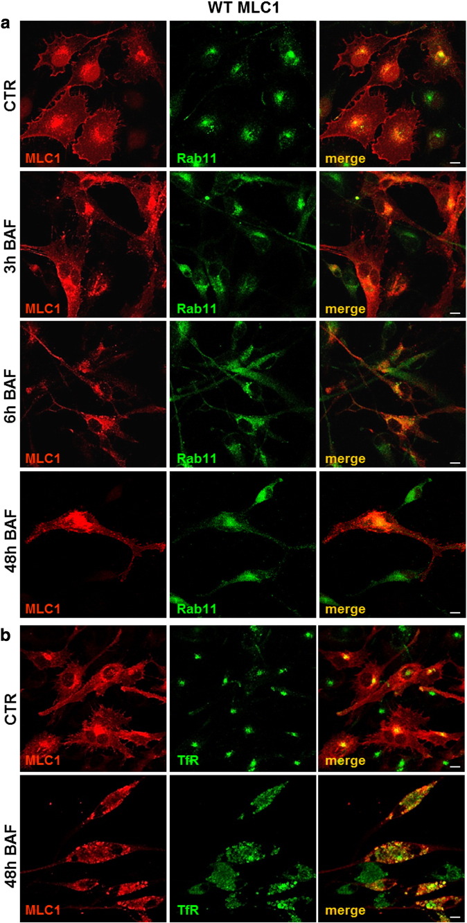
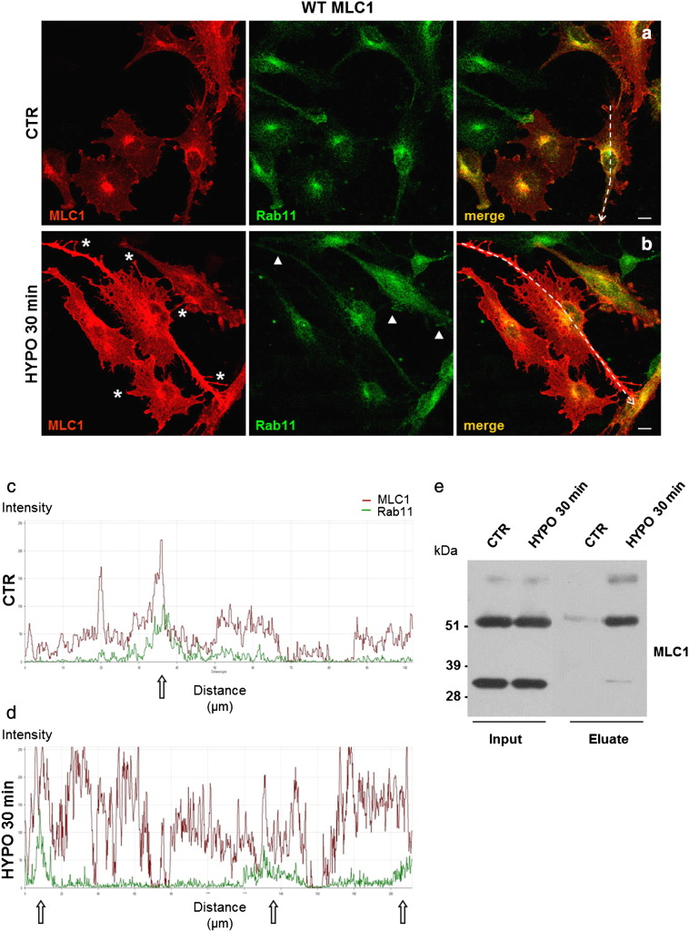
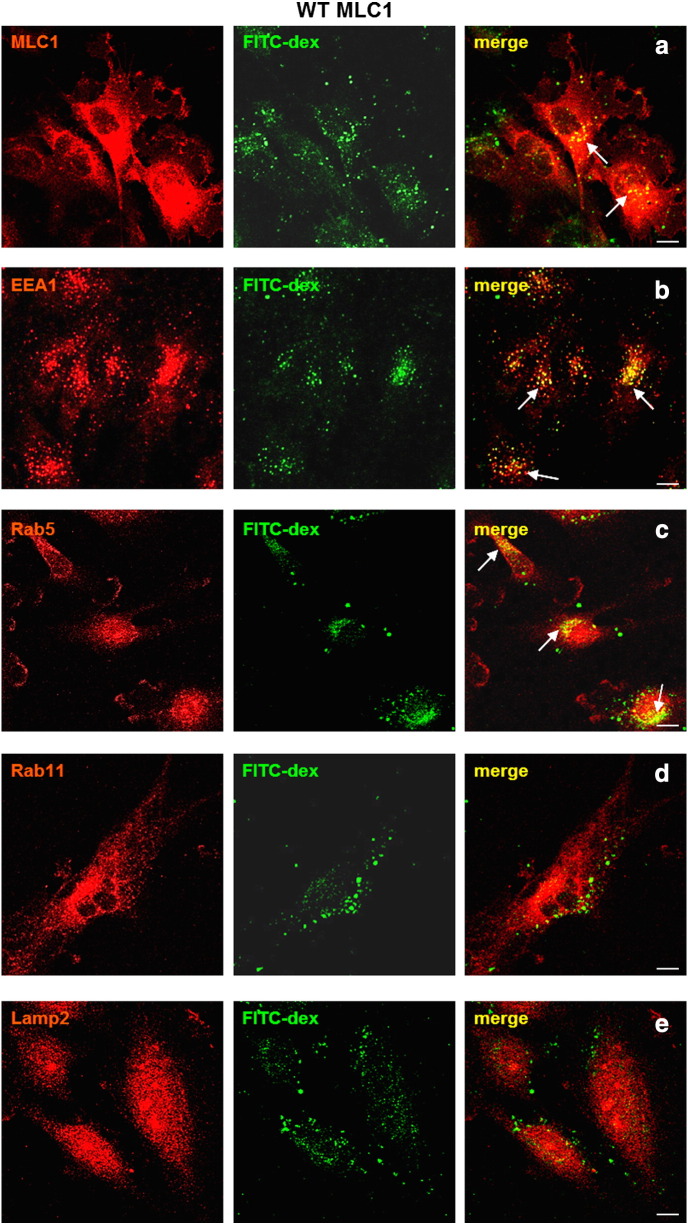
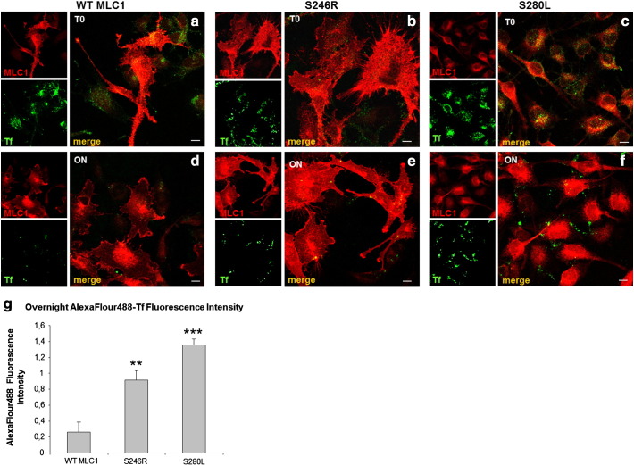
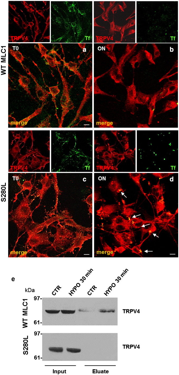
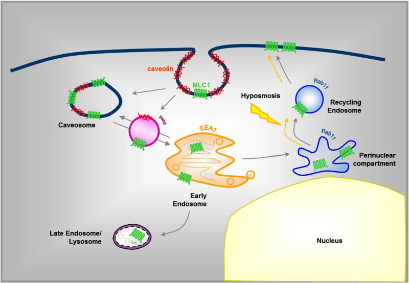
Megalencephalic leukoencephalopathy with subcortical cysts (MLC) is a rare leukodystrophy caused by mutations in the gene encoding MLC1, a membrane protein mainly expressed in astrocytes in the central nervous system. Although MLC1 function is unknown, evidence is emerging that it may regulate ion fluxes. Using biochemical and proteomic approaches to identify MLC1 interactors and elucidate MLC1 function we found that MLC1 interacts with the vacuolar ATPase (V-ATPase), the proton pump that regulates endosomal acidity. Because we previously showed that in intracellular organelles MLC1 directly binds Na, K-ATPase, which controls endosomal pH, we studied MLC1 endosomal localization and trafficking and MLC1 effects on endosomal acidity and function using human astrocytoma cells overexpressing wild-type (WT) MLC1 or MLC1 carrying pathological mutations. We found that WT MLC1 is abundantly expressed in early (EEA1(+), Rab5(+)) and recycling (Rab11(+)) endosomes and uses the latter compartment to traffic to the plasma membrane during hyposmotic stress. We also showed that WT MLC1 limits early endosomal acidification and influences protein trafficking in astrocytoma cells by stimulating protein recycling, as revealed by FITC-dextran measurement of endosomal pH and transferrin protein recycling assay, respectively. WT MLC1 also favors recycling to the plasma-membrane of the TRPV4 cation channel which cooperates with MLC1 to activate calcium influx in astrocytes during hyposmotic stress. Although MLC disease-causing mutations differentially affect MLC1 localization and trafficking, all the mutated proteins fail to influence endosomal pH and protein recycling. This study demonstrates that MLC1 modulates endosomal pH and protein trafficking suggesting that alteration of these processes contributes to MLC pathogenesis.

Keywords: Calcium; Early/recycling endosomes; Hyposmosis; K-ATPase; Leukodystrophy; Na; Rab11; TRPV4; V-ATPase.


Figures

Fig. 1
Fig. 1
Interaction between WT MLC1 and V-ATPase. a. Pull-down assay shows the interaction between MLC1 and V-ATPase (a1 subunit) in rat primary astrocytes. Cytosolic and membrane fractions from primary cultures of rat astrocytes (input) were pulled-down by His-MLC1 and His-empty vector (used as control) bound to NiNTA-agarose and eluted with 0.1 M glycine, pH 3. Western blot (WB) analysis shows that V-ATPase interacts with His-MLC1 in both the cytoplasmic (cyt) and membrane (mem) fractions. A very low level of unspecific binding is observed only in the membrane fraction of the NiNTA-agarose bound with His empty vector. Molecular weight markers are indicated on the left (kDa). b. His co-purification assay was performed to verify the association between MLC1 and V-ATPase in human astrocytoma cells (U251) overexpressing His-tagged WT and mutated MLC1. V-ATPase is co-eluted and enriched with 200 mM imidazole from NiNTA-agarose bound with proteins of WT MLC1 astrocytoma (WT). A decrease in the binding is observed when MLC1 S246R, C125R and S280L NiNTA-agarose samples have been used, while no interaction is detected using samples derived from astrocytoma cells infected with empty vector (used as control). One representative experiment out of 3 performed is shown. Molecular weight markers are indicated on the left (kDa). c. Densitometric analysis of V-ATPase protein bands revealed by WB in (b). d. Double immunofluorescence staining of astrocytoma cells overexpressing WT MLC1 with anti-MLC1 pAb (red) and anti-V-ATPase mAb (green) reveals colocalization of MLC1 and V-ATPase in intracellular vesicles, mainly in perinuclear areas (arrows). e. Immunofluorescence staining of normal human brain tissue with GFAP mAb (green) and anti-V-ATPase pAb (red) shows V-ATPase expression in intracytoplasmic vesicles of GFAP+ astrocytes (arrows in the microscopic field shown at high power magnification on the right). f. Immunofluorescence staining of normal human brain tissue with anti-MLC1 pAb (red) and anti-V-ATPase mAb (green) reveals that MLC1 and V-ATPase colocalize in perivascular astrocyte end-feet (arrows in the microscopic field shown at high power magnification on the right). Scale bars: 10 μm.
Fig. 2
Fig. 2
Intracellular localization of WT and mutated MLC1 in human astrocytoma cell lines. a–d. Double immunofluorescence staining for MLC1 (red) and early endosome antigen (EEA1) (green) shows colocalization of MLC1 and EEA1 in the perinuclear area of MLC1-WT (a) and S246R mutant (b) astrocytoma cells but not of S280L (c) and C125R (d) mutant astrocytoma cells. e–h. Double immunofluorescence for MLC1 (red) and Rab5 (green) reveals protein colocalization in WT (e) and S246R (f) MLC1 astrocytoma cells, but only occasionally in S280L (g) and C125R (h) mutants. i–l. Double immunofluorescence for MLC1 (red) and Rab11 (green) shows that WT (i) and S246R (j) MLC1 proteins are abundantly present in Rab11+ recycling endosomes while colocalization is markedly reduced in S280L (k) and C125R (l) mutants. m–p. Double immunofluorescence for MLC1 (red) and the lysosomal marker Lamp-2 (green) reveals a partial colocalization in WT (m) and mutated MLC1 expressing astrocytoma cells (n–p). Note that Rab5 and Rab11 immunoreactivities have a more diffuse intracellular distribution in WT and S246R MLC1 astrocytoma cells compared to S280L and C125R mutants. Scale bars: 10 μm.
Fig. 3
Fig. 3
WT MLC1 is localized in the perinuclear recycling compartment (PNRC). Double immunofluorescence staining of WT MLC1 astrocytoma cell lines with anti-MLC1 pAb (red) and anti-transferrin receptor mAb (TfR; green), known to localize in PCRN, shows overlap of MLC1 and TfR immunoreactivities in the perinuclear area (arrows). Scale bar: 10 μm.
Fig. 4
Fig. 4
Effect of bafilomycin on WT MLC1 protein localization and expression. a. Double immunofluorescence stainings for MLC1 (red) and Rab11 (green) were performed in WT MLC1 astrocytoma cells that were grown in basal culture conditions (CTR) or treated with 100 nM bafilomycin (BAF) for 3, 6 and 48 h. Note the progressive disappearance of MLC1 from the plasma membrane and concomitant accumulation of the protein in Rab11+ intracellular vacuolar structures. b. Double immunostaining for MLC1 (red) and transferrin receptor (TfR, green) shows that after 48-h treatment with 100 nM bafilomycin, all MLC1 proteins colocalize with TfR in intracellular vacuolar structures. Scale bars: 10 μm.
Fig. 5
Fig. 5
Effects of hyposmotic stress on WT MLC1 intracellular traffic. a,b. Astrocytoma cell lines overexpressing WT MLC1 were incubated in control (CTR) or hyposmotic (HYPO) solution for 30 min and then stained with Abs to MLC1 (red) and Rab11 (green). In control cells (a) MLC1 immunoreactivity is found in the cell membrane and in the perinuclear cytoplasm, where it colocalizes with Rab11+ vesicles (merge). Hyposmotic stress (b) induces a marked increase in MLC1 immunoreactivity throughout the cell body and processes, in the plasma cell membrane and at astrocyte-astrocyte contacts (asterisks), and the redistribution of Rab11+ vesicles along the astrocyte cell body and cytoplasmic extensions (arrowheads). Scale bars: 10 μm. c,d. Immunofluorescence pixel intensity along the white dotted arrows drawn in representative cells in a and b was obtained using the profile analysis tool of the LSM 5 PASCAL. After hyposmotic stress, the fluorescence intensity peaks of MLC1 and Rab11 re-distribute from the central perinuclear area, typically observed in control conditions (arrow in c), to a more peripheral cytoplasmic localization and toward the plasma membrane (arrows in d). A strong increase in MLC1 fluorescence intensity is observed after hyposmotic stress (compare red lines in c and d). e. WB of total cell proteins (Input) and of enriched surface proteins after biotinylation experiments (Eluate) from WT MLC1 astrocytoma cells maintained in control (CTR) and (HYPO) hyposmotic conditions. Although hyposmotic stress does not affect the total amount of WT MLC1 protein (CTR versus HYPO in Input lanes), it strongly increases the amount of the dimeric (60 kDa) membrane-associated MLC1 component (CTR versus HYPO in Eluate lanes). Note that in the surface protein fraction the monomeric (36 kDa) MLC1 component is not detectable anymore. One representative experiment out of three performed is shown. Molecular weight markers are indicated on the left (kDa).
Fig. 6
Fig. 6
WT and mutated MLC1 differently regulate endosomal pH. a,b. Fluorescence images of astrocytoma cells pre-loaded with FITC-dextran (green) and the chromatin selective dye Hoechst 33258 (blue), to depict endosomes and nuclei, respectively. b. Higher magnification of the area selected in (a), depicting the polarized localization of FITC-dextran loaded endosomes. Scale bar: 23 μm. c. The bar graph shows the mean ± SEM pH values in control astrocytoma cells infected with empty vector (CTR) and astrocytoma cells overexpressing WT and mutated MLC1; the number of recorded cells for each cell line ranged between 43 and 83. Significant differences between CTR and MLC1 overexpressing cells were calculated using Student's t test; *P < 0.05. d. The graph shows the time-course of pH changes in labeled endosomes of WT and S280L MLC1 astrocytoma cells recorded in a representative experiment. Note that the pH recorded in the time lag before the application of calibration solutions is more basic in WT compared to S280L MLC1 astrocytoma cells.
Fig. 7
Fig. 7
Characterization of FITC-dextran positive vesicles in WT MLC1 astrocytoma cells. WT MLC1 astrocytoma cells were pre-loaded with FITC-dextran for 30 min to identify endosomes in which pH changes have been recorded (see Fig. 6) and then labeled with anti-MLC1 Ab or Abs specific for endosomal organelles. a,b. MLC1 (red, a) and EEA1 (red, b) immunoreactivities are found in FITC-dextran positive vesicles (green) (arrows). c. Immunostaining for Rab5 (red) reveals a slightly lower degree of localization in the FITC-dextran positive vesicles (green) (arrows). d, e. No overlap is found between FITC-dextran (green) and Rab11 (red, d) or Lamp-2 (red, e) immunoreactivities. Scale bars: 10 μm.
Fig. 8
Fig. 8
Transferrin recycling assay in WT and mutant MLC1 astrocytoma cell lines after hyposmotic stress. Astrocytoma cell lines expressing WT MLC1, S246R or S280L mutated protein were pre-incubated with Alexa Fluor 488-conjugated transferrin (Tf) at 4 °C to allow binding to surface TfR. After washing out labeled Tf (T0), cells were incubated at 37 °C overnight (ON) in the presence of excess unlabeled Tf and then stained with anti-MLC1 Ab (red). a,b,c. Alexa Fluor 488-conjugated Tf (green) shows comparable binding to the surface of WT and mutated MLC1 astrocytoma cells (T0). d,e,f. After overnight (ON) incubation with excess unlabeled Tf, Tf disappears almost completely from the surface of WT MLC1 astrocytoma cells (d), whereas it is still present in S246R and S280L mutant astrocytoma cells (e,f). Scale bars: 10 μm. g. The bar graph shows the mean ± SEM values of the Alexa Fluor 488-conjugated Tf fluorescence intensity in the different astrocytoma cell lines after ON incubation; 10 to 15 random fields (field area = 230 μm2) were analyzed. Significant differences between WT MCL1 and mutated (S246R, S280L) astrocytoma cells were calculated using Student's t test; *P < 0.05. **P < 0.005, ***P < 0.0005.
Fig. 9
Fig. 9
TRPV4 recycling in WT and S280L MLC1 astrocytoma cell lines after hyposmotic stress. Astrocytoma cell lines expressing WT or mutated S280L MLC1 proteins were incubated with Alexa Fluor 488-conjugated Tf at 4 °C to allow binding to surface TfR. After washing out labeled Tf (T0), cells were incubated at 37 °C for 20 min and overnight (ON) in the presence of excess unlabeled Tf and then stained with anti-TRPV4 pAb. a,c. TRPV4 (red) and Alexa Fluor 488-conjugated Tf (green) partially colocalize in intracellular vesicles in WT MLC1 and S280L mutant astrocytoma cell plasma membranes at T0. b,d. After ON incubation with unlabeled Tf, TRPV4-Tf colocalization disappears in WT MLC1 astrocytoma cells (b) but not in S280L astrocytoma cells where TRPV4 still colocalizes with Tf in clustered intracytoplasmic vesicles (d, arrows). Scale Bars:10 μm. e. WB of total cell proteins (input lanes) and of enriched surface proteins after biotinylation (eluates) reveals that 30-min incubation in hyposmotic solution induces an increase in surface expression of TRPV4 in WT MLC1 astrocytoma cells but not in cells expressing S280L mutation. Molecular weight markers are indicated on the left (kDa).
Fig. 10
Fig. 10
Schematic representation of MLC1 intracellular trafficking. A model of MLC1 intracellular trafficking is proposed on the basis of our previous results (Lanciotti et al., 2010) and the data presented in this paper. MLC1 is internalized via caveolae-mediated endocytosis and traffics through Rab5+ and EEA1+ early endosomes where it is sorted to the recycling or degradative pathway. Most of the intracellular MLC1 protein is stored in the perinuclear Rab11+ recycling vesicles from which it is recycled to plasma membrane in stress condition (Hyposmosis).

Similar articles

Cited by

References

    1. Abe K., Puertollano R. Role of TRP channels in the regulation of the endosomal pathway. Physiology (Bethesda) 2011;26:14–22. - PMC - PubMed
    1. Agresti C., Aloisi F., Levi G. Heterotypic and homotypic cellular interactions influencing the growth and differentiation of bipotential oligodendrocyte-type-2 astrocyte progenitors in culture. Dev. Biol. 1991;144:16–29. - PubMed
    1. Ambrosini E., Serafini B., Lanciotti A., Tosini F., Scialpi F., Psaila R., Raggi C., Di Girolamo F., Petrucci T.C., Aloisi F. Biochemical characterization of MLC1 protein in astrocytes and its association with the dystrophin–glycoprotein complex. Mol. Cell. Neurosci. 2008;37:480–493. - PubMed
    1. Aoki T., Hagiwara H., Matsuzaki T., Suzuki T., Takata K. Internalization of caveolae and their relationship with endosomes in cultured human and mouse endothelial cells. Anat. Sci. Int. 2007;82:82–97. - PubMed
    1. Bacac M., Fusco C., Planche A., Santodomingo J., Demaurex N., Leemann-Zakaryan R., Provero P., Stamenkovic I. Securin and separase modulate membrane traffic by affecting endosomal acidification. Traffic. 2011;12:615–626. - PubMed

Publication types

MeSH terms