. 2014 Apr;25(8):1338-54.
doi: 10.1091/mbc.E13-08-0449. Epub 2014 Feb 19.

Interaction of the HOPS complex with Syntaxin 17 mediates autophagosome clearance in Drosophila

Affiliations

Interaction of the HOPS complex with Syntaxin 17 mediates autophagosome clearance in Drosophila

Szabolcs Takáts et al. Mol Biol Cell. 2014 Apr.

Abstract

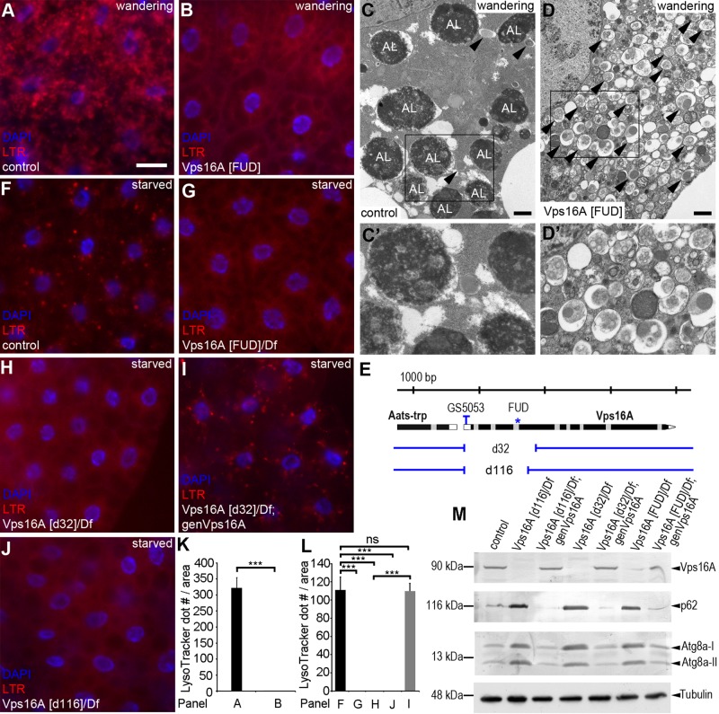
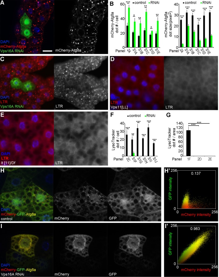
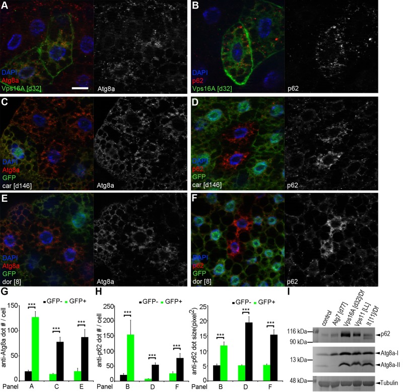
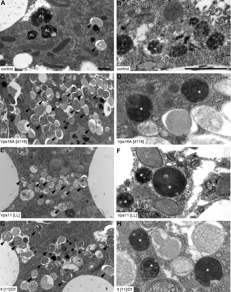
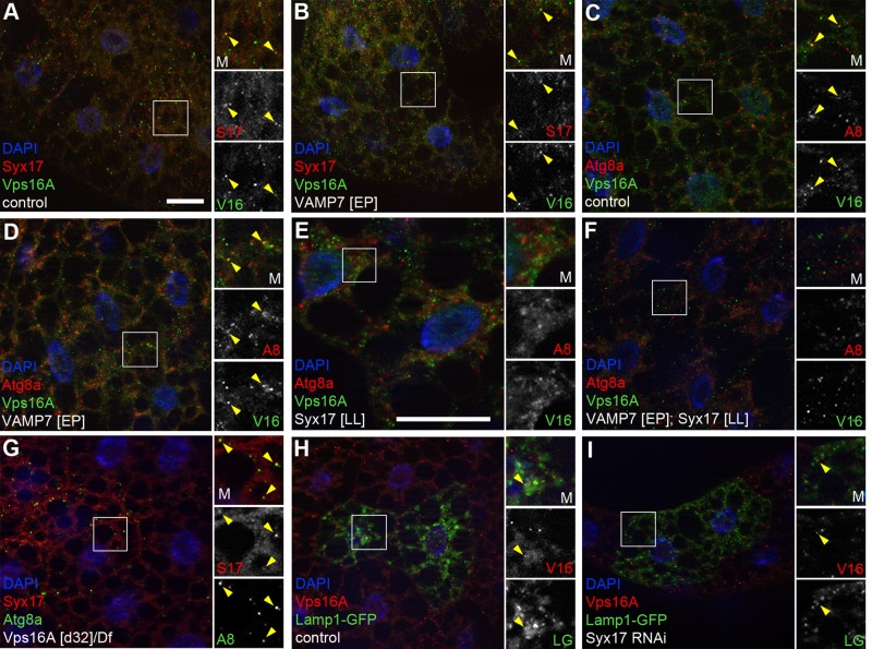
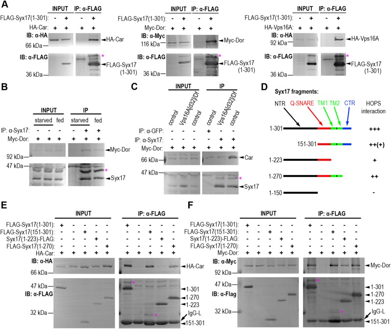
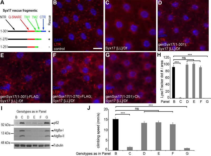
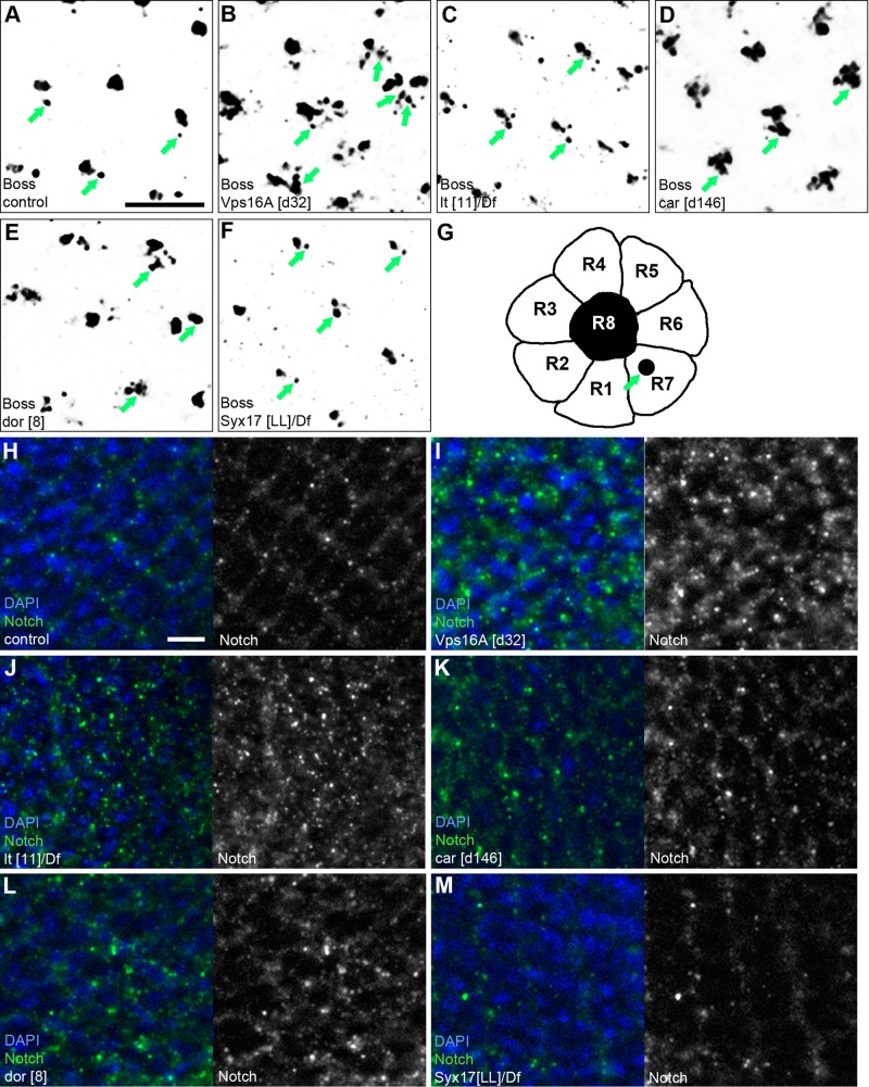
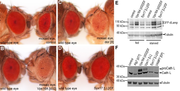
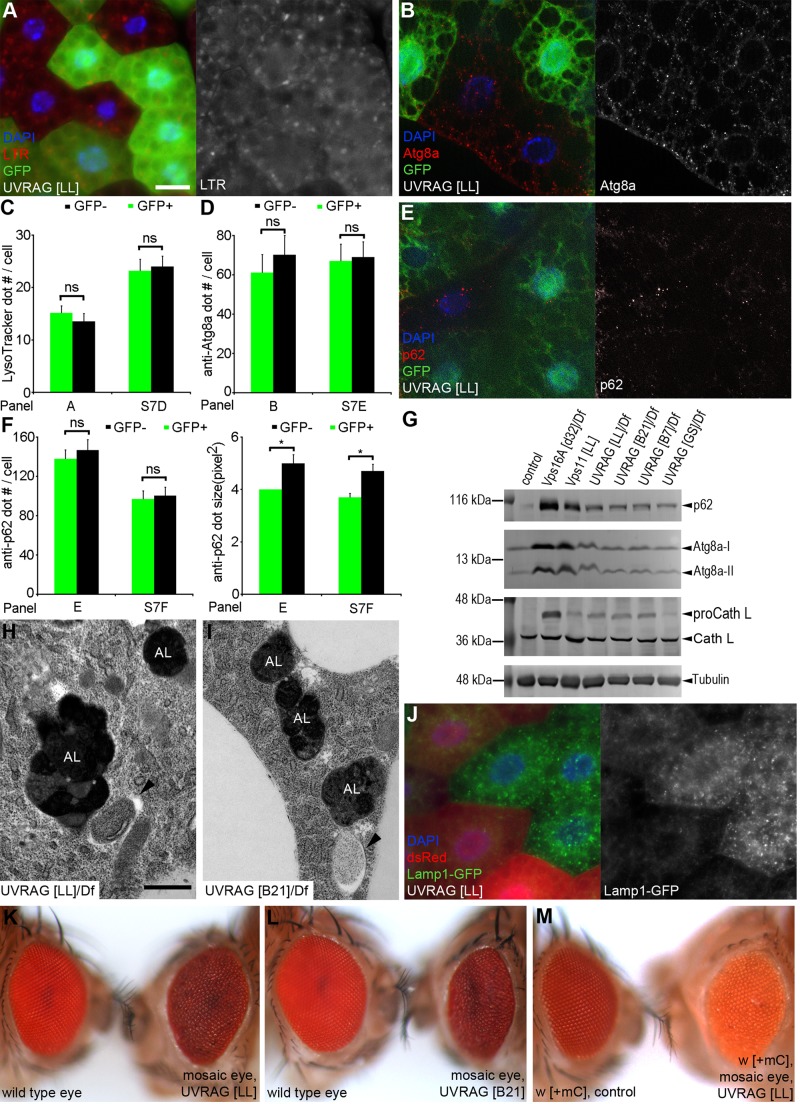
Homotypic fusion and vacuole protein sorting (HOPS) is a tethering complex required for trafficking to the vacuole/lysosome in yeast. Specific interaction of HOPS with certain SNARE (soluble NSF attachment protein receptor) proteins ensures the fusion of appropriate vesicles. HOPS function is less well characterized in metazoans. We show that all six HOPS subunits (Vps11 [vacuolar protein sorting 11]/CG32350, Vps18/Dor, Vps16A, Vps33A/Car, Vps39/CG7146, and Vps41/Lt) are required for fusion of autophagosomes with lysosomes in Drosophila. Loss of these genes results in large-scale accumulation of autophagosomes and blocks autophagic degradation under basal, starvation-induced, and developmental conditions. We find that HOPS colocalizes and interacts with Syntaxin 17 (Syx17), the recently identified autophagosomal SNARE required for fusion in Drosophila and mammals, suggesting their association is critical during tethering and fusion of autophagosomes with lysosomes. HOPS, but not Syx17, is also required for endocytic down-regulation of Notch and Boss in developing eyes and for proper trafficking to lysosomes and eye pigment granules. We also show that the formation of autophagosomes and their fusion with lysosomes is largely unaffected in null mutants of Vps38/UVRAG (UV radiation resistance associated), a suggested binding partner of HOPS in mammals, while endocytic breakdown and lysosome biogenesis is perturbed. Our results establish the role of HOPS and its likely mechanism of action during autophagy in metazoans.


Figures

FIGURE 1:
FIGURE 1:
Autophagosomes accumulate in Vps16A mutants. (A) Large numbers of LTR-positive dots are seen in fat body cells of wandering larvae. (B) No discernible LTR dots are seen in homozygous l(3)S007902 (FUD) mutant fat bodies of the same developmental stage. (C) Large-scale generation of autolysosomes (AL) is observed in fat body cells of wandering larvae. (D) Autolysosome formation is inhibited and autophagosomes (arrowheads) accumulate in large numbers in FUD mutant fat body cells of wandering larvae. Boxed areas in (C) and (D) are shown enlarged in (C′) and (D′). (E) Genomic map of the Vps16A locus. FUD mutants carry a point mutation at the 3′ end of the third intron (blue asterisk). The null alleles d32 and d116 were generated from the P element insertion GS5053 (located in the 5′ untranslated region of Vps16A), and carry 1105– and 926–base pair deletions extending into the coding sequence starting from the original P element insertion site, respectively. (F–J) Three-hour starvation in a 20% sucrose solution induces punctate LTR staining in control larvae (F), unlike in hemizygous Vps16A[FUD] (G), Vps16A[d32] (H), or Vps16A[d116] (J) mutants. Transgenic expression of Vps16A restores starvation-induced LTR dot formation in Vps16A[d32] mutants (I). (K) Quantification of data shown in (A) and (B); n = 10/genotype. (L) Quantification of data shown in (F)–(J); n = 10/genotype. (M) Western blots reveal that Vps16A protein cannot be detected in lysates of starved Vps16A[d32] and Vps16A[d116] mutant larvae, and its level is strongly reduced in Vps16A[FUD] mutants. The autophagic cargo p62 and autophagosome-associated Atg8a-II accumulate to similar levels in all three mutants. Transgenic expression of Vps16A restores wild-type protein levels for Vps16A, p62, and Atg8a in all three mutant backgrounds. Scale bar in (A) = 20 μm for (A), (B), and (F)–(J); scale bars = 1 μm in (C) and (D). Error bars denote SE in (K) and (L); ns, not significant; ***, p < 0.001.
FIGURE 2:
FIGURE 2:
Loss of HOPS complex function leads to impaired starvation-induced autophagy. (A) Expression of a transgenic RNAi construct targeting Vps16A in GFP-marked clones of fat body cells causes the accumulation of small, faint, perinuclear mCherry-Atg8a dots, which are very different from the bigger, brighter puncta observed in neighboring control cells. (B) Quantification of data shown in (A) and in Figure S1, A–E; n = 10/genotype. (C–E) Depletion of Vps16A in cell clones marked by Lamp1-GFP prevents punctate LTR staining (C), compared with neighboring controls cells. Similarly, no discernible LTR structures are seen in starved Vps11[LL] (D) or lt[11] (E) mutant fat cells. (F) Quantification of data shown in (C) and Figure S1, F–J; n = 9/genotype for (F); n = 10/genotype for others. (G) Quantification of data shown in (D), (E), and Figure 1F; n = 10/genotype. (H and I) The double-tagged mCherry-GFP-Atg8a reporter is transported to autolysosomes, which are seen as mCherry-positive dots due to quenching of GFP in starved control cells (H). Silencing of Vps16A (I) results in a block of GFP quenching; thus dots appear yellow in merged images. (H′) and (I′) depict dot plots of intensity and colocalization profiles for the mCherry and GFP channels from (H) and (I). Numbers on top show Pearson correlation coefficients, indicating increased colocalization of GFP and mCherry in (I) compared with (H). Scale bar in (A) = 20 μm for (A), (C), (D), and (E); scale bar = 40 μm for (H) and (I). Error bars denote SE in (B), (F), and (G); ***, p < 0.001, **, p < 0.01, *, p < 0.05.
FIGURE 3:
FIGURE 3:
The autophagic cargo p62 and Atg8a-positive autophagosomes accumulate in starved HOPS mutants. (A–F) Endogenous Atg8a-positive autophagosomes (A, C, and E) and endogenous p62 (B, D, and F) aggregates accumulate in Vps16A (A and B), car (C and D), or dor (E and F) null mutant cells, compared with surrounding control fat cells in starved mosaic larvae. Note that Vps16A mutant cells are marked by GFP in (A) and (B), whereas car or dor mutant cells can be recognized by the lack of GFP in (C)–(F). (G) Quantification of data shown in (A), (C), and (E); n = 10/genotype. (H) Quantification of data shown in (B), (D), and (F); n = 10/genotype. (I) Western blots show that p62 and Atg8a-II are up-regulated in starved Vps16A, Vps11, and lt mutants compared with wild-type controls. Atg7 mutants are used as an additional control. Scale bar in (A) = 20 μm for (A–F). Error bars denote SE in (G) and (H); ***, p < 0.001.
FIGURE 4:
FIGURE 4:
Ultrastructural analysis of starved HOPS mutants. (A) Starvation leads to the formation of autophagosomes (arrowheads) and autolysosomes (AL) in fat body cells of control larvae. (B) This panel illustrates a representative autolysosome containing heterogenous material due to different stages of degradation. Note that distinct parts within this irregularly shaped organelle can be seen, corresponding to multiple autophagosome-lysosome fusion events. (C–H) Autophagosomes accumulate in large numbers in Vps16A (C), Vps11 (E), or lt (G) mutant cells. In addition, numerous dense organelles are seen, which are usually round and smaller than the autolysosomes observed in controls. High-magnification images reveal that a subset of these presumably lytic vesicles (marked by asterisks) in Vps16A (D), Vps11 (F), or lt (H) mutant cells contain recognizable remnants of cytoplasmic material, such as a mitochondrion (m) in (F). Scale bar in (A) = 1 μm for (A), (C), (E), and (G); scale bar in (B) = 1 μm for (B), (D), (F), and (H).
FIGURE 5:
FIGURE 5:
Vps16A colocalizes with the autophagosomal SNARE Syx17, the autophagosome marker Atg8a, and the lysosome reporter Lamp1-GFP in fat body cells of starved larvae. (A and B) Endogenous Syx17 colocalizes with endogenous Vps16A in starved control (A) and Vamp7 mutant (B) fat body cells. (C–F) Vps16A localizes to endogenous Atg8a-positive structures in starved control (C) and Vamp7 mutant (D) fat body cells. Colocalization of Vps16A with Atg8a is not seen in Syx17 mutants (E) and Vamp7, Syx17 double mutants (F). (G) Syx17 colocalizes with Atg8a in Vps16A mutants. (H and I) Vps16A colocalizes with the lysosome marker Lamp1-GFP in both starved control (H) and Syx17 RNAi (I) fat body cells. Boxed areas are shown enlarged as indicated, with representative colocalizations highlighted by yellow arrowheads. M, merged; S17, Syx17; V16, Vps16A; A8, Atg8a; LG, Lamp1-GFP. Scale bar in (A) = 20 μm for (A)–(D) and (F)–(I); scale bar in (E) = 20 μm.
FIGURE 6:
FIGURE 6:
Multiple HOPS subunits interact with Syx17. (A) HA-Car, Myc-Dor, and HA-Vps16A coprecipitate with full-length FLAG-Syx17 in cultured Drosophila cells. (B) The interaction of transiently expressed Myc-Dor increases with endogenous Syx17 in response to starvation in immunoprecipitation experiments from adult flies. (C) Endogenous Car coprecipitates with endogenous Syx17 in starved control larvae, but not in starved Vps16A mutants. (D) An illustration of the truncated Syx17 fragments used in mapping experiments, together with the summary of their interactions with HOPS subunits based on experiments shown in (E), (F), and Figure S6, I and J. Abbreviations are as follows: NTR, N-terminal region; Q-SNARE, Q-SNARE domain; TM1 and 2, transmembrane domains 1 and 2, respectively; CTR, C-terminal region. Numbers refer to amino acid positions in full-length Syx17. (E and F) Immunoprecipitation analysis of HA-Car (E) and Myc-Dor (F) with FLAG-tagged Syx17 constructs shown in (D). Asterisks mark a higher-molecular-weight isoform of endogenous Syx17 in (B) and (C), which likely represents a C-terminally extended 346–amino acid protein isoform as a result of stop codon readthrough (Dunn et al., 2013). Note that a higher-molecular-weight isoform (also marked by asterisks in A, E, and F) is seen in the case of tagged constructs expressing full-length and N-terminally truncated Syx17, both of which contain the full 3′ untranslated region of this gene, but not in the case of C-terminally truncated versions. IP, immunoprecipitate; IgG-L, immunoglobulin light chain.
FIGURE 7:
FIGURE 7:
Genetic rescue experiments using truncated Syx17 fragments. (A) An illustration of the various Syx17 transgenes used in genetic rescue experiments. Please see Figure 6D for abbreviations. (B–G) LTR staining of fat body cells dissected from starved larvae. Punctate LTR staining seen in control cells (B) is missing from Syx17 mutants (C). Both untagged (D) and C-terminally FLAG-tagged (E) full-length transgenes restore LTR dot formation in Syx17 mutants, similar to a Syx17 fragment lacking the C-terminal region (F). A Syx17 construct lacking the second transmembrane region and C-terminus is unable to restore punctate LTR in Syx17 mutants (G). Please note that the C-terminus of this last construct is tagged with mCherry, but the expression of this transgene is under direct control of the Syx17 promoter, similar to the other constructs. This results in a very low expression level; therefore its fluorescence does not interfere with LTR staining. (H) Quantification of data shown in (B)–(G); n = 10/genotype. (I) Western blots of well-fed adult flies also show that a Syx17 fragment lacking the C-terminal region fully rescues the p62 and Atg8a-II accumulation phenotypes of well-fed Syx17 mutant adult flies, similar to full-length transgenes. Truncating Syx17 after the first transmembrane domain prevents the rescue of these defects. (J) Negative geotaxis assays of adult flies reveal that both full-length transgenes and a Syx17 fragment lacking the C-terminal region fully rescue the climbing defects of Syx17 mutant adult flies. Again, truncating the second transmembrane domain and C-terminus of Syx17 prevents rescue; n = 90 for all genotypes. Scale bar in (B) = 20 μm for (B)–(G). Error bars denote SE in (H) and (J); ns, not significant; ***, p < 0.001.
FIGURE 8:
FIGURE 8:
HOPS, but not Syx17, is required for endocytic down-regulation of Notch and Boss in developing eyes. (A–F) Anti-Boss staining reveals pairs of bigger and smaller dots corresponding to R8 and R7 photoreceptor neurons in the differentiating retina in control larvae (A), as indicated in the schematic (G). Accumulation of Boss is obvious in Vps16A (B) and lt (C) mutants, and also in developing eyes entirely composed of car (D) and dor (E) null mutant cells. Boss distribution in Syx17 mutants (F) is similar to controls. (H–M) Punctate anti-Notch immunolabeling reveals a regular pattern of developing ommatidia in eye disks of control larvae (H). Mutation of Vps16A (I), lt (J), car (K), or dor (L) leads to large-scale accumulation of Notch. The level and localization of Notch in Syx17 mutants (M) is similar to controls. Scale bar in (A) = 20 μm for (A)–(F); scale bar in (H) = 20 μm for (H)–(M).
FIGURE 9:
FIGURE 9:
HOPS, but not Syx17, is required for biosynthetic transport to lysosomes and pigment granules. (A–D) Eye pigment granules, a type of lysosome-related organelle, are responsible for the bright red color of wild-type eyes (A). Eye tissue homozygous for one chromosome arm can be generated with the GMR-Hid method by eliminating nonhomozygous tissue due to eye-specific expression of the proapoptotic gene Hid in mosaic eyes. The right side (A) shows the eyes of flies entirely homozygous for a chromosome arm bearing a GFP insertion, which serves as an additional control for mosaic eye experiments. Note that the GMR-Hid method results in a rough eye phenotype, but it does not affect eye color. Eyes composed of entirely mutant cells for Vps16A (B) or dor (C) have a much lighter, orange color. The eye color of homozygous, viable Syx17 mutants (D) is similar to that of wild-type flies. (E) Western blot using a GFP knock-in line of endogenous dLamp reveals that this lysosomal membrane protein accumulates in Vps16A mutant larvae, whereas no difference is seen between Syx17 mutant and control larvae. Asterisk indicates a nonspecific band that serves as an additional loading control. (F) The proform of the resident lysosomal hydrolase cathepsin L accumulates in Vps16A and Vps11 mutant larvae compared with controls, but not in Atg7, Syx17, or Vamp7 mutants.
FIGURE 10:
FIGURE 10:
Null mutation of UVRAG has no effect on autophagosome formation and fusion, while it perturbs biosynthetic transport to lysosomes. (A) Starvation-induced punctate LTR staining in UVRAG[LL] mutant fat cells (marked by the lack of GFP) is similar to neighboring heterozygous (marked by expression of one copy of GFP) and homozygous (marked by two copies of GFP) control cells. (B) Atg8a-positive autophagosome numbers are similar in UVRAG[LL] mutant and control cells of starved larvae. (C) Quantification of data shown in (A) and Figure S7D; n = 10/genotype. (D) Quantification of data shown in (B) and Figure S7E; n = 10/genotype. (E) The autophagic cargo p62 is up-regulated in UVRAG[LL] mutant cells compared with surrounding control cells. (F) Quantification of data shown in (E) and Figure S7F; n = 10/genotype. (G) Western blots show that the levels of Atg8a-II are slightly higher in the lysates of different UVRAG mutant larvae than those seen in controls but are much lower than the levels seen in Vps16A or Vps11 mutants. Similarly, p62 is up-regulated in UVRAG mutants relative to controls, but its levels do not reach those observed in Vps16A or Vps11 mutants. Pro-cathepsin L accumulates in UVRAG mutants. (H and I) Ultrastructural analysis reveals a regular number of autophagosomes and autolysosomes in UVRAG[LL] (H) and UVRAG[B21] (I) mutant larvae in response to starvation. Note that the contents and the irregular shape of autolysosomes indicate that several autophagosomal fusion events have occurred, whereas all autolysosomes appear to contain homogenously dark material, suggesting defects in autolysosomal degradation. (J) Turnover of Lamp1-GFP (expressed in all cells) is perturbed in UVRAG[LL] mutant cells (marked by lack of dsRed expression), which accumulate high levels of this lysosomal reporter. (K and L) Formation of the bright red eye color observed in control flies is perturbed in animals with eyes composed of entirely UVRAG[LL] (K) or UVRAG[B21] (L) mutant tissue. (M) The dark orange eye color of flies expressing lower levels of the pigment transporter white (w) is much lighter in UVRAG[LL] mutant eyes. Scale bar in (A) = 20 μm for (A), (B), (E), and (J); scale bar in (H) = 1 μm for (H) and (I). Error bars denote SE in (C), (D), and (F); ns, not significant; *, p < 0.05.

Similar articles

Cited by

References

    1. Akbar MA, Ray S, Kramer H. The SM protein Car/Vps33A regulates SNARE-mediated trafficking to lysosomes and lysosome-related organelles. Mol Biol Cell. 2009;20:1705–1714. - PMC - PubMed
    1. Akbar MA, Tracy C, Kahr WH, Kramer H. The full-of-bacteria gene is required for phagosome maturation during immune defense in Drosophila. J Cell Biol. 2011;192:383–390. - PMC - PubMed
    1. Balderhaar HJ, Ungermann C. CORVET and HOPS tethering complexes—coordinators of endosome and lysosome fusion. J Cell Sci. 2013;126:1307–1316. - PubMed
    1. Bankaitis VA, Johnson LM, Emr SD. Isolation of yeast mutants defective in protein targeting to the vacuole. Proc Natl Acad Sci USA. 1986;83:9075–9079. - PMC - PubMed
    1. Barth JM, Szabad J, Hafen E, Kohler K. Autophagy in Drosophila ovaries is induced by starvation and is required for oogenesis. Cell Death Differ. 2011;18:915–924. - PMC - PubMed

Publication types

MeSH terms