. 2014 Feb 19;34(8):2910-20.
doi: 10.1523/JNEUROSCI.3714-13.2014.

Glial wingless/Wnt regulates glutamate receptor clustering and synaptic physiology at the Drosophila neuromuscular junction

Affiliations

Glial wingless/Wnt regulates glutamate receptor clustering and synaptic physiology at the Drosophila neuromuscular junction

Kimberly S Kerr et al. J Neurosci. .

Abstract

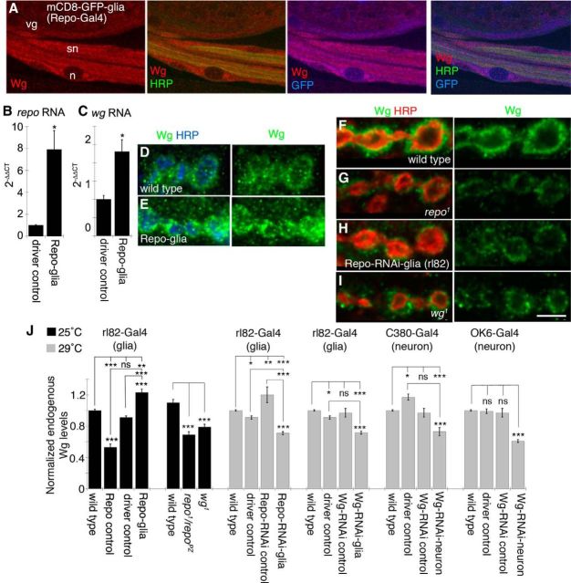
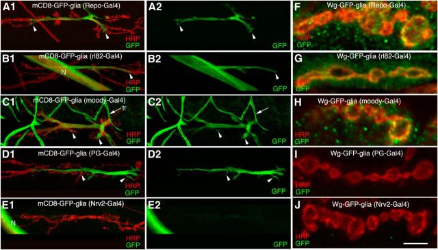
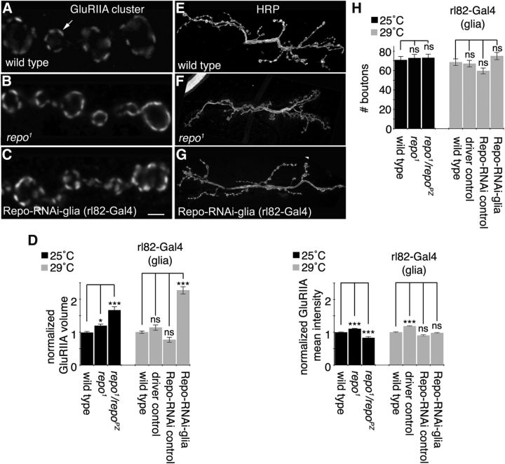
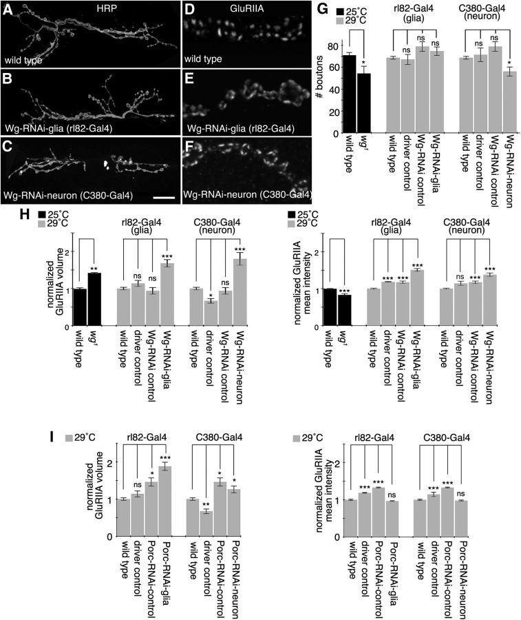
Glial cells are emerging as important regulators of synapse formation, maturation, and plasticity through the release of secreted signaling molecules. Here we use chromatin immunoprecipitation along with Drosophila genomic tiling arrays to define potential targets of the glial transcription factor Reversed polarity (Repo). Unexpectedly, we identified wingless (wg), a secreted morphogen that regulates synaptic growth at the Drosophila larval neuromuscular junction (NMJ), as a potential Repo target gene. We demonstrate that Repo regulates wg expression in vivo and that local glial cells secrete Wg at the NMJ to regulate glutamate receptor clustering and synaptic function. This work identifies Wg as a novel in vivo glial-secreted factor that specifically modulates assembly of the postsynaptic signaling machinery at the Drosophila NMJ.

Keywords: Drosophila; NMJ; glia; synapse; wnt/Wg.


Figures

Figure 1.
Figure 1.
Identification of potential Repo target genes. A, Tiling array data were obtained from two independent Repo ChIP experiments (myc::Repo and Repo::myc) after expression in Drosophila S2 cells. B, Data were analyzed using a MAT algorithm (Johnson et al., 2006) and converted to a linear scale to be viewed using the Affymetrix Integrated Genome Browser. For each gene, the genomic location and isoforms are shown. The relative amplitude of the peaks represents the probability of DNA binding (MAT score). C, Known glial or Wg/Wnt pathway signaling genes identified in Repo-ChIP experiments. D, RNA in situ hybridizations using a gs2 or Cp1 probe to control and repo-null mutant embryos.
Figure 2.
Figure 2.
Repo regulation of Wg in peripheral glia. A, Third instar larval segmental nerves expressing mCD8-GFP in glia and labeled with anti-HRP, anti-GFP, and anti-Wg. n, glial nucleus; sn, segmental nerve; vg, ventral ganglion. B, C, Real-time PCR from larval segmental nerve RNA showing that repo (B) and wg transcript (C) levels are increased when Repo is overexpressed in peripheral glia using repo-Gal4. Transcript fold changes were determined using the Δ-ΔCt method. D–I, Confocal images of third instar larval NMJ branches in preparations double labeled with anti-HRP and anti-Wg in wild-type controls (D, F), upon overexpressing Repo in peripheral glia (E), repo1 mutants (G), upon expressing Repo-RNAi in peripheral glia (H), and in wg1 mutants (I). J, Quantification of total Wg signal intensity divided by bouton volume in each of the indicated genotypes normalized to wild type. Gray and black bars indicate experiments performed at 29°C, to maximize RNAi expression, and 25°C, respectively. Error bars represent SEM. *p ≤ 0.05; **p ≤ 0.01; ***p < 0.001. Scale bar: (in I) A, 8 μm; D–I, 5 μm. The numbers of arbors quantified for normalized endogenous Wg levels are as follows: 25°C, wild type, 10; UAS-Repo/+ (Repo control), 10; rl82-Gal4/+ (driver control), 10; rl82-Gal4>Repo (Repo-glia), 10; repo1/repoPZ, 10; wg1, 10; 29°C, wild type, 47; rl82-Gal4/+ (driver control), 10; UAS-Repo-RNAi/+ (Repo-RNAi control), 10; rl82-Gal4>Repo-RNAi (Repo-RNAi-glia), 10; UAS-Wg-RNAi/+ (Wg-RNAi control), 10; rl82-Gal4>Wg-RNAi (Wg-RNAi-glia), 24; C380-Gal4/+ (driver control), 10; C380-Gal4>Wg-RNAi (Wg-RNAi-neuron), 13; OK6-Gal4/+ (driver control), 10; and OK6-Gal4>Wg-RNAi (Wg-RNAi-neuron), 21.
Figure 3.
Figure 3.
Subperineurial glial membranes invade the NMJ and secrete Wg. A1–E2, Confocal images of third instar NMJs in preparations double labeled with anti-HRP and anti-GFP upon expressing mCD8-GFP in glial cell subtypes using repo-Gal4 (all glia; A1, A2), rl82-Gal4 (subperineurial glia; B1, B2), moody-Gal4 (subperineurial glia; C1, C2), PG-Gal4 (perineurial glia; D1, D2), and Nrv2-Gal4 (wrapping glia; E1, E2). F–J, Confocal images of NMJ branches labeled with anti-HRP and anti-GFP in larvae expressing Wg-GFP in subsets of glia using repo-Gal4 (F), rl82-Gal4 (G), moody-Gal4 (H), PG-Gal4 (I), and Nrv2-Gal4 (J). Scale bar: (in J) A1–J, 5 μm. Arrowheads represent extent of glial membrane infiltration into the NMJ, while arrow denotes tracheal cells (tr). N, nerve.
Figure 4.
Figure 4.
Subperineurial glia are required for normal GluRIIA distribution. A–C, Confocal images of third instar NMJ branches in preparations double labeled with anti-GluRIIA in wild type (A, arrow denotes GluRIIA cluster), repo1 mutants (B), and upon expressing Repo-RNAi RNA in SPGs (C). D, Quantifications of GluRIIA volume divided by bouton volume and mean GluRIIA signal intensity in each of the indicated genotypes normalized to wild type. E–G, Confocal images of third instar larval NMJ arbors labeled with anti-HRP in wild type (E), repo1 mutants (F), and upon expressing Repo-RNAi RNA in SPGs (G). H, Quantification of total bouton number for each of the indicated genotypes. Gray and black bars indicate experiments performed at 29 and 25°C, respectively. Error bars represent SEM. *p ≤ 0.05; ***p < 0.001. Scale bar: (in C) A–C, 2 μm; E–G, 18 μm. The numbers of arbors quantified for GluRIIA parameters are as follows: 25°C, wild type, 10; repo1, 10; repo1/repoPZ, 10; 29°C, wild type, 32; rl82-Gal4/+ (driver control), 10; UAS-Repo-RNAi/+ (Repo-RNAi control), 10; and rl82-Gal4>Repo-RNAi (Repo-RNAi-glia), 10. The numbers of samples for total bouton number are as follows: 25°C, wild type, 42; repo1, 23; repo1/repoPZ, 14; 29°C, wild type, 166; driver control, 15; Repo-RNAi control, 19; and Repo-RNAi-glia, 18.
Figure 5.
Figure 5.
SPG- and motor neuron-derived Wg regulate glutamate receptors. A–C, Confocal images of third instar larval NMJ arbors labeled with anti-HRP in wild type (A), upon expressing Wg-RNAi in SPGs (B), and upon expressing Wg-RNAi in neurons (C). D–F, Confocal images of third instar NMJ branches in preparations double labeled with anti-GluRIIA in wild type (D), upon expressing Wg-RNAi in SPGs (E), and upon expressing Wg-RNAi in motor neurons (F). G, Quantification of total bouton number for each of the indicated genotypes. H, I, Quantifications of GluRIIA volume divided by bouton volume and mean GluRIIA signal intensity in each of the indicated genotypes normalized to wild type. Gray and black bars indicate experiments performed at 29 and 25°C respectively. Error bars represent SEM. *p ≤ 0.05; **p ≤ 0.01; ***p < 0.001. Scale bar: (in C) A–C, 20 μm; D–F, 4 μm. The numbers of animals quantified for total bouton number are as follows: 25°C, wild type, 42; wg1, 13; 29°C, wild type, 166; rl82-Gal4/+ (driver control), 15; UAS-Wg-RNAi/+ (Wg-RNAi control), 9; rl82-Gal4>Wg-RNAi (Wg-RNAi-glia), 27; C380-Gal4/+ (driver control), 13; and C380-Gal4>Wg-RNAi (Wg-RNAi-neuron), 29. The numbers of arbors quantified for GluRIIA parameters are as follows: 25°C, wild type, 10; wg1, 10; 29°C, wild type, 32; rl82 driver control, 10; Wg-RNAi control, 10; Wg-RNAi-glia, 18; C380 driver control, 10; Wg-RNAi-neuron, 10; UAS-Porc-RNAi/+ (Porc-RNAi control), 10; rl82-Gal4>Porc-RNAi (Porc-RNAi-glia), 10; and C380-Gal4>Porc-RNAi (Porc-RNAi-neuron), 10.
Figure 6.
Figure 6.
Synaptic transmission is altered in repo mutants and upon Wg decrease in glia or neurons. A, B, Representative mEJP (A) and evoked EJP traces (B) in the indicated genotypes. C–F, Quantification of mEJP amplitude (C), mEJP frequency (D), evoked EJP amplitude (E), and quantal content (F). Error bars represent SEM. *p ≤ 0.05; **p ≤ 0.01; ***p < 0.001. The numbers of animals quantified are as follows: wild type, 23; repo1/repoPZ, 5; rl82-Gal4/+ (driver control), 8; C380-Gal4/+ (driver control), 7; UAS-Wg-RNAi/+ (Wg-RNAi control), 12; rl82-Gal4>Wg-RNAi (Wg-RNAi-glia), 30; and C380-Gal4>Wg-RNAi (Wg-RNAi-neuron), 11.

Similar articles

Cited by

References

    1. Akiyama Y, Hosoya T, Poole AM, Hotta Y. The gcm-motif: a novel DNA-binding motif conserved in Drosophila and mammals. Proc Natl Acad Sci U S A. 1996;93:14912–14916. doi: 10.1073/pnas.93.25.14912. - DOI - PMC - PubMed
    1. Allen NJ, Bennett ML, Foo LC, Wang GX, Chakraborty C, Smith SJ, Barres BA. Astrocyte glypicans 4 and 6 promote formation of excitatory synapses via GluA1 AMPA receptors. Nature. 2012;486:410–414. - PMC - PubMed
    1. Ashley J, Packard M, Ataman B, Budnik V. Fasciclin II signals new synapse formation through amyloid precursor protein and the scaffolding protein dX11/Mint. J Neurosci. 2005;25:5943–5955. doi: 10.1523/JNEUROSCI.1144-05.2005. - DOI - PMC - PubMed
    1. Ataman B, Ashley J, Gorczyca D, Gorczyca M, Mathew D, Wichmann C, Sigrist SJ, Budnik V. Nuclear trafficking of Drosophila Frizzled-2 during synapse development requires the PDZ protein dGRIP. Proc Natl Acad Sci U S A. 2006;103:7841–7846. doi: 10.1073/pnas.0600387103. - DOI - PMC - PubMed
    1. Awasaki T, Tatsumi R, Takahashi K, Arai K, Nakanishi Y, Ueda R, Ito K. Essential role of the apoptotic cell engulfment genes draper and ced-6 in programmed axon pruning during Drosophila metamorphosis. Neuron. 2006;50:855–867. doi: 10.1016/j.neuron.2006.04.027. - DOI - PubMed

Publication types

MeSH terms