. 2014 Mar 28;289(13):8828-38.
doi: 10.1074/jbc.M113.516237. Epub 2014 Feb 7.

Prolyl isomerase Pin1-mediated conformational change and subnuclear focal accumulation of Runx2 are crucial for fibroblast growth factor 2 (FGF2)-induced osteoblast differentiation

Affiliations

Prolyl isomerase Pin1-mediated conformational change and subnuclear focal accumulation of Runx2 are crucial for fibroblast growth factor 2 (FGF2)-induced osteoblast differentiation

Won-Joon Yoon et al. J Biol Chem. .

Abstract

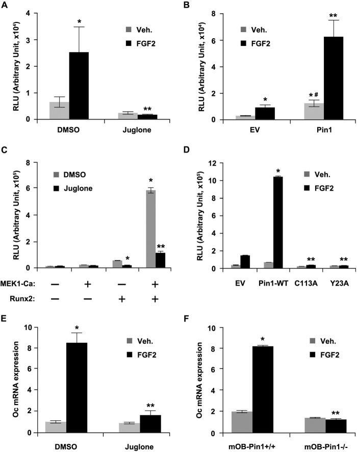
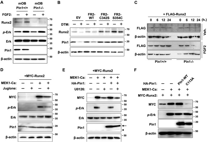
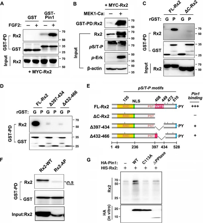
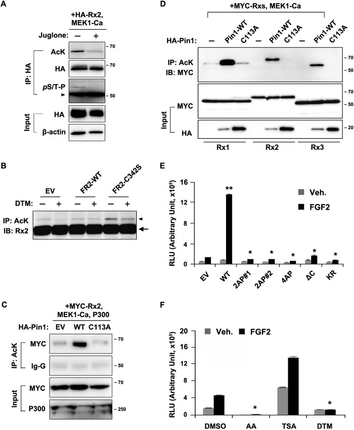
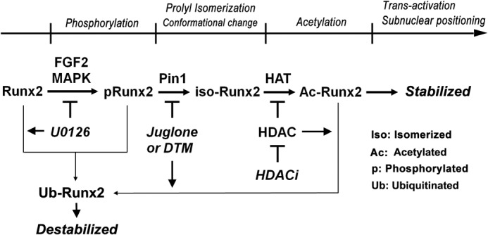
Fibroblast growth factor 2 (FGF2) signaling plays a pivotal role in bone growth/differentiation through the activation of osteogenic master transcription factor Runx2, which is mediated by the ERK/MAPK-dependent phosphorylation and the p300-dependent acetylation of Runx2. In this study, we found that Pin1-dependent isomerization of Runx2 is the critical step for FGF2-induced Runx2 transactivation function. We identified four serine or threonine residues in the C-terminal domain of Runx2 that are responsible for Pin1 binding and structural modification. Confocal imaging studies indicated that FGF2 treatment strongly stimulated the focal accumulation of Pin1 in the subnuclear area, which recruited Runx2. In addition, active forms of RNA polymerase-II also colocalized in the same subnuclear compartment. Dipentamethylene thiuram monosulfide, a Pin1 inhibitor, strongly attenuated their focal accumulation as well as Runx2 transactivation activity. The Pin1-mediated structural modification of Runx2 is an indispensable step connecting phosphorylation and acetylation and, consequently, transcriptional activation of Runx2 by FGF signaling. Thus, the modulation of Pin1 activity may be a target for the regulation of bone formation.

Keywords: Acetylation; Cell Differentiation; FGF Signaling; Osteoblasts; Pin1; Protein Conformation; Protein Phosphorylation; Runx2; Subnuclear Accumulation; Transcription Factors.


Figures

FIGURE 1.
FIGURE 1.
Pin1 is essential for FGF2-induced Runx2 transactivation activity. A, FGF2-induced Oc promoter activation was fully abrogated by the inhibition of Pin1 activity. C2C12 cells were transiently transfected with the Oc-Luc reporter vector. After 18 h of transfection, cells were treated with 20 ng/ml FGF2 in the presence or absence of juglone (10 μm) for 24 h, and the luciferase activity was measured from the harvested cells. Data represent the means ± S.D. (n = 3). *, p < 0.05 (compared with DMSO-treated group); **, p < 0.05 (compared with DMSO/FGF2-treated group). B, Oc-Luc reporter activity was strongly enhanced by Pin1 overexpression. C2C12 cells were transiently transfected with the Oc-Luc reporter vector either with the empty or the Pin1-expression plasmid. After 24 h, cells were treated with 20 ng/ml FGF2 for an additional 24 h, and luciferase activity was measured from the harvested cells. Data represent the means ± S.D. (n = 3). *, p < 0.05 (compared with EV/DMSO group); **, p < 0.05 (compared with Pin1/FGF2 group); #, p < 0.05 (compared with EV/FGF group). C, down-regulation of transactivation activity of Runx2 by the inhibition of Pin1 activity. C2C12 cells stably transfected with the p6×OSE2-Luc reporter vector (OSE2-C2C12) were transiently transfected with Runx2 along with either an empty vector or a MEK1-Ca plasmid. After 18 h of transfection, the cells were incubated with juglone (10 μm) for an additional 24 h, and luciferase activity was measured from the harvested cells. Data represent the means ± S.D. (n = 3). *, p < 0.0005 (compared with DMSO group expressing only Runx2 without MEK1-Ca expression); **, p < 0.0005 (compared with DMSO group expressing both Runx2 and MEK1-Ca). D, FGF2-induced Runx2 transactivation activity was dependent on Pin1 activity. MC3T3-E1 cells were transiently transfected with p6×OSE-Luc and either wild-type Pin1 or mutant Pin1 plasmids (C113A or Y23A) for 24 h. Cells were cultured in the presence or absence of FGF2 for an additional 18 h. Data represent the means ± S.D. (n = 3). *, p < 0.005 (compared with EV/FGF2 group); **, p < 0.005 (compared with Pin1-WT/FGF2 group). E and F, induction of Oc mRNA expression by FGF2 was abrogated by the treatment of the Pin1 inhibitor or the absence of Pin1. C2C12 cells were stimulated with 20 ng/ml FGF2 in the presence or absence of juglone (10 μm) for 24 h. E, equal numbers of primary mOBs from Pin1+/+ and Pin1−/− mice were treated with FGF2 (20 ng/ml) for 24 h after confluence. Data represent the means ± S.D. (n = 3). **, p < 0.001 (compared with DMSO/veh. group); *, p < 0.05 (compared with DMSO/FGF2 group). F, total RNAs were isolated, and the Oc mRNA levels were analyzed by quantitative real time PCR. Data represent the means ± S.D. (n = 3). **, p < 0.001 (compared with DMSO treated mOB-Pin1+/+); *, p < 0.005 (compared with FGF2 treated mOB-Pin1+/+). Veh, vehicle; EV, empty vector; RLU, relative luciferase units.
FIGURE 2.
FIGURE 2.
FGF2-induced increase of Runx2 protein level requires phosphorylation and subsequent structural modification by Pin1. A, determination of Runx2 protein levels depending on the Pin1 activity in mOBs. mOBs isolated from Pin1+/+ or Pin1−/− mice were cultured in the presence or absence of FGF2 for 24 h. B, gain-of-function mutations of FGFR2 increased Runx2 protein levels, which was attenuated by Pin1 inhibitor treatment. C342S and S354C are constitutively active FGFR2 mutants and have been shown to cause the craniosynostosis syndrome in humans. MC3T3-E1 cells were transiently transfected with the indicated constructs and were further incubated for 48 h under physiological culture conditions with or without the Pin1 inhibitor, DTM (1 μm). C, determination of Runx2 protein half-life in the presence or absence of Pin1 activity. Tet-On-inducible Runx2 expression vectors (38) were transiently expressed in MEF-Pin1+/+ and MEF-Pin1−/− cells. Cells were cultured for the induction of Runx2 expression in the presence of doxycycline (1 μg/ml). After 24 h, cells were washed and cultured in the fresh media without doxycycline to determine the remaining Runx2 protein level. The remaining Runx2 protein levels at the indicated time were determined by immunoblot assay. D, Runx2 stabilization requires both ERK/MAPK and Pin1 activation. Primary MEF cells were transiently transfected with Runx2. E, Runx2 stabilization requires ERK/MAPK activation. U0126 was used to inhibit the MEK/ERK pathway. Recombinant and endogenous Pin1 proteins are indicated by an arrow and an arrowhead, respectively. F, absence of Pin1 could not support ERK/MAPK-induced Runx2 stabilization. C2C12 cells were transiently transfected with indicated plasmids for 24 h, and the cells were further exposed to juglone or U0126 for 18 h (D–F). Veh, vehicle; EV, empty vector.
FIGURE 3.
FIGURE 3.
Pin1 recruits Runx2 protein to subnuclear domains that are transcriptionally active. A, FGF2 enhances the colocalization of Runx2 and Pin1 protein in the same subnuclear foci. MC3T3-E1 cells were cultured in serum-free media and treated with vehicle (Veh) (0.2% BSA/PBS) or with 20 ng/ml FGF2. DTM (1 μm) was added to inhibit Pin1 activity, and the cells were cultured for 12 h. Runx2-Pin1 colocalization was detected by indirect immunofluorescence labeling in the cells. B, increase of Runx2-containing foci number in response to FGF2 stimulation was dependent on Pin1 activity. Cells containing at least three enlarged Runx2 foci (>2 μm diameter) were counted by confocal microscopy. Data represent means ± S.D. The number of cells (n) are 131, 145, and 150 in the groups for vehicle, FGF2, and FGF2 + DTM, respectively. **, p < 0.00001, compared with control group (vehicle); *, p < 0.0001, compared with FGF2-treated group. C, FGF2-induced focal accumulation of Runx2 was colocalized with active RNA polymerase II, and the accumulation was attenuated by the Pin1 inhibitor, DTM. D, Pin1-dependent subnuclear accumulation of Runx2. EGFP-Runx2 (green) was transiently expressed in MC3T3-E1 cells with DsRed-Pin1-WT, C113A, or Y23A (red). E, time-lapse analysis of Runx2 foci (green) accumulation in response to Pin1 (red). Dynamic subnuclear accumulation of Runx2 (green) was monitored by time-lapse confocal microscopy (LSM700, Carl Zeiss). The supplemental movie covers 669 min (m) and is composed of pictures taken every 15 min. Subnuclear accumulation of Runx2 foci began ∼213 min after complete formation of Pin1 speckles.
FIGURE 4.
FIGURE 4.
FGF2-sitmulated ERK/MAPK signaling strongly enhanced Pin1 binding to the Runx2 C-terminal domain. A, elevated binding of Pin1 to FGF2-stimulated Runx2. HEK-293 cells were transiently transfected with Runx2 and cultured in the presence or absence of 10 ng/ml FGF2. After 60 min of FGF2 administration, lysates were prepared and used for GST-pulldown assays. B, ERK/MAPK-activated Runx2 strongly enhanced GST-Pin1 binding. For ERK activation, MEK1-Ca was transfected into HEK-293 cells with Runx2. After 24 h of transfection, cells were lysed, and the GST-pulldown assay was performed. C and D, identification of the Pin1 binding domain in Runx2. The ΔC-Runx2 construct was generated by excising amino acids 377–528, as described previously (39). FL-Runx2 indicates full-length Runx2. GST and GST-Pin1 are indicated as G and P, respectively. E, summary of the GST-pulldown assay for Pin1 binding to Runx2 deletion mutants. The functional domains of Runx2 conserved among Runx proteins are abbreviated as follows: RHD, runt-homology domain; NLS, nuclear localization sequence; PST, Pro/Ser/Thr-rich transactivation domain; NMTS, nuclear matrix target sequence. F, GST-pulldown assay of mutant Runx2 (Rx2–4AP) proteins in which the C-terminal Thr-408, Thr-449, Ser-472, and Ser-510 residues were substituted with Ala. A nonspecific band is indicated by n.s. G, determination of the structural alteration of Runx2 by subtilisin assay. HIS-Runx2 and MEK1-Ca were overexpressed in MEF-Pin1−/− cells and isolated by nitrilotriacetic acid affinity purification. For in vitro isomerization reaction, recombinant Pin1 proteins were synthesized in vitro and incubated with affinity-purified Runx2 protein-bead complex.
FIGURE 5.
FIGURE 5.
FGF2-induced Runx2 acetylation and activation requires Pin1. A, increased acetylation of Runx2 protein by MEK-Ca depends on Pin1 activity. Levels of acetylated Runx2 were determined by immunoprecipitation (IP) assay with an anti-acetyl-lysine (AcK) antibody. An arrowhead indicates the heavy chain of the antibody. B, activated FGFR2-induced increase of Runx2 acetylation requires Pin1 activity. MC3T3-E1 cells were transiently transfected with FGFR2 mutant plasmids for 24 h and cultured an additional 24 h with or without the Pin1 inhibitor, DTM. The protein extract from the cultures was immunoprecipitated with an acetyl-lysine antibody and immunoblotted (IB) with anti-Runx2 antibody. Arrowhead, acetylated Runx2; arrow, antibody heavy chain. EV, empty vector. C, Pin1 (WT) overexpression enhances Runx2 acetylation, but catalytically inactive Pin1 mutant (C113A) overexpression could not support Runx2 acetylation. HEK-293 cells were transiently transfected with the indicated plasmids and culture for 24 h. The protein extract from the cultures was immunoblotted with anti-Myc antibody. D, Runx protein acetylation depends on Pin1 activity. HEK-293 cells were transfected with Myc-tagged plasmids for Runx1, Runx2, or Runx3 with either empty, Pin1-WT, or Pin1-C113A vector. Comparable amounts of Runx proteins were used for each immunoprecipitation. E, mutant Runx2 proteins exhibited decreased transcriptional activity due to defective Pin1 binding, subnuclear targeting, or acetylation. 2AP#1 and 2AP#2 indicate substitutions of serine or threonine residues to alanine at Thr-408/Thr-449 or Ser-472/Ser-510, respectively. The KR mutation of Runx2 was previously described (47). The substitution of these four lysine residues with arginine residues (KR) dramatically increased the stability of Runx2 due to the absence of its ubiquitination sites, although the mutants do not have transcriptional activity. Data represent the means ± S.D. (n = 3). **, p < 0.0001, compared with empty vector (EV) transfection; *, p < 0.001, compared with wild-type Runx2 overexpression group. F, MC3T3-E1 cells were transiently transfected with the 6×OSE2-Luc reporter vector. After 24 h, cells were treated with 20 ng/ml FGF2 for an additional 24 h in the presence of DMSO (vehicle), 50 nm trichostatin A (TSA) (HDAC inhibitor), 25 μm anacardic acid (AA) (HAT inhibitor), or 1 μm DTM (Pin1 inhibitor). **, p < 0.0001, compared with control group (DMSO).
FIGURE 6.
FIGURE 6.
Positioning of Pin1-mediated prolyl isomerization in post-phosphorylational Runx2 activation cascade. Functional activation of Runx2 is tightly controlled by multiple post-translational modification cascades that occur in the following sequence: phosphorylation, prolyl isomerization, and acetylation. In these processes, Pin1 has a role as a molecular switch to determine the fate of Runx2 acetylation and further transactivation.

Similar articles

Cited by

References

    1. Ornitz D. M., Marie P. J. (2002) FGF signaling pathways in endochondral and intramembranous bone development and human genetic disease. Genes Dev. 16, 1446–1465 - PubMed
    1. Canalis E., Centrella M., McCarthy T. (1988) Effects of basic fibroblast growth factor on bone formation in vitro. J. Clin. Invest. 81, 1572–1577 - PMC - PubMed
    1. Mayahara H., Ito T., Nagai H., Miyajima H., Tsukuda R., Taketomi S., Mizoguchi J., Kato K. (1993) In vivo stimulation of endosteal bone formation by basic fibroblast growth factor in rats. Growth Factors 9, 73–80 - PubMed
    1. Yoshimura S., Takagi Y., Harada J., Teramoto T., Thomas S. S., Waeber C., Bakowska J. C., Breakefield X. O., Moskowitz M. A. (2001) FGF-2 regulation of neurogenesis in adult hippocampus after brain injury. Proc. Natl. Acad. Sci. U.S.A. 98, 5874–5879 - PMC - PubMed
    1. Zhou D., DiFiglia M. (1993) Basic fibroblast growth factor enhances the growth of postnatal neostriatal GABAergic neurons in vitro. Exp. Neurol. 122, 171–188 - PubMed

Publication types

MeSH terms

Substances

LinkOut - more resources