. 2014 Apr;18(4):656-70.
doi: 10.1111/jcmm.12213. Epub 2014 Feb 4.

The roles of αV integrins in lens EMT and posterior capsular opacification

Affiliations

The roles of αV integrins in lens EMT and posterior capsular opacification

Fahmy A Mamuya et al. J Cell Mol Med. 2014 Apr.

Abstract

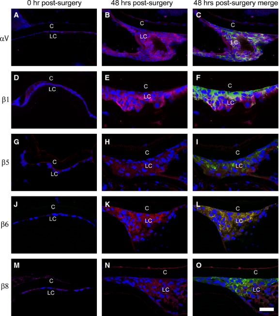
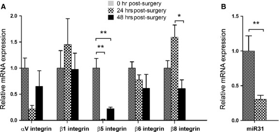
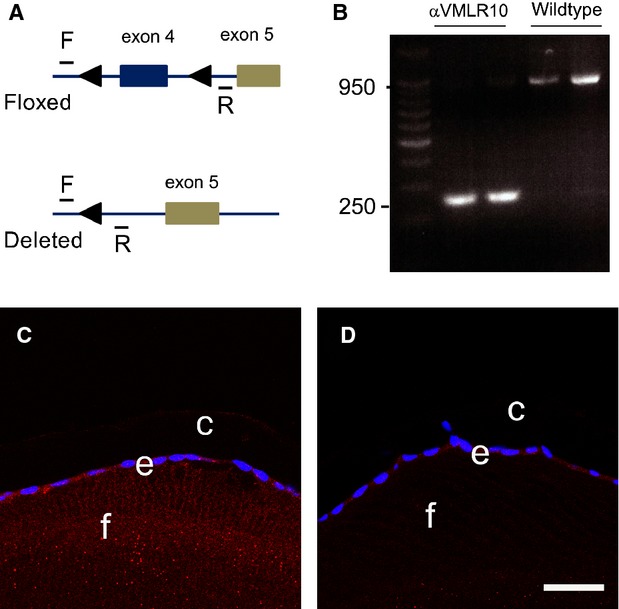
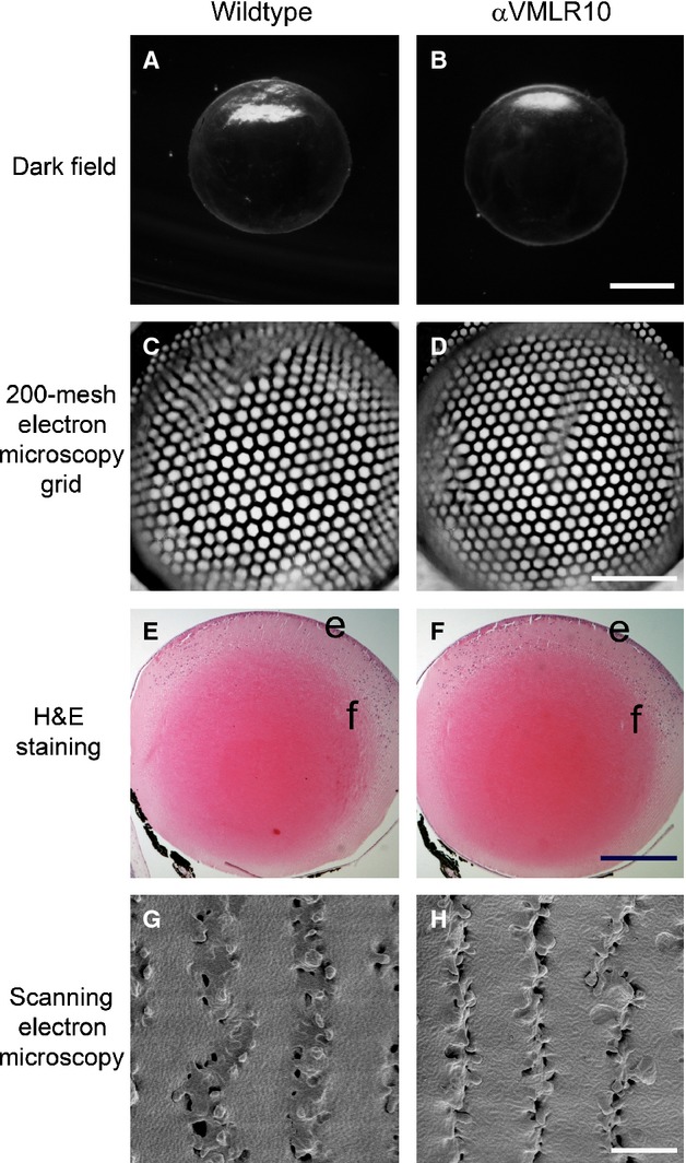
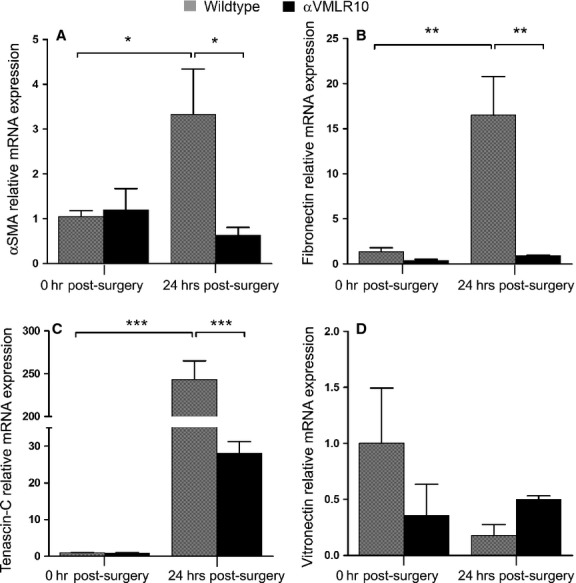
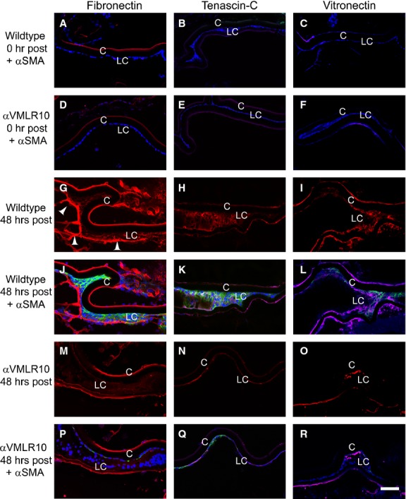
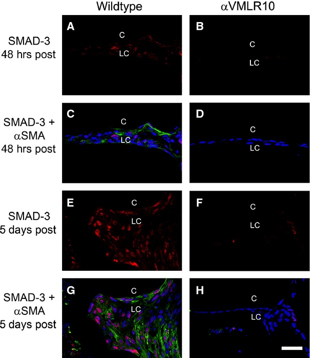
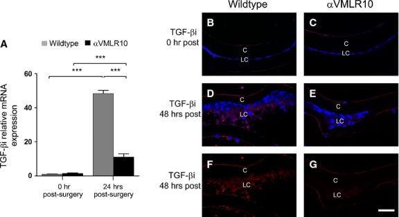
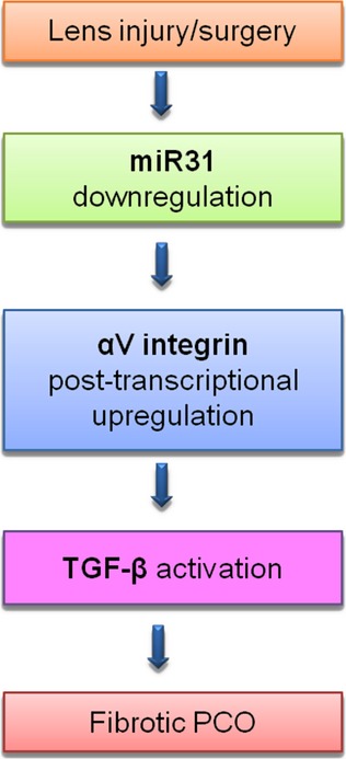
Posterior capsular opacification (PCO) is the major complication arising after cataract treatment. PCO occurs when the lens epithelial cells remaining following surgery (LCs) undergo a wound healing response producing a mixture of α-smooth muscle actin (α-SMA)-expressing myofibroblasts and lens fibre cells, which impair vision. Prior investigations have proposed that integrins play a central role in PCO and we found that, in a mouse fibre cell removal model of cataract surgery, expression of αV integrin and its interacting β-subunits β1, β5, β6, β8 are up-regulated concomitant with α-SMA in LCs following surgery. To test the hypothesis that αV integrins are functionally important in PCO pathogenesis, we created mice lacking the αV integrin subunit in all lens cells. Adult lenses lacking αV integrins are transparent and show no apparent morphological abnormalities when compared with control lenses. However, following surgical fibre cell removal, the LCs in control eyes increased cell proliferation, and up-regulated the expression of α-SMA, β1-integrin, fibronectin, tenascin-C and transforming growth factor beta (TGF-β)-induced protein within 48 hrs, while LCs lacking αV integrins exhibited much less cell proliferation and little to no up-regulation of any of the fibrotic markers tested. This effect appears to result from the known roles of αV integrins in latent TGF-β activation as αV integrin null lenses do not exhibit detectable SMAD-3 phosphorylation after surgery, while this occurs robustly in control lenses, consistent with the known roles for TGF-β in fibrotic PCO. These data suggest that therapeutics antagonizing αV integrin function could be used to prevent fibrotic PCO following cataract surgery.

Keywords: epithelial-to-mesenchymal transition; fibrosis; integrins; lens; posterior capsular opacification; secondary cataract; transforming growth factor beta; wound healing response.


Figures

Figure 1
Figure 1
Immunofluorescent analysis showing that α-smooth muscle actin (α-SMA) and αV-β integrin levels increase in wild-type residual lens cells (LC) at 48 hrs after surgery (A) αV integrin + α-SMA expression, 0 hr after surgery. (B) αV integrin expression at 48 hrs after surgery. (C) αV integrin + α-SMA expression, 48 hrs after surgery. (D) β1 integrin + α-SMA expression, 0 hr after surgery. (E) β1 integrin expression, 48 hrs after surgery. (F) β1 integrin + α-SMA expression, 48 hrs after surgery. (G) β5 integrin + α-SMA 0 hr after surgery. (H) β5 integrin expression at 48 hrs after surgery. (I) β5 integrin + α-SMA expression at 48 hrs after surgery. (J) β6 integrin + α-SMA expression 0 hr after surgery. (K) β6 integrin expression at 48 hrs after surgery. (L) β6 integrin + α-SMA expression 48 hrs after surgery. (M) β8 integrin + α-SMA expression 0 hr after surgery. (N) β8 integrin expression at 48 hrs after surgery. (O) β8 integrin + α-SMA expression, 48 hrs after surgery. Scale bar = 35 μm, red = integrin; blue = nucleus; green = α-SMA, LC = residual lens cells, C = lens capsule.
Figure 2
Figure 2
(A) RT-PCR quantitation of integrin mRNA levels in wild-type residual lens cells (LC) at 0, 24 and 48 hrs after surgery normalized to β2-microglobulin N = 4. αV integrin subunit mRNA expression appeared attenuated at 24 hrs, but this did not reach significance (P = 0.083). β5 integrin subunit mRNA expression was significantly reduced at both 24 and 48 hrs after surgery (**P < 0.0014). No significant changes were observed in β1 or β6 integrin subunit mRNA expression after surgery. β8 integrin subunit mRNA appeared to increase slightly at 24 hrs, although the difference was not significant. Its abundance did significantly fall at 48 hrs after surgery (*P < 0.014). (B) Quantitation of miR-31 mRNA levels in wild-type LCs at 24 hrs after surgery normalized to snoRNA202 levels (**P < 0.0087, N = 5). All fold changes after surgery were calculated by setting values obtained at 0 hr after surgery in each specific group to one. Values are expressed as mean ± SEM. Asterisks (*) indicate statistically significant fold changes from 0 hr after surgery.
Figure 3
Figure 3
αV integrin gene deletion analysis. (A) Diagram of the αV integrin locus showing the position of the PCR primers and the loxP sites . (B) PCR results from DNA obtained from 3-month-old lenses demonstrating successful deletion of exon 4 in mice lacking αV integrin in all lens cells αV [−/flox]; MLR10-cre (αVMLR10). (C) Immunofluorescence showing αV integrin protein expression in a 3-month-old wild-type lens. (D) Immunofluorescence showing αV integrin protein expression in a 3-month-old αVMLR10 lens Key: Scale = 35 μm. Red = αV integrin, blue = nucleus, e = epithelial lens cells, f = lens fibre cells and c = lens capsule.
Figure 4
Figure 4
Morphological analysis of αV integrin null lenses. (A) A dark-field image showing a 3-month-old wild-type lens. (B) A dark-field image showing a 3-month-old αVMLR10 lens. (C) A 200-mesh electron microscopy grid analysis of a 4-month-old wild-type lens. (D) A 200-mesh electron microscopy grid analysis of a 4-month-old αVMLR10 lens. (E) Haematoxylin and eosin staining showing 4-month-old wild-type lens. (F) Haematoxylin and eosin staining showing 4-month-old αVMLR10 lens. (G) SEM analysis of the fibre cell organization of a 4-month-old wild-type lens. (H) SEM analysis of the fibre cell organization of a 4-month-old αVMLR10 lens fibre cell organization. Scale bar for (A, B) = 1.0 mm, (C, D) = 0.5 mm, (E, F) = 0.5 mm and (G, H) = 4.0 μm; e = lens epithelium, f = lens fibre cells.
Figure 5
Figure 5
Immunohistochemistry analysis of α-smooth muscle actin (α-SMA) expression and 5-ethynyl-2′-deoxyuridine (EdU) click-it labelling of wild-type and αVMLR10 residual lens cells (LC) after surgery. (A) EdU labelling + α-SMA expression in wild-type LCs at 0 hr after surgery. (B) EdU + α-SMA expression in αVMLR10 LCs at 0 hr after surgery. (C) EdU staining of proliferating wild-type LCs at 48 hrs after surgery. (D) EdU labelling of αVMLR10 LCs at 48 hrs after surgery. (E) EdU labelling alone in proliferating wild-type LCs at 48 hrs after surgery. (F) EdU labelling alone in αVMLR10 LCs at 48 hrs after surgery. (G) α-SMA expression in wild-type LCs at 48 hrs after surgery. (H) α-SMA expression on αVMLR10 LCs at 48 hrs after surgery. Scale bar (A–F) = 70 μm, (G, H) = 35 μm. Red = EdU positive (Proliferating LCs), blue = nucleus, green = α-SMA, LC = residual lens cells, C = lens capsule.
Figure 6
Figure 6
RT-PCR quantification of mRNA expression levels in wild-type and αVMLR10 residual lens cells on capsular bags collected at 0 and 24 hrs after surgery. For each gene, mRNA expression was normalized to β2M and fold change was calculated based on the mean 0 hr after surgery, wild-type mRNA level equated to 1. (A) α-smooth muscle actin (α-SMA) relative mRNA expression after surgery, *P < 0.02. There was no significant changes in α-SMA mRNA levels between 0 and 24 hrs αVMLR10 post-surgery lenses, P = 0.54 (B) Fibronectin relative mRNA expression after surgery, **P < 0.001. There was no significant changes in fibronectin mRNA levels between 0 and 24 hrs αVMLR10 post-surgery lenses, P = 0.90 (C) Tenascin-C relative mRNA expression after surgery ***P < 0.0001. There was no significant increase in tenascin-C mRNA expression in αVMLR10 24 hrs post-surgery lenses, P = 0.08 (D) No significant changes were observed in vitronectin relative mRNA expression in any group after surgery when compared with wild-type 0 hr, P = 0.21. The decrease in vitronectin mRNA expression in wild-type at 24 hrs after surgery was not significant, P = 0.79; neither was the slight increase in vitronectin mRNA expression in αVMLR10 at 24 hrs after surgery, P = 0.41. All experiments had N = 5. Values are expressed as mean ± SEM. Asterisks (*) indicate statistically significant fold changes from 0 hr after surgery.
Figure 7
Figure 7
Immunofluorescent analysis of fibronectin, tenascin-C and vitronectin deposition in wild-type and αVMLR10 residual lens cells (LC) after surgery. (A) Fibronectin + α-smooth muscle actin (α-SMA) expression in wild-type LCs at 0 hr after surgery. (B) Tenascin-C + α-SMA expression in wild-type LCs at 0 hr after surgery. (C) Vitronectin + α-SMA expression in wild-type LCs at 0 hr after surgery. (D) Fibronectin + α-SMA expression in αVMLR10 LCs at 0 hr after surgery. (E) Tenascin-C + α-SMA expression in αVMLR10 LCs at 0 hr after surgery. (F) Vitronectin + α-SMA expression in αVMLR10 LCs at 0 hr after surgery. (G) Fibronectin expression in wild-type LCs 48 hrs after surgery with arrowheads showing fibronectin deposition on the leading LCs. (H) Tenascin-C expression alone in wild-type LCs at 48 hrs after surgery. (I) Vitronectin expression alone in wild-type LCs at 48 hrs after surgery. (J) Fibronectin + α-SMA expression in wild-type LCs at 48 hrs after surgery. (K) Tenascin-C + α-SMA expression in wild-type LCs at 48 hrs after surgery. (L) Vitronectin + α-SMA expression in wild-type LCs at 48 hrs after surgery. (M) Fibronectin expression in αVMLR10 LCs at 48 hrs after surgery. (N) Tenascin-C expression alone in αVMLR10 LCs at 48 hrs after surgery. (O) Vitronectin expression in αVMLR10 LCs at 48 hrs after surgery. (P) Fibronectin + α-SMA expression in αVMLR10 LCs at 48 hrs after surgery. (Q) Tenascin-C + α-SMA expression in αVMLR10 LCs at 48 hrs after surgery (R) vitronectin + α-SMA expression in αVMLR10 LCs at 48 hrs after surgery. Scale bar = 60 μm. Red = Fibronectin, tenascin-C or Vitronectin, blue = nucleus, green = α-SMA. LC = residual lens cells, C = lens capsule.
Figure 8
Figure 8
Immunohistochemistry of α-smooth muscle actin (α-SMA) and the lens fibre differentiation markers Prox-1 and cMaf in wild-type and αVMLR10 residual lens cells (LC) from capsular bags collected at 5 days after surgery. (A) cMaf expression alone in wild-type LCs at 5 days after surgery. (B) cMaf expression alone in αVMLR10 LCs at 5 days after surgery. (C) cMaf + α-SMA expression in wild-type LCs at 5 days after surgery. (D) cMaf + α-SMA expression in αVMLR10 LCs at 5 days after surgery. (E) Prox-1 expression alone in wild-type LC at 5 days after surgery. (F) Prox-1 expression alone in αVMLR10 LCs at 5 days after surgery. (G) Prox-1 + α-SMA expression in wild-type LCs at 5 days after surgery. (H) Prox-1 + α-SMA expression in αVMLR10 LCs at 5 days after surgery. Scale bar = 35 μm. Red = Prox-1 and cMaf, blue = nucleus, green = α-SMA, LC = residual lens cells, C = lens capsule.
Figure 9
Figure 9
Immunofluorescent analysis of α-smooth muscle actin (α-SMA) and phospho-SMAD-3 in wild-type and αVMLR10 residual lens cells (LC) from capsular bags collected at 48 hrs and 5 days after surgery (A) Phospho-SMAD-3 expression alone in wild-type LCs at 48 hrs after surgery. (B) Phospho-SMAD-3 expression alone in αVMLR10 LCs at 48 hrs after surgery. (C) Phospho-SMAD-3 + α-SMA expression in wild-type LCs at 48 hrs after surgery. (D) Phospho-SMAD-3 + α-SMA expression in αVMLR10 LCs at 48 hrs after surgery. (E) Phospho-SMAD-3 expression alone in wild-type LCs at 5 days aftersurgery. (F) Phospho-SMAD-3 expression alone in αVMLR10 LECs at 5 days after surgery. (G) Phospho-SMAD-3 + α-SMA expression in wild-type LCs at 5 days after surgery. (H) Phospho-SMAD-3 + α-SMA expression in αVMLR10 LCs at 5 days after surgery. Scale bar = 35 μm. Red = phospho-SMAD-3, blue = nucleus, green = α-SMA, LC = residual lens cells, C = lens capsule.
Figure 10
Figure 10
(A) RT-PCR quantification of TGF-βi mRNA levels in wild-type and αVMLR10 mice residual lens epithelial cells (LCs) on capsular bags collected at 0 and 24 hrs after surgery showing almost no expression of TGF-βi mRNA at 0 hr after surgery in both wild-type and αVMLR10 LCs, but a significant increase in mRNA levels in both wild-type and αVMLR10 LCs ***P < 0.0001. TGF-βi mRNA levels in wild-type LCs at 24 hrs after surgery were significantly higher than that of αVMLR10 LCs at 24 hrs after surgery (***P < 0.001). mRNA expression was normalized to β2M and fold change differences were calculated based on 0 hr post-surgery wild-type mRNA expression. All experiments had N = 5. Values are expressed as mean ± SEM. Asterisks (*) indicate statistically significant fold changes from 0 hr after surgery. (B) Immunohistochemistry results showing TGF-βi protein expression 0 hr after surgery in wild-type LCs and (C) αVMLR10 LCs. (D) TGF-βi expression in wild-type LCs at 48 hrs after surgery. (E) TGF-βi expression in αVMLR10 LCs at 48 hrs after surgery. (F) TGF-βi expression alone in wild-type LCs at 48 hrs after surgery. (G) TGF-βi expression alone in αVMLR10 LCs at 48 hrs after surgery. Scale bar 35 μm. Red = TGF-βi, blue = nucleus, LC = residual lens cells, C = lens capsule.
Figure 11
Figure 11
Proposed model of how αV integrin could mediate TGF-β–induced epithelial–mesenchymal transition during fibrotic PCO.

Similar articles

Cited by

References

    1. Asbell PA, Dualan I, Mindel J, et al. Age-related cataract. Lancet. 2005;365:599–609. - PubMed
    1. World Health Organization. Global initiative for the elimination of blindness. WHO/PBL. 2008 ; 97.61. Rev.1.
    1. Steinberg EP, Javitt JC, Sharkey PD, et al. The content and cost of cataract surgery. Arch Ophthalmol. 1993;111:1041–9. - PubMed
    1. Laroche L. actuality in cataract treatment. Rev Prat. 2013;63:43–7. - PubMed
    1. Foster A. Cataract and “vision 2020-the right to sight” initiative. Br J Ophthalmol. 2001;85:635–7. - PMC - PubMed

Publication types

MeSH terms

LinkOut - more resources