Attenuation and restoration of severe acute respiratory syndrome coronavirus mutant lacking 2'-o-methyltransferase activity
- PMID: 24478444
- PMCID: PMC3993736
- DOI: 10.1128/JVI.03571-13
Attenuation and restoration of severe acute respiratory syndrome coronavirus mutant lacking 2'-o-methyltransferase activity
Abstract
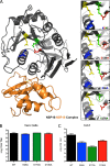
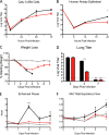
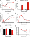
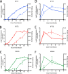
The sudden emergence of severe acute respiratory syndrome coronavirus (SARS-CoV) in 2002 and, more recently, Middle Eastern respiratory syndrome CoV (MERS-CoV) underscores the importance of understanding critical aspects of CoV infection and pathogenesis. Despite significant insights into CoV cross-species transmission, replication, and virus-host interactions, successful therapeutic options for CoVs do not yet exist. Recent identification of SARS-CoV NSP16 as a viral 2'-O-methyltransferase (2'-O-MTase) led to the possibility of utilizing this pathway to both attenuate SARS-CoV infection and develop novel therapeutic treatment options. Mutations were introduced into SARS-CoV NSP16 within the conserved KDKE motif and effectively attenuated the resulting SARS-CoV mutant viruses both in vitro and in vivo. While viruses lacking 2'-O-MTase activity had enhanced sensitivity to type I interferon (IFN), they were not completely restored in their absence in vivo. However, the absence of either MDA5 or IFIT1, IFN-responsive genes that recognize unmethylated 2'-O RNA, resulted in restored replication and virulence of the dNSP16 mutant virus. Finally, using the mutant as a live-attenuated vaccine showed significant promise for possible therapeutic development against SARS-CoV. Together, the data underscore the necessity of 2'-O-MTase activity for SARS-CoV pathogenesis and identify host immune pathways that mediate this attenuation. In addition, we describe novel treatment avenues that exploit this pathway and could potentially be used against a diverse range of viral pathogens that utilize 2'-O-MTase activity to subvert the immune system.
Importance: Preventing recognition by the host immune response represents a critical aspect necessary for successful viral infection. Several viruses, including SARS-CoV, utilize virally encoded 2'-O-MTases to camouflage and obscure their viral RNA from host cell sensing machinery, thus preventing recognition and activation of cell intrinsic defense pathways. For SARS-CoV, the absence of this 2'-O-MTase activity results in significant attenuation characterized by decreased viral replication, reduced weight loss, and limited breathing dysfunction in mice. The results indicate that both MDA5, a recognition molecule, and the IFIT family play an important role in mediating this attenuation with restored virulence observed in their absence. Understanding this virus-host interaction provided an opportunity to design a successful live-attenuated vaccine for SARS-CoV and opens avenues for treatment and prevention of emerging CoVs and other RNA virus infections.
Figures
Similar articles
-
Natural evidence of coronaviral 2'-O-methyltransferase activity affecting viral pathogenesis via improved substrate RNA binding.Signal Transduct Target Ther. 2024 May 29;9(1):140. doi: 10.1038/s41392-024-01860-x. Signal Transduct Target Ther. 2024. PMID: 38811528 Free PMC article.
-
SARS-CoV-2 Uses Nonstructural Protein 16 To Evade Restriction by IFIT1 and IFIT3.J Virol. 2023 Feb 28;97(2):e0153222. doi: 10.1128/jvi.01532-22. Epub 2023 Feb 1. J Virol. 2023. PMID: 36722972 Free PMC article.
-
Middle East Respiratory Syndrome Coronavirus Nonstructural Protein 16 Is Necessary for Interferon Resistance and Viral Pathogenesis.mSphere. 2017 Nov 15;2(6):e00346-17. doi: 10.1128/mSphere.00346-17. eCollection 2017 Nov-Dec. mSphere. 2017. PMID: 29152578 Free PMC article.
-
Coronavirus non-structural protein 16: evasion, attenuation, and possible treatments.Virus Res. 2014 Dec 19;194:191-9. doi: 10.1016/j.virusres.2014.09.009. Epub 2014 Sep 30. Virus Res. 2014. PMID: 25278144 Free PMC article. Review.
-
Coronavirus 2'-O-methyltransferase: A promising therapeutic target.Virus Res. 2023 Oct 15;336:199211. doi: 10.1016/j.virusres.2023.199211. Epub 2023 Sep 4. Virus Res. 2023. PMID: 37634741 Free PMC article. Review.
Cited by
-
SARS-CoV-2 Uses Nonstructural Protein 16 to Evade Restriction by IFIT1 and IFIT3.bioRxiv [Preprint]. 2022 Sep 26:2022.09.26.509529. doi: 10.1101/2022.09.26.509529. bioRxiv. 2022. Update in: J Virol. 2023 Feb 28;97(2):e0153222. doi: 10.1128/jvi.01532-22 PMID: 36203546 Free PMC article. Updated. Preprint.
-
Reverse genetic systems: Rational design of coronavirus live attenuated vaccines with immune sequelae.Adv Virus Res. 2020;107:383-416. doi: 10.1016/bs.aivir.2020.06.003. Epub 2020 Jun 30. Adv Virus Res. 2020. PMID: 32711735 Free PMC article. Review.
-
Mechanisms of innate immune evasion in re-emerging RNA viruses.Curr Opin Virol. 2015 Jun;12:26-37. doi: 10.1016/j.coviro.2015.02.005. Epub 2015 Mar 9. Curr Opin Virol. 2015. PMID: 25765605 Free PMC article. Review.
-
A single amino acid substitution in the mRNA capping enzyme λ2 of a mammalian orthoreovirus mutant increases interferon sensitivity.Virology. 2015 Sep;483:229-35. doi: 10.1016/j.virol.2015.04.020. Epub 2015 May 15. Virology. 2015. PMID: 25985441 Free PMC article.
-
QTQTN motif upstream of the furin-cleavage site plays a key role in SARS-CoV-2 infection and pathogenesis.Proc Natl Acad Sci U S A. 2022 Aug 9;119(32):e2205690119. doi: 10.1073/pnas.2205690119. Epub 2022 Jul 26. Proc Natl Acad Sci U S A. 2022. PMID: 35881779 Free PMC article.
References
-
- Perlman S. 1998. Pathogenesis of coronavirus-induced infections: review of pathological and immunological aspects. Adv. Exp. Med. Biol. 440:503–513 - PubMed
-
- Becker MM, Graham RL, Donaldson EF, Rockx B, Sims AC, Sheahan T, Pickles RJ, Corti D, Johnston RE, Baric RS, Denison MR. 2008. Synthetic recombinant bat SARS-like coronavirus is infectious in cultured cells and in mice. Proc. Natl. Acad. Sci. U. S. A. 105:19944–19949. 10.1073/pnas.0808116105 - DOI - PMC - PubMed
Publication types
MeSH terms
Substances
Grants and funding
LinkOut - more resources
Full Text Sources
Other Literature Sources
Molecular Biology Databases
Research Materials
Miscellaneous

