. 2014 Jan 17;33(1):7.
doi: 10.1186/1756-9966-33-7.

The downregulation of PRDM1/Blimp-1 is associated with aberrant expression of miR-223 in extranodal NK/T-cell lymphoma, nasal type

Affiliations

The downregulation of PRDM1/Blimp-1 is associated with aberrant expression of miR-223 in extranodal NK/T-cell lymphoma, nasal type

Li Liang et al. J Exp Clin Cancer Res. .

Abstract

Background: The mechanism for inactivation of positive regulatory domain containing I (PRDM1), a newly identified tumour suppressor gene in extranodal NK/T-cell lymphoma, nasal type (EN-NK/T-NT) has not been well defined. The aim of the present study was to investigate the expression of PRDM1 in EN-NK/T-NT and analyse its downregulation by miRNAs.

Methods: PRDM1 and miRNA expression were evaluated in EN-NK/T-NT samples by immunohistochemical analysis, qRT-PCR, and in situ hybridisation. Luciferase assays were performed to verify the direct binding of miR-223 to the 3'-untranslated region of PRDM1 mRNA. In addition, the effect of miR-223 on PRDM1 expression was assessed in NK/T lymphoma cell lines by transfecting a miR-223 mimic or inhibitor to increase or decrease the effective expression of miR-223. Overall survival and failure-free survival in EN-NK/T-NT patients were analysed using Kaplan-Meier single-factor analysis and the log-rank test.

Results: Investigation of the downregulation of PRDM1 in EN-NK/T-NT cases revealed that PRDM1-positive staining might be a favourable predictor of overall survival and failure-free survival in EN-NK/T-NT patients. However, the negative staining of PRDM1 usually presented transcripts, suggesting a possible post-transcriptional regulation. miR-223 and its putative target gene, PRDM1, exhibited opposite patterns of expression in EN-NK/T-NT tissues and cell lines. Moreover, PRDM1 was identified as a direct target gene of miR-223 by luciferase assays. The ectopic expression of miR-223 led to the downregulation of the PRDM1 protein in the NK/T-cell lymphoma cell line, whereas a decrease in miR-223 restored the level of PRDM1 protein.

Conclusions: Our findings reveal that the downregulation of the tumour suppressor PRDM1 in EN-NK/T-NT samples is mediated by miR-223 and that PRDM1-positive staining might have prognostic value for evaluating the clinical outcome of EN-NK/T-NT patients.


Figures
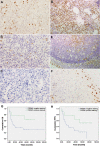
Figure 1
Figure 1
Immunohistochemistry (IHC) and prognostic analysis of PRDM1 in extranodal NK/T-cell lymphoma, nasal type (EN-NK/T-NT) cases. Examples of IHC analysis of PRDM1 in EN-NK/T-NT specimens and control samples. (A) PRDM1 staining in the nuclei of tumour cells was observed in approximately 50% of tumour cells in 1 case of EN-NK/T-NT; most cells had moderate to weak nuclear staining. (B) PRDM1 was expressed in approximately 10% of tumour cells in 1 case of EN-NK/T-NT. (C) No PRDM1 staining was detected in 1 case of EN-NK/T-NT. In the control cases, strong nuclear PRDM1 immunostaining was observed in plasma cell myeloma (D), the epithelium and germinal centre of the tonsil (E), and the squamous epithelium of the nasal mucosa (F) (all by IHC; A, B, C, and F are shown at 400× magnification; D and E are shown at 200× magnification). (G) and (H) Kaplan-Meier survival analysis demonstrated that PRDM1 expression predicted a favourable effect on overall survival (OS) and failure-free survival (FFS) of EN-NK/T-NT patients (P = 0.084 and P = 0.042, respectively).
Figure 2
Figure 2
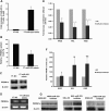
Discrepancy between PRDM1α mRNA and protein expression in extranodal NK/T-cell lymphoma, nasal type (EN-NK/T-NT). (A) The relative levels of PRDM1α mRNA by qRT-PCR and the corresponding PRDM1 protein by immunohistochemistry (IHC) were analysed in 16 EN-NK/T-NT cases, one plasma cell myeloma, and one tonsil case. Case #1 is plasma cell myeloma. Case #2 is tonsil, and cases #3 to #18 are 16 EN-NK/T-NT cases. Levels of PRDM1α mRNA in the tonsil and EN-NK/T-NT cases were estimated relative to that in plasma cell myeloma (arbitrarily set as 100%), which showed strong expression of PRDM1 protein. The data of PRDM1α mRNA by qRT-PCR are presented as mean ± SE of 3 independent experiments. Expression of PRDM1 protein in formalin-fixed paraffin-embedded sections of EN-NK/T-NT specimens, plasma cell myeloma, and one tonsil case was determined by immunostaining and assessed by the percentage of PRDM1 positive cells. Of 16 EN-NK/T-NT cases, 9 cases (#3, 6, 7, 8, 10, 11, 14, 15, and 16) showed high level of PRDM1α mRNA relative to plasma cell myeloma by qRT-PCR but low or absent percentage of PRDM1 protein positive tumor cells by IHC. (B) PRDM1α mRNA was determined by qRT-PCR in NK/T-cell lymphoma cell lines YT, NK92, and NKL, and the human chronic myelogenous leukaemia cell line K562 (mean ± SE of 3 independent experiments). The level of PRDM1α transcript was assessed relative to that in YT cells (arbitrarily considered as 100%). PRDM1α mRNA levels in NK92, NKL, and K562 cells were 15.0%, 73.0%, and 40.1% of those in YT cells, respectively. (C) The expression of PRDM1α protein was detected in cell lines by western blot. The density of PRDM1α in YT cells was set as 100%, and the levels in NK92, NKL, and K562 cells were calculated as 5.0%, 6.0%, and 9.3% of YT cells, respectively.
Figure 3
Figure 3
Representative cases of miRNA expression identified by in situ hybridisation (ISH). ISH analysis revealed characteristic upregulation of miR-223 in the cytoplasm of extranodal NK/T-cell lymphoma, nasal type (EN-NK/T-NT) tumour cells (A1), whereas no signal was detected in peripheral T-cell lymphoma (A2) and inflammatory nasal mucosa (A3). In addition, miR-886-3p was also overexpressed in the cytoplasm of EN-NK/T-NT tumour cells (B1) but was negative in peripheral T-cell lymphoma (B2) and inflammatory nasal mucosa (B3). There were no miR-34c-5p signals in EN-NK/T-NT samples (C1), peripheral T-cell lymphoma samples (C2), or inflammatory nasal mucosa samples (C3). All images show ISH at 400x magnification.
Figure 4
Figure 4
Statistical analysis of miR-223, miR-886-3p, and miR-34c-5p expression by in situ hybridisation (ISH). The expression percentage of miR-223, miR-886-3p, and miR-34c-5p was statistically analysed in extranodal NK/T-cell lymphoma, nasal type (EN-NK/T-NT), peripheral T-cell lymphoma and inflammatory nasal mucosa cases by ISH. Statistically, ISH results revealed that the expression level of miR-223 was significantly higher in EN-NK/T-NT cases than in peripheral T-cell lymphoma (A, P = 0.013) and in inflammatory nasal mucosa (A, ※※P = 0.043). Similarly, miR-886-3p expression upregulated in EN-NK/T-NT cases compared to peripheral T-cell lymphoma (B, #P = 0.028) and inflammatory nasal mucosa (B, ##P = 0.022). However, the expression level of miR-34c-5p (C, P = 1.000 and ∆∆P = 0.254) did not differ significantly between these 3 groups.
Figure 5
Figure 5
Verification of PRDM1 as a direct target gene of miR-223. (A) The complementarity between miR-223 and its 3 conserved putative binding sites in the PRDM1 3′-untranslated region (UTR) is highlighted in bold between different species. (B) Luciferase reporter assays were performed in 293 T cells that were co-transfected with miR-223 mimic and wild type pmirGLO expression-PRDM1-3′UTR (WT) reporter plasmid or mutant pmirGLO expression-PRDM1-3′UTR reporter plasmids harbouring point mutations in the target sites for miR-223 (Mut1, Mut2, Mut3, Mut1 + 2, Mut1 + 3, Mut2 + 3, and Mut1 + 2 + 3). Mimic Negative Control was used as a negative control (NC). Firefly luciferase activity was normalised relative to Renilla luciferase activity. Transfection of the miR-223 mimic resulted in a marked decrease in luciferase activity in the WT group compared to the NC group (48.08%). Mutations in each of the putative target sites or combined mutations restored luciferase activity to varying degrees: 74.87% for Mut1, 85.21% for Mut2, 74.84% for Mut3, 90.76% for Mut1 + 2, 87.55% for Mut1 + 3, 81.15% for Mut2 + 3, and 94.51% for Mut1 + 2 + 3. Data are presented as mean ± SE of 4 independent experiments. (C) Two nucleotides in the middle of each target site were mutated to generate different mutant luciferase reporters.
Figure 6
Figure 6
Correlation of the expression of PRDM1 and miR-223 in extranodal NK/T-cell lymphoma, nasal type (EN-NK/T-NT). (A) The expression of PRDM1 and miR-223 in EN-NK/T-NT cases were analysed by immunohistochemistry (IHC) and in situ hybridisation (ISH), respectively, and the result is shown as a scatter diagram. As described in the Materials and Methods section, these results were semi-quantitatively scored into 3 grades according to the number of positive tumour cells. In this figure, the numbers of ordinate are as follows: “1” indicates negative (0% to <10% positive cells), “2” indicates weak (10% to ≤50% positive cells), and “3” indicates strong (>50% to 100% positive cells). Statistically, a significantly opposing correlation was observed between the levels of PRDM1 protein and miR-223 expression in 31 EN-NK/T-NT cases (P < 0.001); only 2 cases had the same relative expression levels of PRDM1 and miR-223. (B) One representative case of EN-NK/T-NT was negative for PRDM1 by IHC but strongly positive for miR-223 by ISH (400×). (C) qRT-PCR analysis revealed much lower levels of miR-223 in YT cells than in NK92, NKL, and K562 cells (mean ± SE of 3 independent experiments). (D) Western blotting revealed markedly higher levels of PRDM1α protein in YT cells than in NK92, NKL, and K562 cells.
Figure 7
Figure 7
Endogenous PRDM1 protein expression is affected by increased miR-223 or decreased miR-223. A miR-223 mimic or mimic negative control (NC) was transfected into YT cells by electroporation. (A) qRT-PCR analysis revealed a significantly increased level of miR-223 in YT cells transfected with miR-223 mimic compared to NC. The results were confirmed in 3 independent experiments with data presented as mean ± SE (P < 0.001). (B) Western blot showed that PRDM1α protein level was markedly diminished (54.44% relative to YT-NC, normalised to β-actin) in YT cells transfected with miR-223 mimic. YT-NC was adjusted to 100%. Results were quantified by densitometry in 3 independent experiments (mean ± SD) (#P = 0.008). (C) A representative image of PRDM1α protein expression in YT cells as detected by western blot. (D) RT-PCR and agarose gel electrophoresis showed ectopic expression of miR-223 with no effect on PRDM1α transcript. NK92, NKL, and K562 cells were transfected with miR-223 inhibitor or NC with HiPerFect Transfection Reagent. (E) Compared to NC, the level of endogenous miR-223 was significantly decreased in NK92, NKL, and K562 cells by qRT-PCR analysis. The data are presented as mean ± SE of 4 independent experiments (P = 0.026, ∆∆P = 0.017, and ∆∆∆P = 0.044). (F) Semi-quantitative analysis by densitometry demonstrated that the PRDM1α protein was restored to 220% and 234% by miR-223 inhibition in NKL and K562 cells, respectively, compared to NC, but the level of PRDM1α protein in NK92 cells was not significantly affected. The data are presented as mean ± SD of 4 independent experiments (§P = 1.000, §§P = 0.040, and §§§P = 0.022). (G) Representative western blot images of PRDM1α protein levels in NK92, NKL, and K562 cells are shown.
Figure 8
Figure 8
Kaplan-Meier survival analysis of miR-223 in extranodal NK/T-cell lymphoma, nasal type (EN-NK/T-NT) patients. According to Kaplan-Meier survival analysis, no correlation was investigated between the expression status of miR-223 and on overall survival (OS) (A, P = 0.784) and failure-free survival (FFS) (B, P = 0.691) of EN-NK/T-NT patients.

Similar articles

Cited by

References

    1. Aozasa K, Takakuwa T, Hongyo T, Yang WI. Nasal NK/T-cell lymphoma: epidemiology and pathogenesis. Int J Hematol. 2008;87:110–117. doi: 10.1007/s12185-008-0021-7. - DOI - PMC - PubMed
    1. Ren YL, Nong L, Zhang S, Zhao J, Zhang XM, Li T. Analysis of 142 Northern Chinese patients with peripheral T/NK-Cell lymphomas: subtype distribution, clinicopathologic features, and prognosis. Am J Clin Pathol. 2012;138:435–447. doi: 10.1309/AJCPWKJ3GPFRT7GA. - DOI - PubMed
    1. Huang Y, de Reynies A, de Leval L, Ghazi B, Martin-Garcia N, Travert M, Bosq J, Briere J, Petit B, Thomas E. et al.Gene expression profiling identifies emerging oncogenic pathways operating in extranodal NK/T-cell lymphoma, nasal type. Blood. 2010;115:1226–1237. doi: 10.1182/blood-2009-05-221275. - DOI - PMC - PubMed
    1. Coppo P, Gouilleux-Gruart V, Huang Y, Bouhlal H, Bouamar H, Bouchet S, Perrot C, Vieillard V, Dartigues P, Gaulard P. et al.STAT3 transcription factor is constitutively activated and is oncogenic in nasal-type NK/T-cell lymphoma. Leukemia. 2009;23:1667–1678. doi: 10.1038/leu.2009.91. - DOI - PMC - PubMed
    1. Zhang S, Li T, Zhang B, Nong L, Aozasa K. Transcription factors engaged in development of NK cells are commonly expressed in nasal NK/T-cell lymphomas. Hum Pathol. 2011;42:1319–1328. doi: 10.1016/j.humpath.2009.11.022. - DOI - PubMed

Publication types

MeSH terms