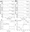
Connexin hemichannel and pannexin channel electrophysiology: how do they differ?
- PMID: 24434538
- PMCID: PMC3989408
- DOI: 10.1016/j.febslet.2013.12.023
Connexin hemichannel and pannexin channel electrophysiology: how do they differ?
Abstract
Connexin hemichannels are postulated to form a cell permeabilization pore for the uptake of fluorescent dyes and release of cellular ATP. Connexin hemichannel activity is enhanced by low external [Ca(2+)]o, membrane depolarization, metabolic inhibition, and some disease-causing gain-of-function connexin mutations. This paper briefly reviews the electrophysiological channel conductance, permeability, and pharmacology properties of connexin hemichannels, pannexin 1 channels, and purinergic P2X7 receptor channels as studied in exogenous expression systems including Xenopus oocytes and mammalian cell lines such as HEK293 cells. Overlapping pharmacological inhibitory and channel conductance and permeability profiles makes distinguishing between these channel types sometimes difficult. Selective pharmacology for Cx43 hemichannels (Gap19 peptide), probenecid or FD&C Blue #1 (Brilliant Blue FCF, BB FCF) for Panx1, and A740003, A438079, or oxidized ATP (oATP) for P2X7 channels may be the best way to distinguish between these three cell permeabilizing channel types. Endogenous connexin, pannexin, and P2X7 expression should be considered when performing exogenous cellular expression channel studies. Cell pair electrophysiological assays permit the relative assessment of the connexin hemichannel/gap junction channel ratio not often considered when performing isolated cell hemichannel studies.
Keywords: Channels; Connexin; Gap junction; Hemichannel; P2X(7) receptor; Pannexin.
Copyright © 2014 Federation of European Biochemical Societies. All rights reserved.
Figures


Similar articles
-
Emerging issues of connexin channels: biophysics fills the gap.Q Rev Biophys. 2001 Aug;34(3):325-472. doi: 10.1017/s0033583501003705. Q Rev Biophys. 2001. PMID: 11838236 Review.
-
Activation, permeability, and inhibition of astrocytic and neuronal large pore (hemi)channels.J Biol Chem. 2014 Sep 19;289(38):26058-26073. doi: 10.1074/jbc.M114.582155. Epub 2014 Aug 1. J Biol Chem. 2014. PMID: 25086040 Free PMC article.
-
Connexin and pannexin hemichannels in inflammatory responses of glia and neurons.Brain Res. 2012 Dec 3;1487:3-15. doi: 10.1016/j.brainres.2012.08.042. Epub 2012 Sep 10. Brain Res. 2012. PMID: 22975435 Free PMC article. Review.
-
The food dye FD&C Blue No. 1 is a selective inhibitor of the ATP release channel Panx1.J Gen Physiol. 2013 May;141(5):649-56. doi: 10.1085/jgp.201310966. Epub 2013 Apr 15. J Gen Physiol. 2013. PMID: 23589583 Free PMC article.
-
P2X7 receptor-pannexin 1 hemichannel association: effect of extracellular calcium on membrane permeabilization.J Mol Neurosci. 2012 Mar;46(3):585-94. doi: 10.1007/s12031-011-9646-8. Epub 2011 Sep 20. J Mol Neurosci. 2012. PMID: 21932038
Cited by
-
The connexin hemichannel inhibitor D4 produces rapid antidepressant-like effects in mice.J Neuroinflammation. 2023 Aug 20;20(1):191. doi: 10.1186/s12974-023-02873-z. J Neuroinflammation. 2023. PMID: 37599352 Free PMC article.
-
Acute activation of hemichannels by ethanol leads to Ca2+-dependent gliotransmitter release in astrocytes.Front Cell Dev Biol. 2024 Jun 21;12:1422978. doi: 10.3389/fcell.2024.1422978. eCollection 2024. Front Cell Dev Biol. 2024. PMID: 38974144 Free PMC article.
-
Pattern of cell-to-cell transfer of microRNA by gap junction and its effect on the proliferation of glioma cells.Cancer Sci. 2019 Jun;110(6):1947-1958. doi: 10.1111/cas.14029. Cancer Sci. 2019. PMID: 31012516 Free PMC article.
-
Epithelial tissue geometry directs emergence of bioelectric field and pattern of proliferation.Mol Biol Cell. 2020 Jul 21;31(16):1691-1702. doi: 10.1091/mbc.E19-12-0719. Epub 2020 Jun 10. Mol Biol Cell. 2020. PMID: 32520653 Free PMC article.
-
Astroglial Hemichannels and Pannexons: The Hidden Link between Maternal Inflammation and Neurological Disorders.Int J Mol Sci. 2021 Sep 1;22(17):9503. doi: 10.3390/ijms22179503. Int J Mol Sci. 2021. PMID: 34502412 Free PMC article. Review.
References
-
- Beyer EC, Paul DL, Goodenough DA. Connexin family of gap junction proteins. J Membr Biol. 1990;116:187–194. - PubMed
Publication types
MeSH terms
Substances
Grants and funding
LinkOut - more resources
Full Text Sources
Other Literature Sources
Miscellaneous

