

High expression of IGFBP7 in fibroblasts induced by colorectal cancer cells is co-regulated by TGF-β and Wnt signaling in a Smad2/3-Dvl2/3-dependent manner
- PMID: 24427302
- PMCID: PMC3888407
- DOI: 10.1371/journal.pone.0085340
High expression of IGFBP7 in fibroblasts induced by colorectal cancer cells is co-regulated by TGF-β and Wnt signaling in a Smad2/3-Dvl2/3-dependent manner
Abstract
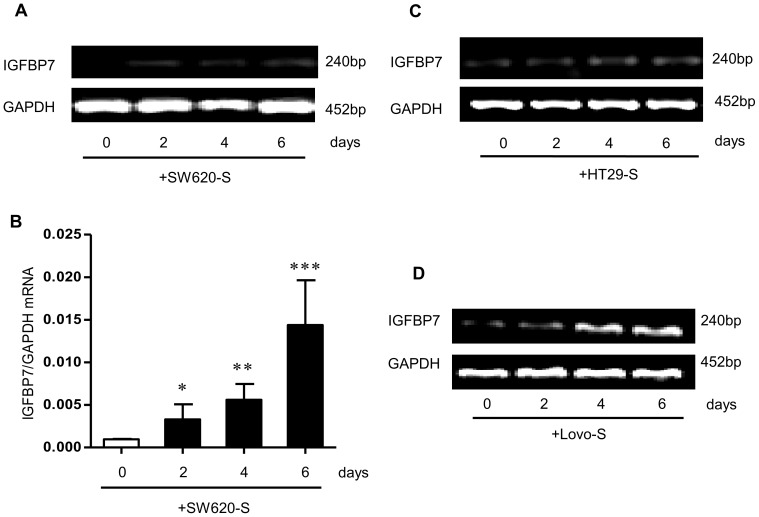
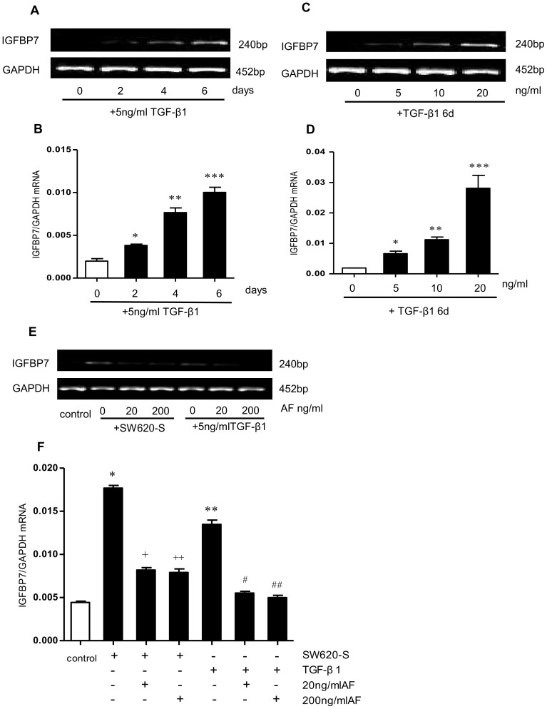
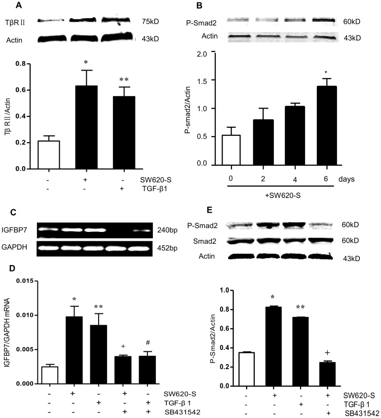
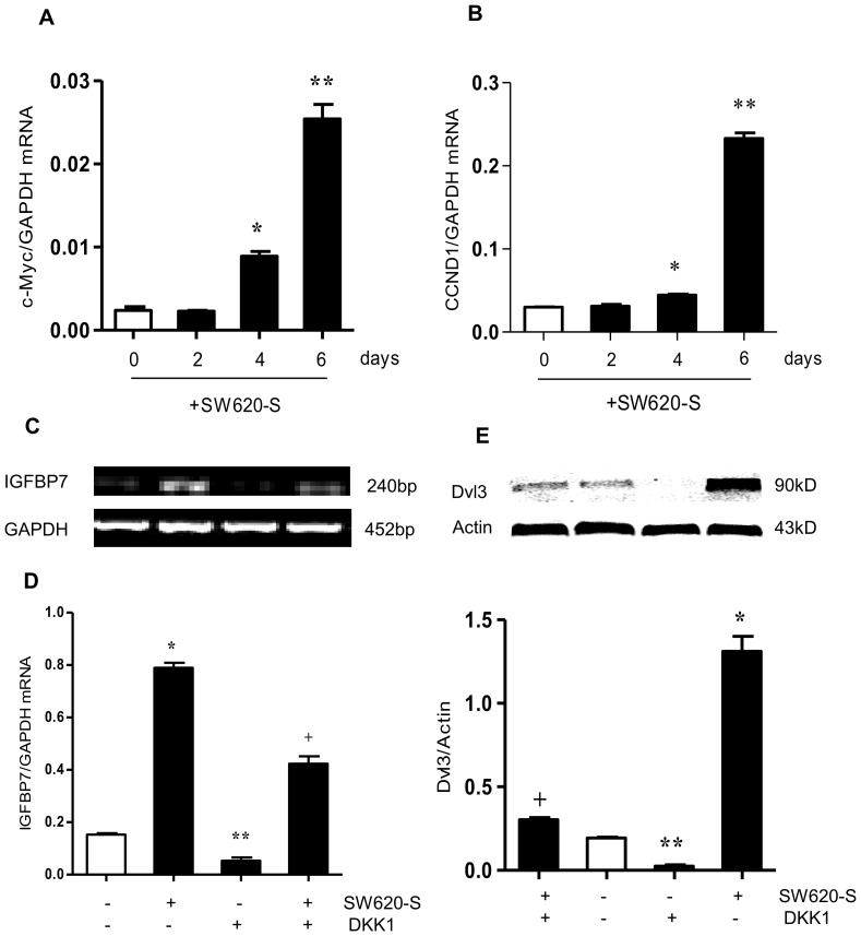
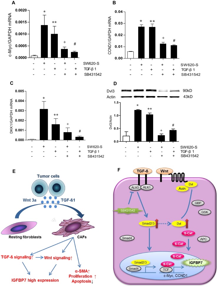
Fibroblasts in the tumor microenvironment are a key determinant in cancer progression and may be a promising target for cancer therapy. Insulin-like growth factor binding protein 7 (IGFBP7) is known as a tumor suppressor in colorectal cancer (CRC). The present study investigated the inductive mechanism of IGFBP7 expression in fibroblasts by supernatant from the CRC cell line, SW620. The results showed that the expression of IGFBP7 was up-regulated in the fibroblasts when treated with SW620 supernatant and exogenous TGF-β1. The IGFBP7 induced by SW620 supernatant or TGF-β1 was partially inhibited by the TGF-β1 specific antibody AF and TGF-β1 receptor antagonist SB431542. The Wnt signaling-targeted genes, c-Myc, CCND1 and the proteins Dvl2/3, were all up-regulated in fibroblasts expressing high levels of IGFBP7, and the up-regulation could be inhibited both by the Wnt signaling antagonist Dickkopf-1 (DKK1) and by the TGF-β1 receptor antagonist SB431542. In conclusion, CRC cells promote the high expression of IGFBP7 in fibroblasts, most likely through the co-regulation of TGF-β and Wnt signaling in a Smad2/3-Dvl2/3 dependent manner. Taken together, these data suggest that the fibroblasts could be a novel therapeutic target in tumor therapy.
Conflict of interest statement
Figures
Similar articles
-
Glioblastoma-secreted factors induce IGFBP7 and angiogenesis by modulating Smad-2-dependent TGF-beta signaling.Oncogene. 2008 Nov 20;27(54):6834-44. doi: 10.1038/onc.2008.287. Epub 2008 Aug 18. Oncogene. 2008. PMID: 18711401
-
Constitutive Smad signaling and Smad-dependent collagen gene expression in mouse embryonic fibroblasts lacking peroxisome proliferator-activated receptor-gamma.Biochem Biophys Res Commun. 2008 Sep 19;374(2):231-6. doi: 10.1016/j.bbrc.2008.07.014. Epub 2008 Jul 15. Biochem Biophys Res Commun. 2008. PMID: 18627765 Free PMC article.
-
Differential regulation of TGF-β-induced, ALK-5-mediated VEGF release by SMAD2/3 versus SMAD1/5/8 signaling in glioblastoma.Neuro Oncol. 2015 Feb;17(2):254-65. doi: 10.1093/neuonc/nou218. Epub 2014 Aug 27. Neuro Oncol. 2015. PMID: 25165192 Free PMC article.
-
A tale of two proteins: differential roles and regulation of Smad2 and Smad3 in TGF-beta signaling.J Cell Biochem. 2007 May 1;101(1):9-33. doi: 10.1002/jcb.21255. J Cell Biochem. 2007. PMID: 17340614 Review.
-
TGFβ and the Tumor Microenvironment in Colorectal Cancer.Cells. 2023 Apr 12;12(8):1139. doi: 10.3390/cells12081139. Cells. 2023. PMID: 37190048 Free PMC article. Review.
Cited by
-
Kidney and lung tissue modifications after BDL-induced liver injury in mice are associated with increased expression of IGFBPrP1 and activation of the NF-κB inflammation pathway.Int J Clin Exp Pathol. 2020 Feb 1;13(2):192-202. eCollection 2020. Int J Clin Exp Pathol. 2020. PMID: 32211099 Free PMC article.
-
IGFBP7 remodels the tumor microenvironment of esophageal squamous cell carcinoma by activating the TGFβ1/SMAD signaling pathway.Oncol Lett. 2022 Jun 10;24(2):251. doi: 10.3892/ol.2022.13371. eCollection 2022 Aug. Oncol Lett. 2022. PMID: 35761941 Free PMC article.
-
Protective effect of fucoidan from Fucus vesiculosus on liver fibrosis via the TGF-β1/Smad pathway-mediated inhibition of extracellular matrix and autophagy.Drug Des Devel Ther. 2016 Feb 12;10:619-30. doi: 10.2147/DDDT.S98740. eCollection 2016. Drug Des Devel Ther. 2016. PMID: 26929597 Free PMC article.
-
PAK4 methylation by the methyltransferase SETD6 attenuates cell adhesion.Sci Rep. 2020 Oct 13;10(1):17068. doi: 10.1038/s41598-020-74081-1. Sci Rep. 2020. PMID: 33051544 Free PMC article.
-
Serum Insulin-Like Growth Factor Binding Protein 7 as a Potential Biomarker in the Diagnosis and Prognosis of Esophagogastric Junction Adenocarcinoma.Gut Liver. 2020 Nov 15;14(6):727-734. doi: 10.5009/gnl19135. Gut Liver. 2020. PMID: 31822054 Free PMC article.
References
-
- Siegel R, Naishadham D, Jemal A (2013) Cancer statistics, 2013. CA Cancer J Clin 63: 11–30. - PubMed
-
- Dvorak HF (1986) Tumors: wounds that do not heal. Similarities between tumor stroma generation and wound healing. N Engl J Med 315: 1650–9. - PubMed
-
- Kalluri R, Zeisberg M (2006) Fibroblasts in cancer. Nat Rev Cancer 6: 392–401. - PubMed
-
- Liu M, Xu JJ, Deng H (2011) Tangled fibroblasts in tumor-stroma interactions. Int J Cancer 129: 1795–805. - PubMed
-
- Mueller MM, Fusenig NE (2004) Friends or foes - bipolar effects of the tumour stroma in cancer. Nat Rev Cancer 4: 839–49. - PubMed
Publication types
MeSH terms
Substances
Grants and funding
LinkOut - more resources
Full Text Sources
Other Literature Sources
Medical
Research Materials
Miscellaneous
