

doi: 10.1038/leu.2014.16.
Epub 2014 Jan 13.
WTAP is a novel oncogenic protein in acute myeloid leukemia
Affiliations
- PMID: 24413322
- PMCID: PMC4369791
- DOI: 10.1038/leu.2014.16
Item in Clipboard
WTAP is a novel oncogenic protein in acute myeloid leukemia
Leukemia.
2014 May.
Erratum in
- Leukemia. 2014 Dec;28(12):2427. Proia, D [Corrected to Proia, D A]
No abstract available
Conflict of interest statement
The remaining authors declare no conflict of interest.
Figures
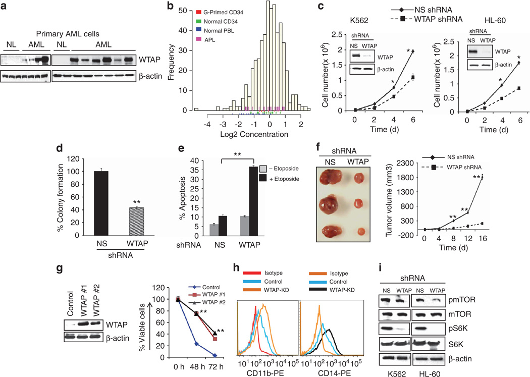
Expression of WTAP in AML and effect of WTAP silencing on AML cell behavior. (a) Peripheral blood mononuclear cells from normal donors (NL) and AML patients (AML) were obtained by Ficoll–Paque density centrifugation, and protein extracts were examined for WTAP expression by western blotting. (b) Histogram of WTAP expression measured by RPPA in bulk AML cells relative to normal CD34+ cells. WTAP levels were above normal CD34+ cells in 32% of AML patients and below normal in 9%. Expression is shown in log 2 scale. The genetic and clinical information on AML patients is described in Supplementary Table 1. (c) K562 and HL-60 cells were transfected with scrambled (NS) or WTAP shRNA, and selected by puromycin. The degree of WTAP knockdown was analyzed by western blotting (inset). Stable downregulation of WTAP expression by shRNA inhibited proliferation of K562 and HL-60 cells. *P ≤ 0.05. (d) HL-60 cells stably expressing NS or WTAP shRNA were plated in methylcellulose medium, and colony formation was scored after 10 days. The values were expressed as % of colonies formed by WTAP-shRNA cells compared with NS shRNA cells taken as 100%. **P ≤ 0.01. (e) Stable K562 cells with NS or WTAP shRNA were untreated or treated with etoposide (10 µm ) for 48 h. The percentage of apoptotic cells was measured by the flow cytometric annexin-V/PI staining method. **P ≤ 0.01. (f) Control (NS shRNA) or WTAP-shRNA K562 cells were subcutaneously implanted into flanks of nude mice and tumor formation and progression was monitored. Tumors originating from the control and WTAP-knockdown cells were excised and photographed. WTAP knockdown significantly reduced tumor growth in vivo. **P ≤ 0.01. (g) Ba/F3 cells were transfected with WTAP or an empty vector and G418-selected stable clones (#1 and #2) were analyzed for WTAP overexpression by western blotting (left panel). Control and stable WTAP-expressing Ba/F3 cells were grown in an IL-3-free medium for indicated time and viable cells were assessed by annexin-V/PI staining followed by FACS analysis (right panel). **P ≤ 0.01. (h) Control and stable WTAP knockdown (WTAP-KD) HL-60 cells were treated with 20 nm of PMA, and 96 h later, cells were labeled with anti-CD11b, anti-CD14 or an isotype-matched control. The expression of myeloid surface markers CD11b and CD14 was analyzed by flow cytometry. (i) WTAP knockdown inhibits the mTOR pathway. Control (NS) and stable WTAP knockdown K562 and HL-60 cell extracts were analyzed by western blot with the indicated antibodies.

Hsp90 associates with WTAP and is necessary for its stability. (a) The K562 cells were treated with vehicle (−) or Hsp90 inhibitor, ganetespib (1 µm for 6 h) followed by immunoprecipitation (IP) with IgG and WTAP antibody. The immunoprecipitates and cell lysates were immunoblotted (IB) with Hsp90, Hsp70 and WTAP antibodies. (b) The GST pull-down assay. In vitro-translated and 35S-methionine-labeled full-length WTAP was incubated with GST or GST-Hsp90 protein immobilized on glutathione–sepharose beads, and bound WTAP was detected by fluorography. Twenty percent of the in vitro-translated protein was used for pull-downs. (c) K562, MV4-11 and Kasumi-1 leukemia cells were treated with the Hsp90 inhibitor ganetespib (1 µm ) for 24 h and analyzed for WTAP expression by western blotting. (d) AML blasts were isolated from peripheral blood or bone marrow of AML patients using Ficoll gradient separation followed by treatment with ganetespib (1 µm ) for 24 h; WTAP expression was analyzed by western blotting. (e) MV4–11 tumor-bearing animals were given single doses of vehicle and ganetespib as described previously, and after 6 h, tumors were removed for western blot analysis using WTAP and β-actin antibody. (f) K562 cells were treated with vehicle (−) or proteasomal inhibitor (bortezomib; 100 nm ) for 2 h and then treated with additional vehicle or ganetespib (1 µm ) for 24 h, and cell lysates were subjected to western blot analysis using anti-WTAP and anti-β-actin antibodies. (g) K562 cells were treated as above for 6 h, and proteins extracts were immunoprecipitated (IP) with anti-WTAP followed by immunoblotting with anti-ubiquitin (top panel) and anti-WTAP (bottom panel) to detect the ubiquitinated WTAP (Ub-WTAP) and WTAP protein levels, respectively.
Similar articles
-
High Wilms' tumor 1 associating protein expression predicts poor prognosis in acute myeloid leukemia and regulates m6A methylation of MYC mRNA.J Cancer Res Clin Oncol. 2021 Jan;147(1):33-47. doi: 10.1007/s00432-020-03373-w. Epub 2020 Sep 3. J Cancer Res Clin Oncol. 2021. PMID: 32880751
-
[The Relationship between HIF1α and WTAP Expression Level in t(8;21) Acute Myeloid Leukemia].Zhongguo Shi Yan Xue Ye Xue Za Zhi. 2021 Oct;29(5):1424-1428. doi: 10.19746/j.cnki.issn.1009-2137.2021.05.009. Zhongguo Shi Yan Xue Ye Xue Za Zhi. 2021. PMID: 34627420 Chinese.
-
METTL3 regulates WTAP protein homeostasis.Cell Death Dis. 2018 Jul 23;9(8):796. doi: 10.1038/s41419-018-0843-z. Cell Death Dis. 2018. PMID: 30038300 Free PMC article.
-
Role of nucleophosmin in acute myeloid leukemia.Expert Rev Anticancer Ther. 2009 Sep;9(9):1283-94. doi: 10.1586/era.09.84. Expert Rev Anticancer Ther. 2009. PMID: 19761432 Review.
-
Therapy-related acute myeloid leukaemia with mutated NPM1: treatment induced or de novo in origin?Leukemia. 2008 May;22(5):891-2. doi: 10.1038/leu.2008.44. Leukemia. 2008. PMID: 18478020 Review. No abstract available.
Cited by
-
Systematic analysis of molecular characterization and clinical relevance of m6A regulators in digestive system pan-cancers.Exp Biol Med (Maywood). 2021 Sep;246(18):2007-2018. doi: 10.1177/15353702211019681. Epub 2021 Jun 8. Exp Biol Med (Maywood). 2021. PMID: 34102905 Free PMC article.
-
Context-Dependent Roles of RNA Modifications in Stress Responses and Diseases.Int J Mol Sci. 2021 Feb 16;22(4):1949. doi: 10.3390/ijms22041949. Int J Mol Sci. 2021. PMID: 33669361 Free PMC article. Review.
-
Circular RNAs, Noncoding RNAs, and N6-methyladenosine Involved in the Development of MAFLD.Noncoding RNA. 2024 Feb 5;10(1):11. doi: 10.3390/ncrna10010011. Noncoding RNA. 2024. PMID: 38392966 Free PMC article. Review.
-
im6A-TS-CNN: Identifying the N6-Methyladenine Site in Multiple Tissues by Using the Convolutional Neural Network.Mol Ther Nucleic Acids. 2020 Sep 4;21:1044-1049. doi: 10.1016/j.omtn.2020.07.034. Epub 2020 Jul 31. Mol Ther Nucleic Acids. 2020. PMID: 32858457 Free PMC article.
-
The Emerging Role of Epitranscriptomics in Cancer: Focus on Urological Tumors.Genes (Basel). 2018 Nov 13;9(11):552. doi: 10.3390/genes9110552. Genes (Basel). 2018. PMID: 30428628 Free PMC article. Review.
References
-
- Estey E, Dohner H. Acute myeloid leukaemia. Lancet. 2006;368:1894–1907. - PubMed
-
- Tallman MS, Gilliland DG, Rowe JM. Drug therapy for acute myeloid leukemia. Blood. 2005;106:1154–1163. - PubMed
-
- Sugiyama H. WT1 (Wilms’ tumor gene 1): biology and cancer immunotherapy. Jpn J Clin Oncol. 2010;40:377–387. - PubMed
-
- Little NA, Hastie ND, Davies RC. Identification of WTAP, a novel Wilms’ tumour 1-associating protein. Hum Mol Genet. 2000;9:2231–2239. - PubMed
Publication types
MeSH terms
Substances
Grants and funding
LinkOut - more resources
Full Text Sources
Other Literature Sources
Medical
Molecular Biology Databases
