Review
. 2014 Feb;15(2):81-94.
doi: 10.1038/nrm3735. Epub 2014 Jan 8.

Self-consumption: the interplay of autophagy and apoptosis

Affiliations
Review

Self-consumption: the interplay of autophagy and apoptosis

Guillermo Mariño et al. Nat Rev Mol Cell Biol. 2014 Feb.

Abstract

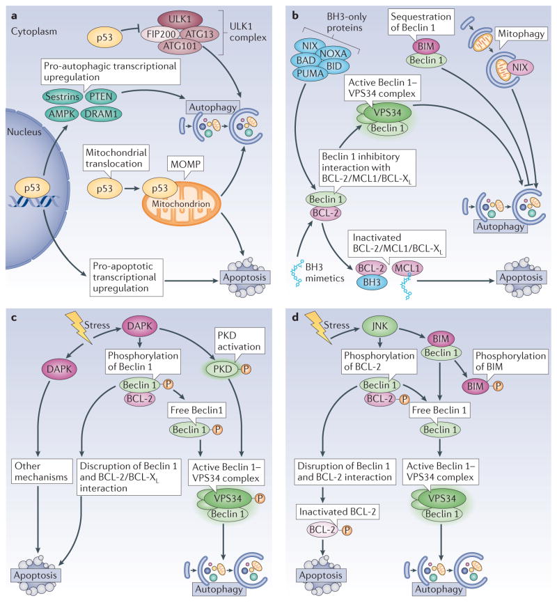
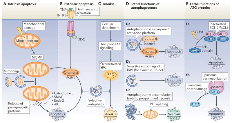
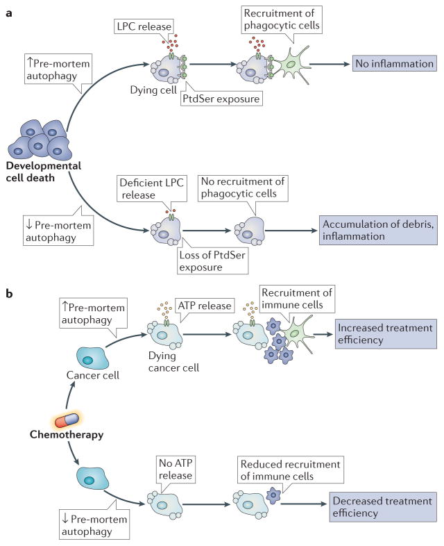
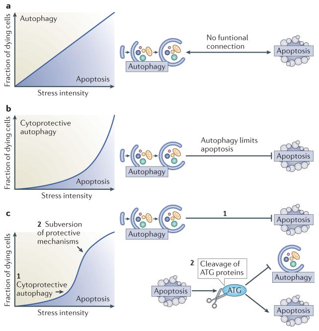
Autophagy and apoptosis control the turnover of organelles and proteins within cells, and of cells within organisms, respectively, and many stress pathways sequentially elicit autophagy, and apoptosis within the same cell. Generally autophagy blocks the induction of apoptosis, and apoptosis-associated caspase activation shuts off the autophagic process. However, in special cases, autophagy or autophagy-relevant proteins may help to induce apoptosis or necrosis, and autophagy has been shown to degrade the cytoplasm excessively, leading to 'autophagic cell death'. The dialogue between autophagy and cell death pathways influences the normal clearance of dying cells, as well as immune recognition of dead cell antigens. Therefore, the disruption of the relationship between autophagy and apoptosis has important pathophysiological consequences.


Conflict of interest statement

Competing interests statement

The authors declare no competing interests.

Figures

Figure 1
Figure 1. Signals that induce both apoptosis and autophagy
a | p53. The tumour suppressor p53 exerts both pro- and anti-autophagic functions, which depends on its subcellular localization and activation status. When present in the cytoplasm, p53 inhibits autophagy by interacting with FIP200 (FAK family kinase-interacting protein of 200 kDa) and thus interfering with ULK1 (UNC-51-like kinase 1) complex activity. In response to stress, p53 translocates to the nucleus, where it binds to the promoter region of multiple pro-autophagic genes, including the β1, β2 and γ-subunits of AMP-activated protein kinase (AMPK), damage-regulated autophagy modulator 1 (DRAM1), sestrin 1, sestrin 2 and phosphatase and tensin homologue (PTEN). Nuclear p53 also induces transcription of multiple pro-apoptotic genes. Under certain circumstances, p53 translocates to mitochondria, where it triggers mitochondrial outer membrane permeabilization (MOMP), and thus induces both mitophagy (at first) and apoptosis (when the intensity of the pro-apoptotic stimuli overcomes the protective ability of mitophagy). b | BH3 (BCL-2 homology 3)-only proteins and BH3 mimetics. Through its BH3 domain, Beclin 1 interacts with anti-apoptotic proteins from the BCL-2 family. This inhibits the pro-autophagic function of Beclin 1, but does not interfere with the anti-apoptotic activity of the BCL-2 family proteins. BH3-only proteins, as well as BH3 mimetics, disrupt this interaction, which enables Beclin 1 to increase autophagic activity. One particular BH3-only protein, BCL-2-interacting mediator of cell death (BIM), has a peculiar effect on autophagy. BIM interacts with Beclin 1 and mislocalizes it to dynein light chain 1 (DLC1; not shown), thereby preventing autophagy. Moreover, another BH3-only protein, NIX (NIP3-like protein X), which localizes to mitochondria, favours mitophagy. c | Death associated protein kinase (DAPK). This kinase can stimulate autophagy by phosphorylating Beclin 1, which enables its dissociation from BCL-2 and its association with vacuolar protein sorting 34 (VPS34). DAPK can also activate a second kinase, protein kinase D (PKD), which phosphorylates and activates VPS34. d | JUN N-terminal kinase (JNK). JNK induces autophagy by phosphorylating BCL-2 or BIM, which disrupts their inhibitory interaction with Beclin 1. This leads to an active Beclin 1–VPS34 complex and autophagy and also prevents BCL-2 from inhibiting pro-apoptotic proteins, thus promoting apoptosis. BAD, BCL-2 antagonist of cell death; BCL-XL, BCL extra large; BID, BH3-interacting domain death agonist; PUMA, p53 upregulated modulator of apoptosis.
Figure 2
Figure 2. Effects of autophagy on lethal signalling
AC | Inhibition of apoptosis and anoikis by autophagy. A | Distinct types of proteins, lipids and metabolites may induce mitochondrial damage. Once established, mitochondrial outer membrane permeabilization (MOMP) leads to the release of catabolic hydrolases (apoptosis-inducing factor (AIF) or endonuclease G (EndoG)) and caspase activators (such as cytochrome c and SMAC (second mitochondria-derived activator of caspase)) into the cytoplasm. These changes usually mark the ‘point-of-no-return’ of intrinsic apoptosis. The selective autophagy of damaged mitochondria (mitophagy) delays intrinsic apoptosis by limiting the release of pro-apoptotic factors. B | The extrinsic pathway of apoptosis is triggered by the binding of trimeric ligands to so-called death receptors, which include CD95, the tumour necrosis factor receptor 1 (TNFR1, depicted in the figure) and the TRAIL (TNF-related apoptosis-inducing ligand) receptors. One key event in this pathway is the activation of caspase 8. Selective clearance of active caspase 8 by autophagy may delay the onset of extrinsic apoptosis after death receptor stimulation. As caspase 8-induced apoptosis often relies on the cleavage and activation of the BH3-only protein BID (BH3-interacting domain death agonist), which is a potent MOMP inducer, mitophagy may also inhibit lethal signalling via death receptors. C | The selective clearance of the SRC protein by autophagy, which is overactivated in response to disrupted focal adhesion kinase (FAK) signalling, delays the onset of anoikis, which is induced by cell detachment from the extracellular matrix. D, E | Role of autophagy proteins in cell death. Although it generally constitutes a cytoprotective process, autophagosomes or ATG proteins may participate in lethal signalling in some specific circumstances. Da | Autophagosomes may act as platforms for caspase 8 activation, although the precise mechanism (or mechanisms) underlying this event remain poorly understood (indicated by the question mark). Db | In Drosophila melanogaster, the selective autophagy of anti-apoptotic proteins, such as Bruce (BIR-containing ubiquitin-conjugating enzyme; an orthologue of the mammalian inhibitor of apoptosis proteins (IAPs)), contributes to the induction of apoptosis. Dc | Autophagosome accumulation, due to the saturation of the degradative capacity of lysosomes, may facilitate PTP opening and thus lethal MOMP by hitherto unknown mechanisms (indicated by the question mark). Ea | Certain pro-apoptotic stimuli, such as C6 ceramide or tunicamycin, induce the interaction of ATG12 with anti-apoptotic BCL-2 or MCL1 (myeloid cell leukaemia sequence 1) proteins, thereby inhibiting their function and increasing apoptosis. Eb | ATG7 facilitates lysosomal membrane permeabilization, one of the entry points of the apoptotic pathway, after lysosomal photodamage. DISC, death-inducing signalling complex; PTP, permeability transition pore.
Figure 3
Figure 3. Impact of autophagy on the removal of dead cells in tissues
a | During the course of developmental cell death, pre-mortem autophagy is required for the release of lysophosphatidylcholine (LPC), a potent chemotactic signal, and for the exposure of the ‘eat-me’ signal phosphatidylserine (PtdSer), on the cell surface. Without these two events, recruitment of phagocytic cells is impaired, which results in the defective clearance of dead cells, increased inflammation and compromised development. b | Pre-mortem autophagy induction is essential for ATP release from dying cancer cells. Inhibition of ATP release compromises the recruitment of immune cells, and thus the activation of the immune system. This prevents the immune system from recognizing tumour-specific antigens, a process that is essential for the effectiveness of anti-tumour therapies.
Figure 4
Figure 4. Functional relationship between autophagy and apoptosis
a | Hypothetical scenario of an absent interplay between autophagy and apoptosis. If no functional connection between apoptosis and autophagy existed, the percentage of cells activating the autophagic pathway or instead succumbing to apoptosis would be solely determined by stress intensity over time. b | Effect of cytoprotective autophagy on a dose-response curve. If autophagy inhibits apoptosis, the cell population becomes relatively resistant to cell death, unless the intensity of the stressor overcomes the protective barrier of autophagy. c | Cytoprotective autophagy, followed by lethal subversion of the apoptotic machinery. In this scenario, autophagy would constitute an initial barrier against apoptosis when stress intensity is low (1). As stress intensity increases (2), the induction of apoptosis results in the subversion of cytoprotective mechanisms, including autophagy, and the conversion of cytoprotective molecules into cytotoxic ones. For example, cleavage of essential ATG proteins does not only inactivate the autophagic machinery but also leads to the generation of protein fragments with novel pro-apoptotic properties.

Similar articles

Cited by

References

    1. Maiuri MC, Zalckvar E, Kimchi A, Kroemer G. Self-eating and self-killing: crosstalk between autophagy and apoptosis. Nature Rev Mol Cell Biol. 2007;8:741–752. - PubMed
    1. Yang Z, Klionsky DJ. Mammalian autophagy: core molecular machinery and signaling regulation. Curr Opin Cell Biol. 2010;22:124–131. - PMC - PubMed
    1. Mizushima N, Komatsu M. Autophagy: renovation of cells and tissues. Cell. 2011;147:728–741. - PubMed
    1. Choi AM, Ryter SW, Levine B. Autophagy in human health and disease. N Engl J Med. 2013;368:1845–1846. - PubMed
    1. Thumm M, et al. Isolation of autophagocytosis mutants of Saccharomyces cerevisiae. FEBS Lett. 1994;349:275–280. - PubMed

Publication types