. 2014 Jan 8:2:5.
doi: 10.1186/2051-5960-2-5.

A new animal model of spontaneous autoimmune peripheral polyneuropathy: implications for Guillain-Barré syndrome

Affiliations

A new animal model of spontaneous autoimmune peripheral polyneuropathy: implications for Guillain-Barré syndrome

Mu Yang et al. Acta Neuropathol Commun. .

Abstract

Background: Spontaneous autoimmune peripheral neuropathy including Guillain-Barré Syndrome (GBS) represents as one of the serious emergencies in neurology. Although pathological changes have been well documented, molecular and cellular mechanisms of GBS are still under-explored, partially due to short of appropriate animal models. The field lacks of spontaneous and translatable models for mechanistic investigations. As GBS is preceded often by viral or bacterial infection, a condition can enhance co-stimulatory activity; we sought to investigate the critical role of T cell co-stimulation in this autoimmune disease.

Results: Our previous study reported that transgene-derived constitutive expression of co-stimulator B7.2 on antigen presenting cells of the nervous tissues drove spontaneous neurological disorders. Depletion of CD4+ T cells in L31 mice accelerated the onset and increased the prevalence of the disease. In the current study, we further demonstrated that L31/CD4-/- mice exhibited both motor and sensory deficits, including weakness and paresis of limbs, numbness to mechanical stimuli and hypersensitivity to thermal stimulation. Pathological changes were characterized by massive infiltration of macrophages and CD8+ T cells, demyelination and axonal damage in peripheral nerves, while changes in spinal cords could be secondary to the PNS damage. In symptomatic L31/CD4-/- mice, the disruption of the blood neural barriers was observed mainly in peripheral nerves. Interestingly, the infiltration of immune cells was initiated in pre-symptomatic L31/CD4-/- mice, prior to the disease onset, in the DRG and spinal roots where the blood nerve barrier is virtually absent.

Conclusions: L31/CD4-/- mice mimic most parts of clinical and pathological signatures of GBS in human; thus providing an unconventional opportunity to experimentally explore the critical events that lead to spontaneous, autoimmune demyelinating disease of the peripheral nervous system.


Figures

Figure 1
Figure 1
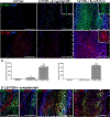
Transgene-derived B7.2 constitutive expression primed macrophages and microglia in the nervous tissues. The number of CD11b+ CD45+ macrophages in the sciatic nerves and microglia in the spinal cord, and the expression of B7.2 among these targeted cells was quantified using fluorescent-activated cell sorting (FACS) analysis. The histogram represents the FACS analysis obtained from 3 animals per group. Percentage of either macrophages (A) or microglia (C) expressing B7.2 is increased in L31/CD4-/- mice, compared with wild type animals. The absolute number of macrophages in the nerves and microglia in the spinal cord is also higher in L31/CD4-/- mice than that of wild type mice (A, C) (*: p < 0.05, ***: p < 0.001). Representative examples of immunohistochemistry analysis of macrophages in the sciatic nerves and microglia in the spinal cords were depicted in (B) and (D). Note that in L31 /CD4-/- mice, many Iba-1+ macrophages and microglia were colocalized with B7.2 which was almost undetectable in wild type mice. Scale bars: B, 50 μm; D, 500 μm.
Figure 2
Figure 2
L31/CD4-/- mice developed spontaneous motor and sensory neurological disorders. The incidence of motor deficits in L31/CD4-/- mice evaluated with established clinical scores was presented in (A). Rotarod test was performed to examine coordination ability. Compared with wild type mice, the latency of fall dropped in both pre-symptomatic and symptomatic L31 mice (B) (n = 6/group, **: p < 0.01, ***: p < 0.001), indicative of motor impairment. Pain responses to mechanical, cold and heat stimuli were assessed in wild type, pre-symptomatic and symptomatic L31/CD4-/- mice. Animals that had their clinical score ≥4 were excluded from sensory tests. Only symptomatic L31/CD4-/- mice demonstrated numbness to von Frey hair stimulation (C) (n = 6/group, ***: p < 0.001). However, thermal hypersensitivity was observed in both pre-symptomatic and symptomatic L31 mice, as the duration of withdrawal in acetone test increased (D) (n = 6/group, *: p < 0.05, ***: p < 0.001), and the hotplate latency decreased (E) (n = 6/group, **: p < 0.01, ***: p < 0.001).
Figure 3
Figure 3
Demyelination and axonal damage were found in the peripheral nerves of symptomatic L31/CD4-/- mice. Myelin in the sciatic nerves was stained with FluoroMyelin. Representative illustrations from longitudinal (A-I) and cross (J-N) sections showed an intact myelin structures in wild type (A, E, J) and pre-symptomatic L31 mouse sciatic nerves (B, F, K), and a focal (C, H) or diffused, severely damaged myelin (D, G) in symptomatic L31 mice. In parallel with demyelination, the axonal damage evidenced by the loss of PGP9.5 staining was detected in symptomatic L31 mice (G, H, L, M, N). Some naked PGP9.5+ axonal fibers were found in the area where myelin disappeared or fragmented (G, H, L, M, N). I: Myelin labelling was found in a cell-shaped structure containing a DAPI+ nucleus, which suggests a macrophage with engulfed myelin fragments. Two naked axonal fibers were located nearby. ATF3 positive signals were found in the cell bodies of both DRG sensory neurons (O) and spinal cord ventral horn (VH) motor neurons (P). Scale bar: 50 μm.
Figure 4
Figure 4
Massive infiltration of immune cells in sciatic nerves of L31/CD4-/- mice. Cell infiltration in the diseased sciatic nerves was first evidenced by the significant increase of DAPI labelled cell number (A-B). Immunohistochemistry analysis demonstrated that while there was a slight increase of elongated (insert) Iba-1+ cells in the pre-symptomatic L31/CD4-/- mice, nerves from the symptomatic L31/CD4-/- mice were submerged with round-shaped (insert) infiltrated Iba-1+ macrophages (A). While there were abundant CD8+ T cells found in the sciatic nerves of symptomatic L31 mice, no CD8+ T cell infiltration was detected in pre-symptomatic L31/CD4-/- mice (B). Quantitative analysis with FACS (n = 3/group) confirmed the immunohistochemistry observation (**: p < 0.01, ***: p < 0.001) (C). In symptomatic L31 mouse sciatic nerves, infiltrates could either group in small foci (D-left) or be distributed diffusely (A/B-right). Fragmented myelin were found within Iba-1+macrophages (yellow signals within Iba-1+ cells). There were less infiltrates in the area where myelin remained in healthy shape (D-left). Majority of infiltrated Iba-1+ macrophages had high levels of B7.2 expression (D-middle) and CD8+ T cells were in close apposition with Iba-1+ macrophages (D-right). Scale bar: 200 μm.
Figure 5
Figure 5
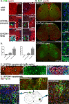
Limited demyelination and immune cell infiltration was detected in the spinal cords of L31/CD4-/- mice. Only isolated myelin loss was found in the spinal white matter of symptomatic L31/CD4-/- mice together with few CD8+ T cell clusters (A). CD8+ T cells were also found in the grey matter of some symptomatic L31/CD4-/- mice, gathered near motor neurons (A). Occasionally very few isolated CD8+ T cells were found even in pre-symptomatic L31/CD4-/- mouse spinal cords (A). In symptomatic L31/CD4-/- mice, robust spinal microglia activation was detected, mainly in the grey matter. Iba-1+ microglia were clustered at the dorsal and ventral horns (B). Morphological changes were revealed with confocal microscope (B-left panels). In the white matter of diseased spinal cord, some demyelinated spots (C-arrows) were found in the dorsal column where Iba-1+ microglia gathered (C). Quantitative analysis of CD11b+CD45+ microglia and CD8+ T cells using FACS (n = 3/group) confirmed the immunohistochemistry observation (*: p < 0.05, **: p < 0.01) (D). Increased B7.2 expression was found in symptomatic spinal cords, on ramified Iba-1/B7.2 double labelled microglia, or on round B7.2 single labelled cells, presumably CD8+ T cells (E). Scale bars: A, 100 μm; B, left, 500 μm, right, 10 μm; C, 100 μm; E, 50 μm.
Figure 6
Figure 6
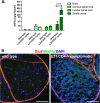
Blood nerve barrier in the sciatic nerves of diseased L31/CD4-/- mice was disrupted. The integrity of neural barriers (BBB, BSCB and BNB) was examined with a fluorescent dye NaFlu (MW, 376 Da) injected intravenously. Quantification of NaFlu content in the brain, the spinal cord and the sciatic nerve of wild type and the symptomatic L31/CD4-/- mice revealed that a significant increase of the dye within the nervous tissues was detected only in the nerve (n = 3/group, ***: p < 0.001 ) (A). Histological analysis on the cross sections of the sciatic nerves confirmed the dye extravasation in the symptomatic L31 mice. Glut1 was used to label blood vessels, perinurium and epineuriums (B). Scale bar: 100 μm.
Figure 7
Figure 7
Focal inflammatory reaction appeared in the DRG and the roots of pre-symptomatic L31/CD4-/- mice, preceding the disease onset. While there were only few resident macrophages and no CD8+ T cells in the DRG and the roots of wild type mice (A), both types of immune cells were found in these two organs in pre-symptomatic L31/CD4-/- mice (B). Some mice showed a slightest degree of infiltration, presumably the earliest pathologic changes (B-upper panels), some had significant amount of both Iba1+ macrophages and CD8+ T cells with increased B7.2 expression (B-bottom panels) suggesting the status close to the disease onset (B). In symptomatic L31/CD4-/- mice, the DRG and the roots were fulfilled with infiltrated Iba1+ macrophages and CD8+ T cells. Both expressed high levels of B7.2 (C). Scale bar: 100 μm.

Similar articles

Cited by

References

    1. Yuki N, Hartung HP. Guillain-Barre syndrome. N Engl J Med. 2012;2:2294–2304. doi: 10.1056/NEJMra1114525. 10.1056/NEJMra1114525. - DOI - PubMed
    1. Brostoff SW, Levit S, Powers JM. Induction of experimental allergic neuritis with a peptide from myelin P2 basic protein. Nature. 1977;2:752–753. doi: 10.1038/268752a0. - DOI - PubMed
    1. Waksman BH, Adams RD. Allergic neuritis: an experimental disease of rabbits induced by the injection of peripheral nervous tissue and adjuvants. J Exp Med. 1955;2:213–236. doi: 10.1084/jem.102.2.213. - DOI - PMC - PubMed
    1. Wekerle H, Flugel A, Fugger L, Schett G, Serreze D. Autoimmunity’s next top models. Nat Med. 2012;2:66–70. doi: 10.1038/nm.2635. 10.1038/nm.2635. - DOI - PubMed
    1. Salomon B, Rhee L, Bour-Jordan H, Hsin H, Montag A, Soliven B, Arcella J, Girvin AM, Padilla J, Miller SD, Bluestone JA. Development of spontaneous autoimmune peripheral polyneuropathy in B7-2-deficient NOD mice. J Exp Med. 2001;2:677–684. doi: 10.1084/jem.194.5.677. - DOI - PMC - PubMed

Publication types

MeSH terms