

Superficial esophageal lesions detected by endoscopic ultrasound enhanced with submucosal edema
- PMID: 24379628
- PMCID: PMC3870556
- DOI: 10.3748/wjg.v19.i47.9034
Superficial esophageal lesions detected by endoscopic ultrasound enhanced with submucosal edema
Abstract
Aim: To determine if there is consistency between endoscopic ultrasound (EUS) findings and pathological results for detecting lesions of different depth in the esophageal mucosa.
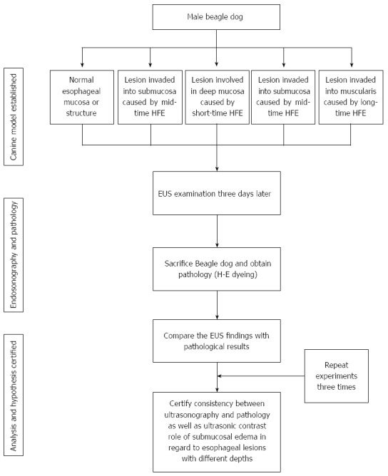
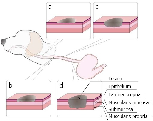
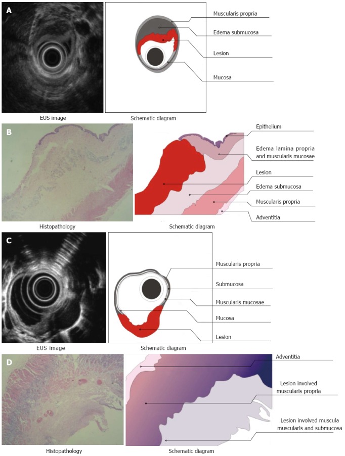
Methods: A canine (Beagle) model was established in which lesions of different depths were created in the esophageal mucosa by thermal burning. Seventy-two hours later, these lesions and adjacent tissue in the esophagus were examined by EUS. EUS findings including infiltrating depth, strength of echogenicity and homogeneity were recorded. Dogs were sacrificed and tissue specimens were obtained. We then compared the EUS findings with the pathology reports.
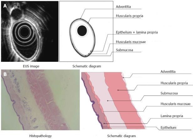
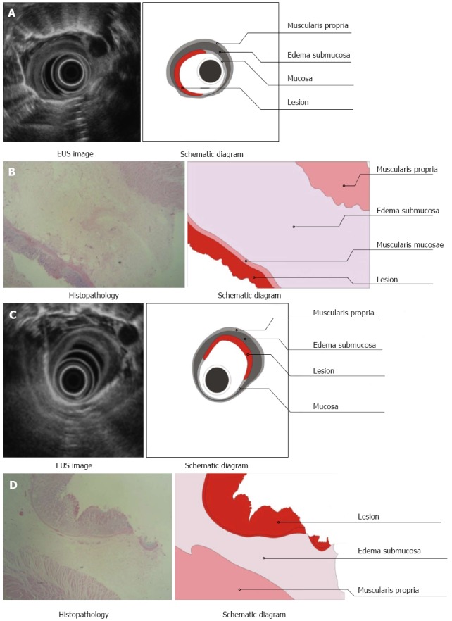
Results: Thermal burns created at different power settings caused lesions of different depth in the esophageal mucosa. When the echo strength was shifted from high, medium, to low echogenicity, an increase in the infiltrating depth of the lesion was noted, which coincided with results of the pathology examination. Obvious submucosal edema visualized by EUS was also detected by pathology. Furthermore, because of the enhancement caused by the submucosal edema, the lesions invading into the submucosa were easily visualized by EUS.
Conclusion: There is consistency between EUS findings and pathological results of esophageal lesions with different depths. Submucosal edema can serve as an ultrasonic contrast agent.
Keywords: Canine; Endoscopic ultrasound; Esophagus; Lesion; Pathology.
Figures
Similar articles
-
Extraesophageal saline enhances endoscopic ultrasonography to differentiate esophagus and adjacent organs.World J Gastroenterol. 2014 Sep 21;20(35):12551-8. doi: 10.3748/wjg.v20.i35.12551. World J Gastroenterol. 2014. PMID: 25253957 Free PMC article.
-
Endoscopic ultrasound: accuracy in staging superficial carcinomas of the esophagus.Ann Thorac Surg. 2008 Jan;85(1):251-6. doi: 10.1016/j.athoracsur.2007.08.021. Ann Thorac Surg. 2008. PMID: 18154819
-
Early esophageal carcinoma: endoscopic ultrasonography using the Sonoprobe.Abdom Imaging. 2003 Jul-Aug;28(4):477-85. doi: 10.1007/s00261-002-0076-5. Abdom Imaging. 2003. PMID: 14580090
-
Barrett's esophagus: current and future role of endosonography and optical coherence tomography.Dis Esophagus. 2004;17(2):118-23. doi: 10.1111/j.1442-2050.2004.00406.x. Dis Esophagus. 2004. PMID: 15230723 Review.
-
Diagnostic accuracy of EUS in differentiating mucosal versus submucosal invasion of superficial esophageal cancers: a systematic review and meta-analysis.Gastrointest Endosc. 2012 Feb;75(2):242-53. doi: 10.1016/j.gie.2011.09.016. Epub 2011 Nov 23. Gastrointest Endosc. 2012. PMID: 22115605 Review.
Cited by
-
Speckle reduction in ultrasound endoscopy using refraction based elevational angular compounding.Sci Rep. 2021 Sep 15;11(1):18370. doi: 10.1038/s41598-021-97717-2. Sci Rep. 2021. PMID: 34526594 Free PMC article.
References
-
- Mariette C, Piessen G, Triboulet JP. Therapeutic strategies in oesophageal carcinoma: role of surgery and other modalities. Lancet Oncol. 2007;8:545–553. - PubMed
-
- Lightdale CJ. Esophageal cancer. American College of Gastroenterology. Am J Gastroenterol. 1999;94:20–29. - PubMed
-
- Moretó M. Diagnosis of esophagogastric tumors. Endoscopy. 2003;35:36–42. - PubMed
-
- Maish MS, DeMeester SR. Endoscopic mucosal resection as a staging technique to determine the depth of invasion of esophageal adenocarcinoma. Ann Thorac Surg. 2004;78:1777–1782. - PubMed
-
- Sobin LH, Gospodarowicz MK, Wittekind Ch. TNM Classification of Malignant Tumors. 7th edn. New York: Wiley-Blackwell; 2010.
Publication types
MeSH terms
LinkOut - more resources
Full Text Sources
Other Literature Sources
Medical
