

The Rac-specific exchange factors Dock1 and Dock5 are dispensable for the establishment of the glomerular filtration barrier in vivo
- PMID: 24365888
- PMCID: PMC4011817
- DOI: 10.4161/sgtp.27430
The Rac-specific exchange factors Dock1 and Dock5 are dispensable for the establishment of the glomerular filtration barrier in vivo
Abstract
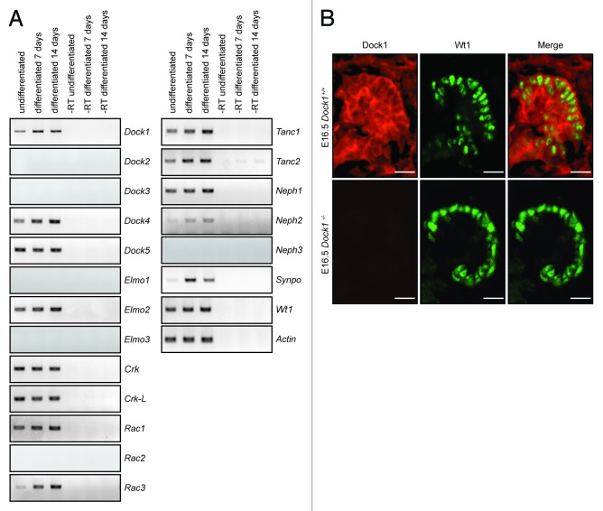
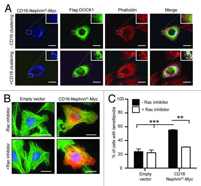
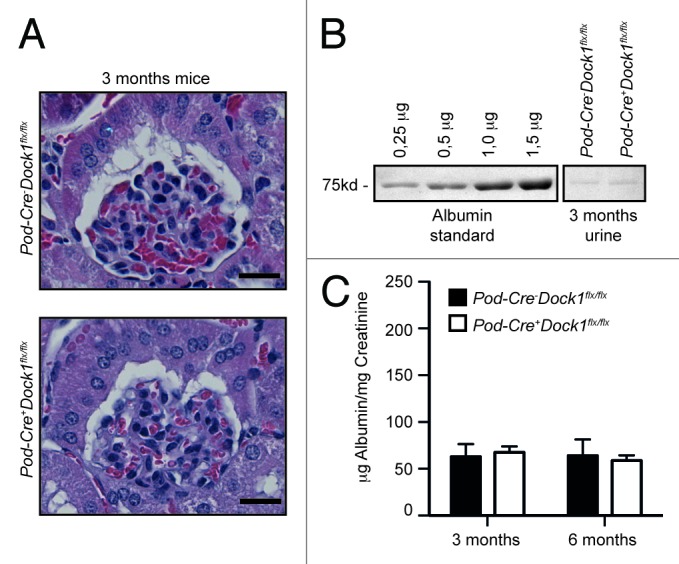
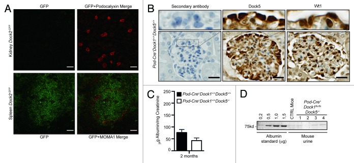
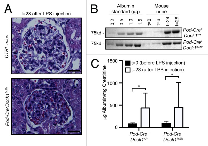
Podocytes are specialized kidney cells that form the kidney filtration barrier through the connection of their foot processes. Nephrin and Neph family transmembrane molecules at the surface of podocytes interconnect to form a unique type of cell-cell junction, the slit diaphragm, which acts as a molecular sieve. The cytoplasmic tails of Nephrin and Neph mediate cytoskeletal rearrangement that contributes to the maintenance of the filtration barrier. Nephrin and Neph1 orthologs are essential to regulate cell-cell adhesion and Rac-dependent actin rearrangement during Drosophila myoblast fusion. We hypothesized here that molecules regulating myoblast fusion in Drosophila could contribute to signaling downstream of Nephrin and Neph1 in podocytes. We found that Nephrin engagement promoted recruitment of the Rac exchange factor Dock1 to the membrane. Furthermore, Nephrin overexpression led to lamellipodia formation that could be blocked by inhibiting Rac1 activity. We generated in vivo mouse models to investigate whether Dock1 and Dock5 contribute to the formation and maintenance of the kidney filtration barrier. Our results indicate that while Dock1 and Dock5 are expressed in podocytes, their functions are not essential for the development of the glomerular filtration barrier. Furthermore, mice lacking Dock1 were not protected from LPS-induced podocyte effacement. Our data suggest that Dock1 and Dock5 are not the important exchange factors regulating Rac activity during the establishment and maintenance of the glomerular barrier.
Keywords: Dock1; Dock180; Dock5; Elmo; Rac; Rho GTPase; podocyte.
Figures

Similar articles
-
The atypical Rac activator Dock180 (Dock1) regulates myoblast fusion in vivo.Proc Natl Acad Sci U S A. 2008 Oct 7;105(40):15446-51. doi: 10.1073/pnas.0805546105. Epub 2008 Sep 26. Proc Natl Acad Sci U S A. 2008. PMID: 18820033 Free PMC article.
-
Phosphorylation of slit diaphragm proteins NEPHRIN and NEPH1 upon binding of HGF promotes podocyte repair.J Biol Chem. 2021 Sep;297(3):101079. doi: 10.1016/j.jbc.2021.101079. Epub 2021 Aug 13. J Biol Chem. 2021. PMID: 34391780 Free PMC article.
-
Phosphatidic acid-dependent recruitment and function of the Rac activator DOCK1 during dorsal ruffle formation.J Biol Chem. 2013 Mar 22;288(12):8092-8100. doi: 10.1074/jbc.M112.410423. Epub 2013 Jan 29. J Biol Chem. 2013. PMID: 23362269 Free PMC article.
-
Actin Up: An Overview of the Rac GEF Dock1/Dock180 and Its Role in Cytoskeleton Rearrangement.Cells. 2022 Nov 11;11(22):3565. doi: 10.3390/cells11223565. Cells. 2022. PMID: 36428994 Free PMC article. Review.
-
Nephrin-signature molecule of the glomerular podocyte?J Pathol. 2010 Feb;220(3):328-37. doi: 10.1002/path.2661. J Pathol. 2010. PMID: 19950250 Review.
Cited by
-
Downregulation of DOCK1 sensitizes bladder cancer cells to cisplatin through preventing epithelial-mesenchymal transition.Drug Des Devel Ther. 2016 Sep 8;10:2845-2853. doi: 10.2147/DDDT.S101998. eCollection 2016. Drug Des Devel Ther. 2016. PMID: 27660415 Free PMC article.
-
The Role of Trio, a Rho Guanine Nucleotide Exchange Factor, in Glomerular Podocytes.Int J Mol Sci. 2018 Feb 6;19(2):479. doi: 10.3390/ijms19020479. Int J Mol Sci. 2018. PMID: 29415466 Free PMC article.
-
DNA Hypomethylation of DOCK1 Leading to High Expression Correlates with Neurologic Deterioration and Poor Function Outcomes after Spontaneous Intracerebral Hemorrhage.Evid Based Complement Alternat Med. 2021 Sep 27;2021:1186458. doi: 10.1155/2021/1186458. eCollection 2021. Evid Based Complement Alternat Med. 2021. Retraction in: Evid Based Complement Alternat Med. 2023 Jun 21;2023:9785782. doi: 10.1155/2023/9785782 PMID: 34616473 Free PMC article. Retracted.
-
TBOPP enhances the anticancer effect of cisplatin by inhibiting DOCK1 in renal cell carcinoma.Mol Med Rep. 2020 Aug;22(2):1187-1194. doi: 10.3892/mmr.2020.11243. Epub 2020 Jun 16. Mol Med Rep. 2020. PMID: 32626999 Free PMC article.
-
Podocyte-actin dynamics in health and disease.Nat Rev Nephrol. 2016 Nov;12(11):692-710. doi: 10.1038/nrneph.2016.127. Epub 2016 Aug 30. Nat Rev Nephrol. 2016. PMID: 27573725 Review.
References
-
- Kestilä M, Lenkkeri U, Männikkö M, Lamerdin J, McCready P, Putaala H, Ruotsalainen V, Morita T, Nissinen M, Herva R, et al. Positionally cloned gene for a novel glomerular protein--nephrin--is mutated in congenital nephrotic syndrome. Mol Cell. 1998;1:575–82. doi: 10.1016/S1097-2765(00)80057-X. - DOI - PubMed
Publication types
MeSH terms
Substances
Grants and funding
LinkOut - more resources
Full Text Sources
Other Literature Sources
Molecular Biology Databases
Research Materials
Miscellaneous