. 2013 Dec 11;8(12):e81811.
doi: 10.1371/journal.pone.0081811. eCollection 2013.

A RabGAP regulates life-cycle duration via trimeric G-protein cascades in Dictyostelium discoideum

Affiliations

A RabGAP regulates life-cycle duration via trimeric G-protein cascades in Dictyostelium discoideum

Hidekazu Kuwayama et al. PLoS One. .

Abstract

Background: The life-cycle of cellular slime molds comprises chronobiologically regulated processes. During the growth phase, the amoeboid cells proliferate at a definite rate. Upon starvation, they synthesize cAMP as both first and second messengers in signalling pathways and form aggregates, migrating slugs, and fruiting bodies, consisting of spores and stalk cells, within 24 h. In Dictyostelium discoideum, because most growth-specific events cease during development, proliferative and heterochronic mutations are not considered to be interrelated and no genetic factor governing the entire life-cycle duration has ever been identified.

Methodology/principal findings: Using yeast 2-hybrid library screening, we isolated a Dictyostelium discoideum RabGAP, Dd Rbg-3, as a candidate molecule by which the Dictyostelium Gα2 subunit directs its effects. Rab GTPase-activating protein, RabGAP, acts as a negative regulator of Rab small GTPases, which orchestrate the intracellular membrane trafficking involved in cell proliferation. Deletion mutants of Dd rbg-3 exhibited an increased growth rate and a shortened developmental period, while an overexpression mutant demonstrated the opposite effects. We also show that Dd Rbg-3 interacts with 2 Gα subunits in an activity-dependent manner in vitro. Furthermore, both human and Caenorhabditis elegans rbg-3 homologs complemented the Dd rbg-3-deletion phenotype in D. discoideum, indicating that similar pathways may be generally conserved in multicellular organisms.

Conclusions/significance: Our findings suggest that Dd Rbg-3 acts as a key element regulating the duration of D. discoideum life-span potentially via trimeric G-protein cascades.


Conflict of interest statement

Competing Interests: The authors have declared that no competing interests exist.

Figures

Figure 1
Figure 1. Sequence analyses and expression of Dictyostelium discoideum Rbg-3 (Dd Rbg-3).
(A) Deduced amino acid sequence of Dd Rbg-3. (B) Schematic diagram of the Dd Rbg-3 domain structure. (C) Multiple sequence alignment of the TBC domains of Rbg-3 homologs. Residues identical among the 3 proteins compared are indicated in yellow, conserved residues in green, and semi-conserved residues in blue. (D) Northern blot analysis of Dd Rbg-3 during development. Ten micrograms of total RNA obtained from the organism at different developmental time-points were loaded in each gel lane, and the blots hybridized with an Dd Rbg-3-specific probe. (E) Western blot analysis of Dd Rbg-3 during development. One hundred micrograms of total protein, obtained from the organism at different developmental time-points, were loaded in each gel lane, and the blots probed with a Dd Rbg-3-specific antibody.
Figure 2
Figure 2. Phenotypes of Dd rbg-3-null and Dd rbg-3-overexpression mutant cells.
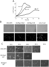
(A) Cell growth in AX2 (filled circles, •), Dd rbg-3–null (filled triangles, ▴), and Dd rbg-3-overexpression (filled squares, ▪) strains. Cells were cultured in HL5 liquid medium, at 21°C, with shaking at 125 rpm. (B) Vegetative cell shape of AX2, Dd rbg-3-null, and Dd rbg-3-overexpression cells. To observe nuclei, vegetative cells were incubated with 0.1 µg/ml DAPI, after fixation with 1% formaldehyde in cold methanol, and 3 washes with PB. Scale bar, 20 µm. (C) Development in the parental AX2 strain, Dd rbg-3-null, and Dd rbg-3-overexpression cells were assessed on non-nutrient agar plates; cells were plated at a density of 1×10⁶ cells/cm2. In AX2 cells, mounds formed at 12 h after plating; slugs were visible at 16 h; culmination occurred by 20 h, and fruiting bodies were observed at 24 h. Development concluded faster in the Dd rbg-3-null strain, and slower in the Dd rbg-3-overexpression strain. Scale bar, 1 mm.
Figure 3
Figure 3. Endocytosis and lysosomes in AX2, Dd rbg-3-null, and Dd rbg-3-overexpression strains.
(A) Fluorescence microscopy images of endocytosis of rhodamine-dextran. Vegetative cells were incubated with rhodamine-dextran [0.4 mg/ml] for 10 min, and then washed 3 times with PB. Scale bar, 10 µm. (B) Fluorescence microscopy images of lysosomes visualized using LysoTracker® Green. Vegetative cells were incubated with 75 nM LysoTracker® Green (Invitrogen, USA) for 30 min, and then washed 3 times with PB. Arrowheads indicate swollen lysosomes in the overexpression cells. Scale bar, 10 µm. (C) Relative fluorescent intensity of cell suspension treated with rhodamine-dextran [0.4 mg/ml] in 5×10⁷ cells/ml. (D) Relative fluorescent intensity of cell suspension stained with LysoTracker® Green in 5×10⁷ cells/ml. **P<0.05 between AX2 and Dd rbg-3 OE (t-test).
Figure 4
Figure 4. Adenylyl cyclase activity and cellular localization of Dd Rbg-3.
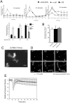
(A) Adenylyl cyclase activity was measured in intact AX2 (filled circles, •), Dd rbg-3-null (filled triangles, ▴), and Dd rbg-3-overexpression (filled squares, ▪) cells following cAMP stimulation. Starved cells were stimulated with 10 µM deoxy-cAMP. At specific time-points, aliquots of cells were lysed and the cAMP levels measured. (B) Adenylyl cyclase activity in cell lysates of the parental and mutant strains measured in the presence of 30 µM GTPγS. (C) Fluorescent microscopy image of AX2 cells expressing Dd Rbg-3-Venus. Scale bar, 10 µm. (D) Fluorescent microscopy images of migration of Dd Rbg-3-Venus in AX2 cells toward a micropipette releasing 1 µM cAMP. Stars represent the position of the tip of the micropipette. Scale bar, 10 µm. (E) Translocation of CRAC. Aggregating cells were pretreated with 5 µM latrunculin A. Fluorescence intensity of CRAC-GFP in the cytosol was measured at the indicated time after stimulation with 1 µM cAMP. Cells were excited at 440 nm and viewed through a cut-off filter of 500–550 nm to assess emission before and after cAMP stimulation. In each experiment, fluorescence intensity of an area in the cytosol was measured at the indicated time-point and was normalized to the average intensity measured 18 s before cAMP stimulation (n = 102 for AX2, n = 102 for the null mutant, n = 143 for the overexpression mutant).
Figure 5
Figure 5. Phenotype of a R364A mutant in Dd rbg-3–null cells.
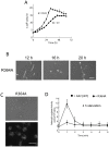
(A) Cell growth in AX2 (filled circles, •) and the R364A (open triangles, △) strains. Cells were cultured in HL5 liquid medium, at 21°C, with shaking at 125 rpm. (B) Development in the R364A was assessed on non-nutrient agar plates; cells were plated at a density of 1×10⁶ cells/cm2. Scale bar, 1 mm.(C) Fluorescence microscopy images of lysosomes in AX2 and the R364A cells using LysoTracker® Green. Scale bar, 10 µm. (D) Adenylyl cyclase activity was measured in intact AX2 (filled circles, •) and R364A (filled diamonds, ♦) cells starved for 4 h following cAMP stimulation. Starved cells for 4 h were stimulated with 10 µM deoxy-cAMP.
Figure 6
Figure 6. Phenotype of rab7A–null mutant.
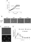
(A) Cell growth in AX2 (filled circles, •) and the rab7A–null (open triangles, △) strains. Cells were cultured in HL5 liquid medium, at 21°C, with shaking at 125 rpm. (B) Development in rab7A-null was assessed on non-nutrient agar plates; cells were plated at a density of 1×10⁶ cells/cm2. Scale bar, 1 mm. (C) Fluorescence microscopy images of lysosomes in AX2 and the rab7A-null cells using LysoTracker® Green. Scale bar, 10 µm. (D) Adenylyl cyclase activity was measured in intact AX2 (filled circles, •) and rab7A-null (inverted filled triangle, ▾) cells starved for 8 h following cAMP stimulation. Starved cells for 4 h were stimulated with 10 µM deoxy-cAMP.
Figure 7
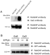
Figure 7. In vivo interaction of Dd Rbg-3 with activated Gα2 and Gα9.
(A) Co-immunoprecipitation of Gα2 with Dd Rbg-3. Lysates of AX2 cells and cells overexpressing dominant gα2 were used. (B) Co-immunoprecipitation of Gα8 and Gα9 with Dd Rbg-3. Lysates of AX2 cells and cells expressing eGFP-fused Gα8 and Gα9 were used. In each experiment, cell lysates were incubated with or without 30 µM GTPγS before the pull-down experiments.
Figure 8
Figure 8. Complementation of Dd rbg-3–null cells by expressing human Rbg-3 homolog.
(A) Growth in AX2 cells (filled circles, •), Dd rbg-3-null cells (open circles, ○), and Dd rbg-3-null cells expressing the human homologue, TBC1D5 gene (open triangles, △), cultured in HL5 liquid medium with shaking. (B) Development of Dd rbg-3-null cells expressing human Rbg-3 incubated on non-nutrient agar plates, at a density of 1×10⁶ cells/cm2. Scale bar, 1 mm.
Figure 9
Figure 9. Caffeine does not inhibit the development, or adenylyl cyclase activity, of Dd rbg-3-null mutant cells and cells expressing the R364A-mutant Dd rbg-3.
(A) Development of AX2 and Dd rbg-3-null cells starved at a density of 1×10⁶ cells/cm2 on non-nutrient agar plates containing 4 mM caffeine. In the parental cells, slugs and fruiting bodies were not observed. Scale bar, 10 µm. (B) Adenylyl cyclase activity was measured, following stimulation with 10 µM deoxy-cAMP, in intact cells grown under starvation conditions for 8 h in the presence of 4 mM caffeine. (C) Adenylyl cyclase activity was measured in the presence of 30 µM GTPγS, in cell lysates grown in medium containing 4 mM caffeine.

Similar articles

Cited by

References

    1. Frasa MA, Koessmeier KT, Ahmadian MR, Braga VM (2012) Illuminating the functional and structural repertoire of human TBC/RABGAPs. Nat Rev Mol Cell Biol 13: 67–73. - PubMed
    1. Williams JG (2010) Dictyostelium finds new roles to model. Genetics 185: 717–726. - PMC - PubMed
    1. Konijn TM, van de Meene JG, Chang YY, Barkley DS, Bonner JT (1969) Identification of adenosine-3′,5′-monophosphate as the bacterial attractant for myxamoebae of Dictyostelium discoideum . J Bacteriol 99: 510–512. - PMC - PubMed
    1. Ginsburg GT, Gollop R, Yu Y, Louis JM, Saxe CL, et al. (1995) The regulation of Dictyostelium development by transmembrane signaling. J Eukaryot Microbiol 42: 200–205. - PubMed
    1. Swaney KF, Huang CH, Devreotes PN (2010) Eukaryotic chemotaxis: a network of signaling pathways controls motility, directional sensing, and polarity. Annu Rev Biophys 39: 265–289. - PMC - PubMed

MeSH terms

Substances

Grants and funding

This work was supported by a Grant-in-Aid for Scientific Research (C) (no. 22510202) from the Japan Society for the Promotion of Science (HK) and by a special fund for tenure-track faculty members of the Institute of Biological Sciences at the University of Tsukuba, Japan (HK). The funders had no role in study design, data collection and analysis, decision to publish, or preparation of the manuscript.

LinkOut - more resources