. 2014 Feb;88(4):2157-67.
doi: 10.1128/JVI.03055-13. Epub 2013 Dec 11.

Innate immune response induced by baculovirus attenuates transgene expression in mammalian cells

Affiliations

Innate immune response induced by baculovirus attenuates transgene expression in mammalian cells

Chikako Ono et al. J Virol. 2014 Feb.

Abstract

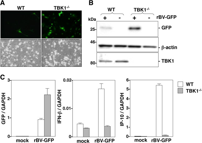
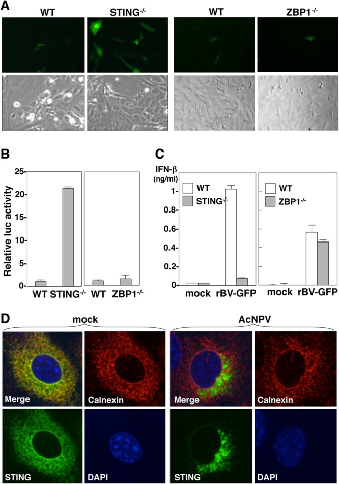
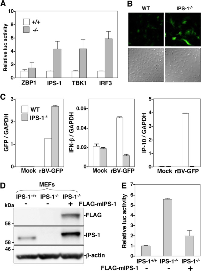
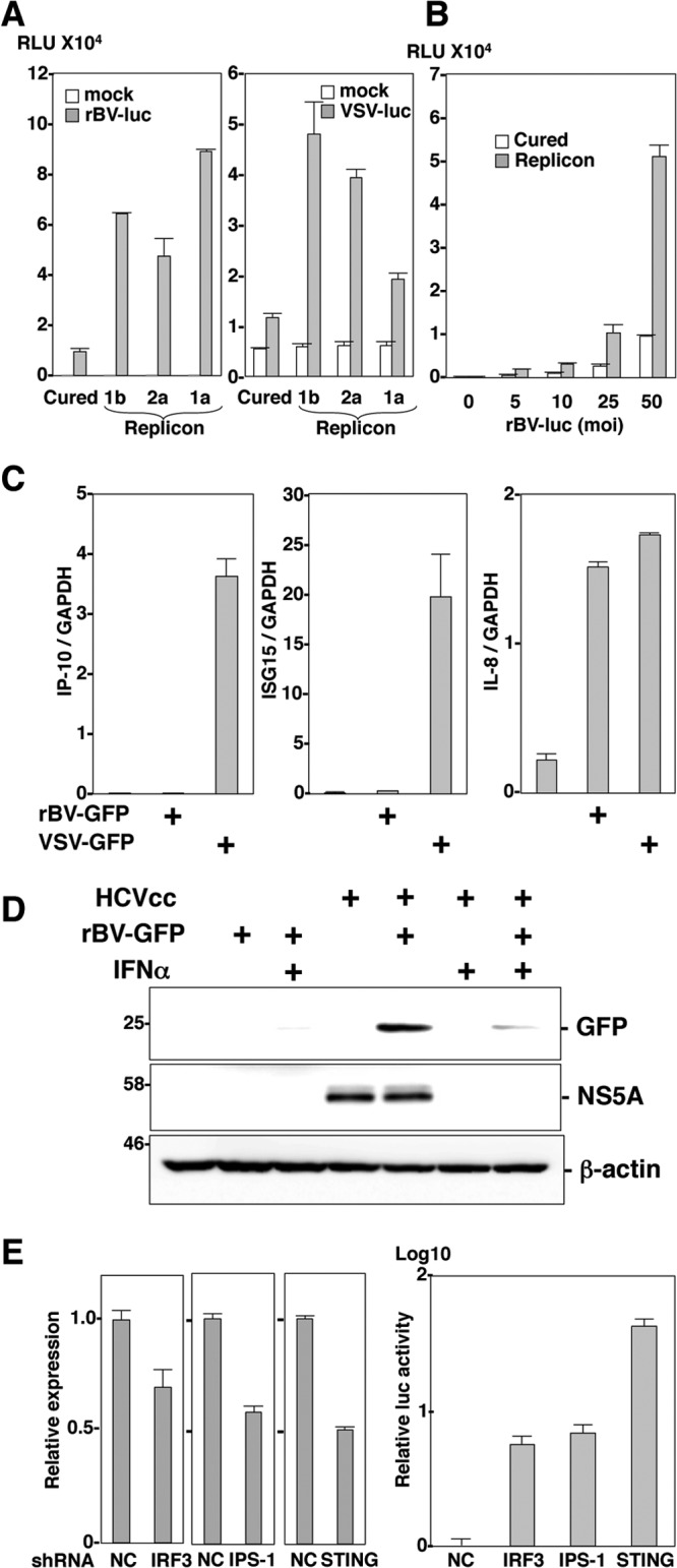
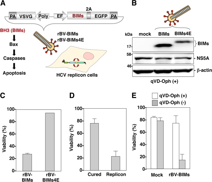
The baculovirus Autographa californica nucleopolyhedrovirus (AcNPV) has been widely used to achieve a high level of foreign gene expression in insect cells, as well as for efficient gene transduction into mammalian cells without any replication. In addition to permitting efficient gene delivery, baculovirus has been shown to induce host innate immune responses in various mammalian cells and in mice. In this study, we examined the effects of the innate immune responses on gene expression by recombinant baculoviruses in cultured cells. The reporter gene expression in IRF3-deficient mouse embryonic fibroblasts (MEFs) infected with the recombinant baculovirus was shown to be enhanced in accordance with the suppression of beta interferon (IFN-β) production. Furthermore, efficient gene transduction by the recombinant baculovirus was achieved in MEFs deficient for stimulator of interferon genes (STING), TANK binding kinase 1 (TBK1), IFN regulatory factor 3 (IRF3), or IFN-β promoter stimulator 1 (IPS-1), but not in those deficient for IRF7, MyD88, or Z-DNA binding protein 1 (ZBP1)/DAI. Enhancement of gene expression by the recombinant baculovirus was also observed in human hepatoma cell lines replicating hepatitis C virus (HCV), in which innate immunity was impaired by the cleavage of IPS-1 by the viral protease. In addition, infection with the recombinant baculovirus expressing the BH3-only protein, BIMS, a potent inducer of apoptosis, resulted in a selective cell death in the HCV replicon cells. These results indicate that innate immune responses induced by infection with baculovirus attenuate transgene expression, and this characteristic might be useful for a selective gene transduction into cells with impaired innate immunity arising from infection with various viruses.


Figures

FIG 1
FIG 1
Efficient gene transduction by recombinant baculovirus in the IRF3−/− MEFs. (A) MEFs derived from wild-type (WT) and IRF3−/−, IRF7−/−, or MyD88−/− mice were inoculated with rBV-GFP (MOI of 100). At 24 h after inoculation, GFP signal was determined by microscopic observation after fixation in 4% paraformaldehyde. (B) MEFs derived from WT and IRF3−/− mice were inoculated with rBV-GFP at an MOI of 100, incubated at 4°C for 30 min, and washed three times with PBS. The total cellular DNA was extracted, and the amounts of baculovirus genome were quantified by real-time PCR. Data represent means ± SD from 2 independent experiments. (C) MEFs derived from WT and IRF3−/− mice were inoculated with 2 doses of rBV-GFP (MOI of 100 or 20). At 24 h after inoculation, total RNA was extracted, and the expression of GFP, IFN-β, and IP-10 mRNAs was determined by real-time PCR. (D) MEFs derived from WT and IRF3-deficient mice were inoculated with 2 doses of rBV-GFP (MOI of 100 and 20). At 24 h after inoculation, cell extracts were subjected to SDS-PAGE and immunoblotted with antibodies against GFP, IRF3, or β-actin, respectively.
FIG 2
FIG 2
IRF3-dependent enhancement of gene transduction by recombinant baculovirus. (A) Cell extracts of MEFs derived from WT or IRF3−/− mice and FLAG-mIRF3-transduced IRF3−/− MEFs were subjected to SDS-PAGE and immunoblotted with antibodies against FLAG, IRF3, or β-actin, respectively. (B) MEFs derived from WT or IRF3−/− mice and FLAG-mIRF3-transduced IRF3-deficient MEFs were inoculated with rBV-luc at an MOI of 100. At 24 h after inoculation, the luciferase activity was determined. Data represent the means ± SD from 3 independent experiments. (C) MEFs were inoculated with rBV-luc at an MOI of 100. At 6 h after inoculation, cells were fixed in 50% methanol–50% acetone for 10 min. IRF3 (green) was stained with the appropriate antibodies, followed by staining with Alexa Fluor 488-conjugated secondary antibodies. Nuclei were stained by DAPI.
FIG 3
FIG 3
Involvement of TBK1 in efficient transgene expression by recombinant baculovirus. (A) MEFs derived from TBK1+/− IKKβ+/− (WT) and TBK1−/− IKKβ+/− (TBK1−/−) mice were inoculated with rBV-GFP (MOI of 100). At 24 h after inoculation, the GFP signal was determined by microscopic observation after fixation in 4% paraformaldehyde. (B) MEFs derived from WT and TBK1−/− mice were inoculated with rBV-GFP at an MOI of 100. At 24 h after inoculation, cell extracts were subjected to SDS-PAGE and immunoblotted with antibodies against GFP, TBK1, or β-actin, respectively. (C) MEFs derived from WT and TBK1-deficient mice were inoculated with rBV-GFP (MOI of 100). At 24 h after inoculation, total RNA was extracted, and the expression of GFP, IFN-β, and IP-10 mRNA was determined by real-time PCR. Data from the real-time PCR were normalized to the amount of GAPDH mRNA.
FIG 4
FIG 4
Efficient gene transduction by recombinant baculovirus vectors through the STING/TBK1/IRF3 axis in MEFs. (A) MEFs derived from wild-type (WT) and STING- or ZBP1-deficient mice were inoculated with rBV-GFP (MOI of 100). At 24 h after inoculation, the GFP signal was determined by microscopic observation after fixation in 4% paraformaldehyde. (B and C) MEFs derived from WT and STING- or ZBP1-deficient mice were inoculated with rBV-luc (MOI of 100) or rBV-GFP (MOI of 100). At 24 h after inoculation, the luciferase activity of cell lysates was determined (B), and the production of IFN-β in the culture supernatant was determined by sandwich ELISA (C). (D) The MEFs were inoculated with wild-type baculovirus (AcNPV) (MOI of 100). At 6 h after inoculation, cells were fixed in 50% methanol–50% acetone for 10 min. STING (green) and ER-calnexin (red) were stained with the appropriate antibodies, followed by staining with Alexa Fluor 488- or Alexa Fluor 555-conjugated second antibodies, respectively. Nuclei were stained by DAPI.
FIG 5
FIG 5
RLR signaling pathways participate in the suppression of gene transduction in MEFs upon infection with recombinant baculovirus. (A) MEFs derived from wild-type (WT) and IPS-1-, TBK1-, ZBP1-, or IRF3-deficient mice were inoculated with rBV-luc at an MOI of 100. At 24 h after inoculation, the luciferase activity was determined. Data represent means ± SD from 3 independent experiments. (B) MEFs derived from WT and IPS-1−/− mice were inoculated with rBV-GFP at an MOI of 100. At 24 h after inoculation, GFP expression was detected by microscopic observation after fixation in 4% paraformaldehyde. (C) MEFs derived from WT or IPS-1-deficient mice were inoculated with rBV-GFP at an MOI of 100. At 24 h after inoculation, total RNA was extracted, and the expression of GFP, IFN-β, and IP-10 mRNAs was determined by real-time PCR. Data from the real-time PCR were normalized to the amount of GAPDH mRNA. (D) Cell extracts of MEFs derived from WT or IPS-1−/− mice and FLAG-mIPS-1-transduced IPS-1−/− MEFs were subjected to SDS-PAGE and immunoblotted with antibodies against FLAG, IPS-1, or β-actin, respectively. (E) MEFs derived from WT or IPS-1−/− mice and FLAG-mIPS-1-transduced IPS-1-deficient MEFs were inoculated with rBV-luc at an MOI of 100. At 24 h after inoculation, the luciferase activity was determined. Data represent means ± SD from 3 independent experiments.
FIG 6
FIG 6
Efficient gene transduction by recombinant baculovirus in HCV replicon-harboring cells. (A) Huh7OK1 cells (cured) and the HCV replicon-harboring cells derived from genotype 1a (RMT strain), 1b (con1 strain), and 2a (JFH1 strain) were inoculated with rBV-luc (MOI of 100) or VSV-luc at an MOI of 5 and (B) with rBV-luc at MOI of 5, 10, 25, and 50. At 24 h after inoculation, the luciferase activity of cell lysates was determined. (C) Huh7 cells were inoculated with rBV-GFP (MOI of 100) or VSV-GFP (NCP mutant) at an MOI of 0.05. At 24 h after inoculation, total RNA was extracted, and the expression of IP-10, ISG15, and IL-8 mRNAs was determined by real-time PCR. Data from the real-time PCR were normalized to the amount of GAPDH mRNA. (D) Huh7 cells infected with HCVcc at an MOI of 1 and incubated for 72 h were inoculated with rBV-GFP (MOI of 100) in the presence or absence of human recombinant IFN-α (rIFN-α) (100 U/ml). At 24 h after inoculation, the cell extracts were subjected to SDS-PAGE and immunoblotted with antibodies against GFP, NS5A, or β-actin, respectively. (E) Relative luciferase activity in Huh7 cells with IRF3, IPS-1, or STING knocked down. Cells were inoculated with rBV-luc at an MOI of 100. At 24 h after inoculation, the luciferase activity of cell lysates was determined (right panel). Luciferase activity is normalized to control shNC cells, and data represent the means ± SD from 2 independent experiments. Total RNA was extracted, and the expression of IRF3, IPS-1, or STING mRNA was determined by real-time PCR (left panel). Data from the real-time PCR were normalized to the amount of GAPDH mRNA.
FIG 7
FIG 7
Induction of apoptosis in cells replicating HCV RNA by a recombinant baculovirus expressing a proapoptotic protein. (A) Structure of recombinant baculoviruses, rBV-BIMS, and rBV-BIMS4E carrying cDNAs of either BIMs or BIMs4E 2A peptides, and enhanced GFP under the control of the EF promoter. The apoptosis pathway was induced by BIMs. (B) HCV replicon cells were infected with either rBV-BIMS or rBV-BIMS4E at an MOI of 500 in the presence of the caspase inhibitor qVD-Oph (20 μM). Cells were subjected to immunoblotting by appropriate antibodies. (C) HCV replicon cells were infected with either rBV-BIMS or rBV-BIMS4E at an MOI of 500, and cell viability was determined by PI exclusion at 24 h postinfection. (D) HCV replicon cells and cured cells were infected with rBV-BIMS at an MOI of 500, and cell viability was determined by PI exclusion at 24 h postinfection. (E) HCV replicon cells and cured cells were infected with rBV-BIMS at an MOI of 500 in the presence or absence of 20 μM qVD-Oph, and cell viability was determined by PI exclusion at 24 h postinfection. Data represent means ± SD from 2 independent experiments.

Similar articles

Cited by

References

    1. Luckow VA, Summers MD. 1988. Signals important for high-level expression of foreign genes in Autographa californica nuclear polyhedrosis virus expression vectors. Virology 167:56–71. 10.1016/0042-6822(88)90054-2 - DOI - PubMed
    1. Matsuura Y, Possee RD, Overton HA, Bishop DH. 1987. Baculovirus expression vectors: the requirements for high level expression of proteins, including glycoproteins. J. Gen. Virol. 68:1233–1250. 10.1099/0022-1317-68-5-1233 - DOI - PubMed
    1. Tani H, Nishijima M, Ushijima H, Miyamura T, Matsuura Y. 2001. Characterization of cell-surface determinants important for baculovirus infection. Virology 279:343–353. 10.1006/viro.2000.0699 - DOI - PubMed
    1. Tani H, Limn CK, Yap CC, Onishi M, Nozaki M, Nishimune Y, Okahashi N, Kitagawa Y, Watanabe R, Mochizuki R, Moriishi K, Matsuura Y. 2003. In vitro and in vivo gene delivery by recombinant baculoviruses. J. Virol. 77:9799–9808. 10.1128/JVI.77.18.9799-9808.2003 - DOI - PMC - PubMed
    1. Hofmann C, Sandig V, Jennings G, Rudolph M, Schlag P, Strauss M. 1995. Efficient gene transfer into human hepatocytes by baculovirus vectors. Proc. Natl. Acad. Sci. U. S. A. 92:10099–10103. 10.1073/pnas.92.22.10099 - DOI - PMC - PubMed

Publication types