

The cytokine and chemokine expression profile of nucleus pulposus cells: implications for degeneration and regeneration of the intervertebral disc
- PMID: 24325988
- PMCID: PMC3979161
- DOI: 10.1186/ar4408
The cytokine and chemokine expression profile of nucleus pulposus cells: implications for degeneration and regeneration of the intervertebral disc
Abstract
Introduction: The aims of these studies were to identify the cytokine and chemokine expression profile of nucleus pulposus (NP) cells and to determine the relationships between NP cell cytokine and chemokine production and the characteristic tissue changes seen during intervertebral disc (IVD) degeneration.
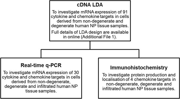
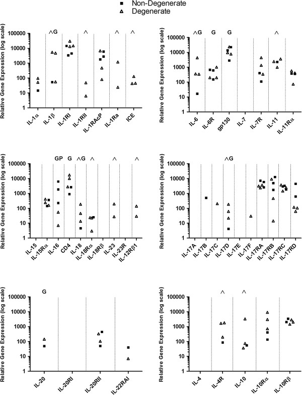
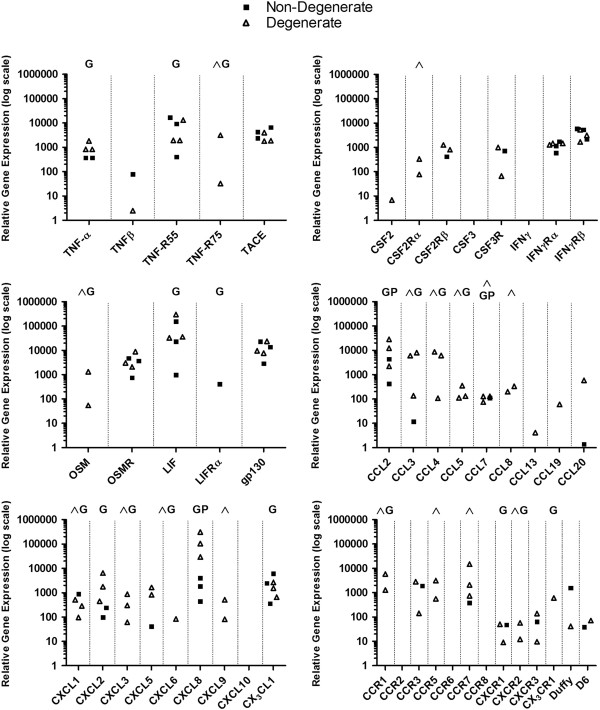
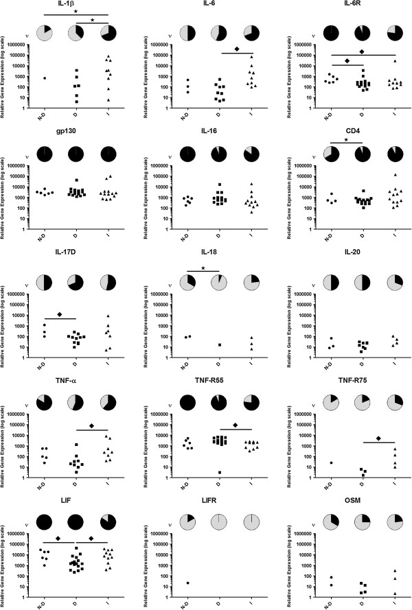
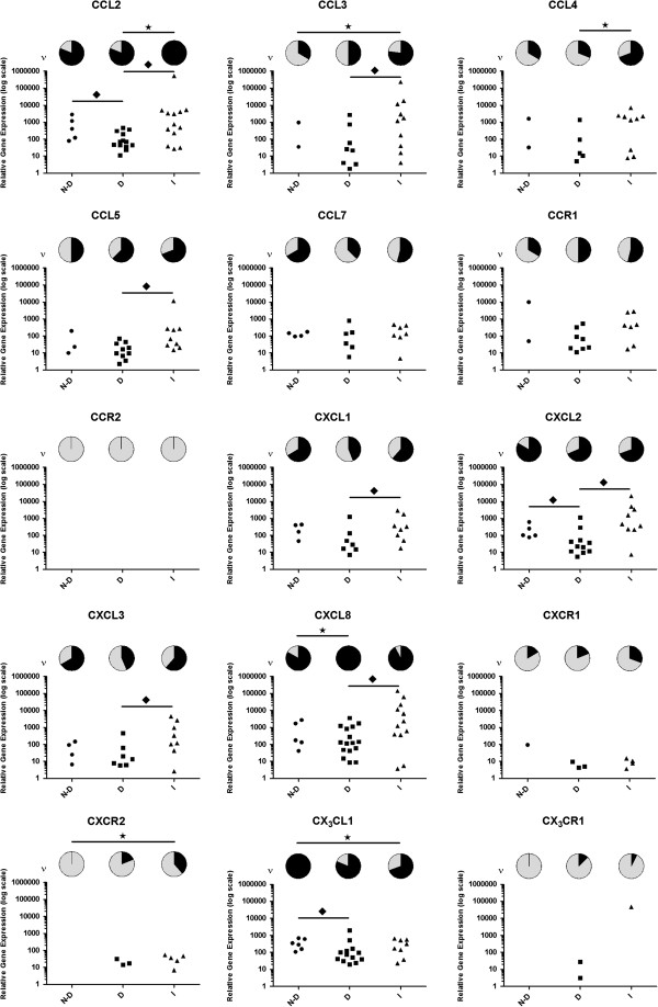
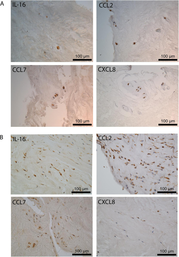
Methods: Real-time q-PCR cDNA Low Density Array (LDA) was used to investigate the expression of 91 cytokine and chemokine associated genes in NP cells from degenerate human IVDs. Further real-time q-PCR was used to investigate 30 selected cytokine and chemokine associated genes in NP cells from non-degenerate and degenerate IVDs and those from IVDs with immune cell infiltrates (‘infiltrated’). Immunohistochemistry (IHC) was performed for four selected cytokines and chemokines to confirm and localize protein expression in human NP tissue samples.
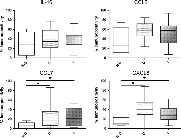
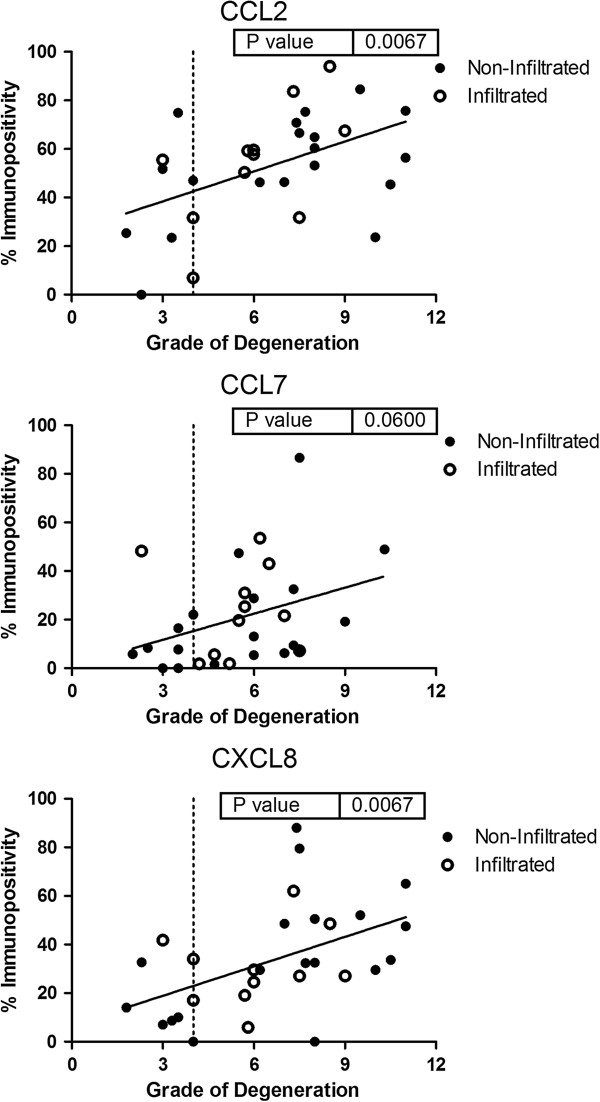
Results: LDA identified the expression of numerous cytokine and chemokine associated genes including 15 novel cytokines and chemokines. Further q-PCR gene expression studies identified differential expression patterns in NP cells derived from non-degenerate, degenerate and infiltrated IVDs. IHC confirmed NP cells as a source of IL-16, CCL2, CCL7 and CXCL8 and that protein expression of CCL2, CCL7 and CXCL8 increases concordant with histological degenerative tissue changes.
Conclusions: Our data indicates that NP cells are a source of cytokines and chemokines within the IVD and that these expression patterns are altered in IVD pathology. These findings may be important for the correct assessment of the ‘degenerate niche’ prior to autologous or allogeneic cell transplantation for biological therapy of the degenerate IVD.
Figures
Similar articles
-
Potential roles of cytokines and chemokines in human intervertebral disc degeneration: interleukin-1 is a master regulator of catabolic processes.Osteoarthritis Cartilage. 2015 Jul;23(7):1165-77. doi: 10.1016/j.joca.2015.02.017. Epub 2015 Mar 4. Osteoarthritis Cartilage. 2015. PMID: 25748081
-
Expression and regulation of neurotrophic and angiogenic factors during human intervertebral disc degeneration.Arthritis Res Ther. 2014 Aug 20;16(5):416. doi: 10.1186/s13075-014-0416-1. Arthritis Res Ther. 2014. PMID: 25209447 Free PMC article.
-
Transcriptional profiling of bovine intervertebral disc cells: implications for identification of normal and degenerate human intervertebral disc cell phenotypes.Arthritis Res Ther. 2010;12(1):R22. doi: 10.1186/ar2929. Epub 2010 Feb 11. Arthritis Res Ther. 2010. PMID: 20149220 Free PMC article.
-
Cell Clusters Are Indicative of Stem Cell Activity in the Degenerate Intervertebral Disc: Can Their Properties Be Manipulated to Improve Intrinsic Repair of the Disc?Stem Cells Dev. 2018 Feb 1;27(3):147-165. doi: 10.1089/scd.2017.0213. Epub 2018 Jan 16. Stem Cells Dev. 2018. PMID: 29241405 Review.
-
Immune cascades in human intervertebral disc: the pros and cons.Int J Clin Exp Pathol. 2013 May 15;6(6):1009-14. Print 2013. Int J Clin Exp Pathol. 2013. PMID: 23696917 Free PMC article. Review.
Cited by
-
SSR1 and CKAP4 as potential biomarkers for intervertebral disc degeneration based on integrated bioinformatics analysis.JOR Spine. 2023 Dec 20;7(1):e1309. doi: 10.1002/jsp2.1309. eCollection 2024 Mar. JOR Spine. 2023. PMID: 38222802 Free PMC article.
-
Identification of Core Genes and Screening of Potential Targets in Intervertebral Disc Degeneration Using Integrated Bioinformatics Analysis.Front Genet. 2022 May 30;13:864100. doi: 10.3389/fgene.2022.864100. eCollection 2022. Front Genet. 2022. PMID: 35711934 Free PMC article.
-
Mesenchymal stem cells: potential application in intervertebral disc regeneration.Transl Pediatr. 2014 Apr;3(2):71-90. doi: 10.3978/j.issn.2224-4336.2014.03.05. Transl Pediatr. 2014. PMID: 26835326 Free PMC article. Review.
-
Biomaterials for intervertebral disc regeneration and repair.Biomaterials. 2017 Jun;129:54-67. doi: 10.1016/j.biomaterials.2017.03.013. Epub 2017 Mar 15. Biomaterials. 2017. PMID: 28324865 Free PMC article. Review.
-
Inflammation in intervertebral disc degeneration and regeneration.J R Soc Interface. 2015 Mar 6;12(104):20141191. doi: 10.1098/rsif.2014.1191. J R Soc Interface. 2015. PMID: 25673296 Free PMC article. Review.
References
-
- Antoniou J, Steffen T, Nelson F, Winterbottom N, Hollander A, Poole R, Aebi M, Alini M. The human lumbar intervertebral disc - evidence for changes in the biosynthesis and denaturation of the extracellular matrix with growth, maturation, ageing, and degeneration. J Clin Invest. 1996;15:996–1003. doi: 10.1172/JCI118884. - DOI - PMC - PubMed
Publication types
MeSH terms
Substances
LinkOut - more resources
Full Text Sources
Other Literature Sources
Research Materials
Miscellaneous
