. 2014 Jan;16(1):38-46.
doi: 10.1038/ncb2885. Epub 2013 Dec 8.

The bacterial cell division proteins FtsA and FtsZ self-organize into dynamic cytoskeletal patterns

Affiliations

The bacterial cell division proteins FtsA and FtsZ self-organize into dynamic cytoskeletal patterns

Martin Loose et al. Nat Cell Biol. 2014 Jan.

Abstract

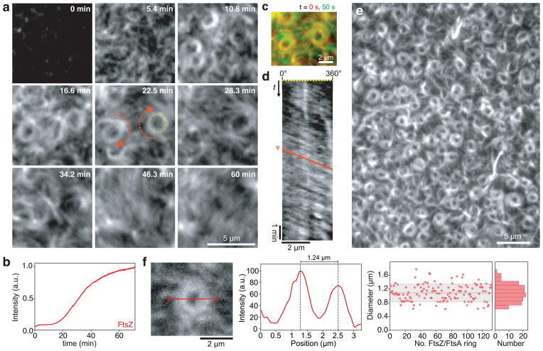
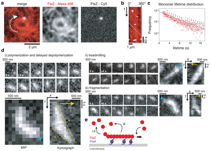
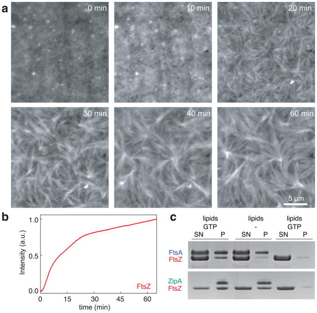
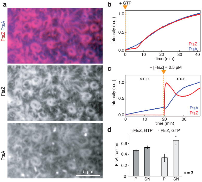
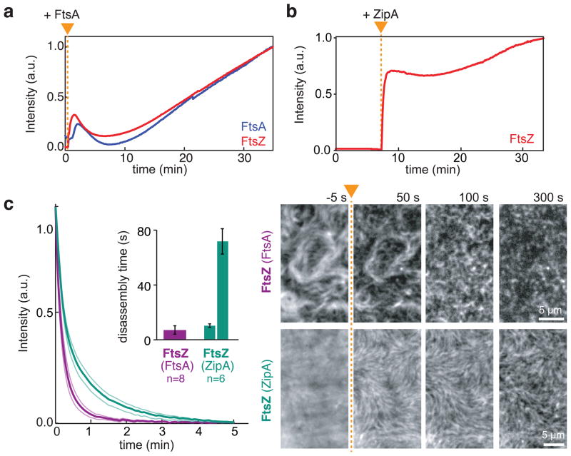
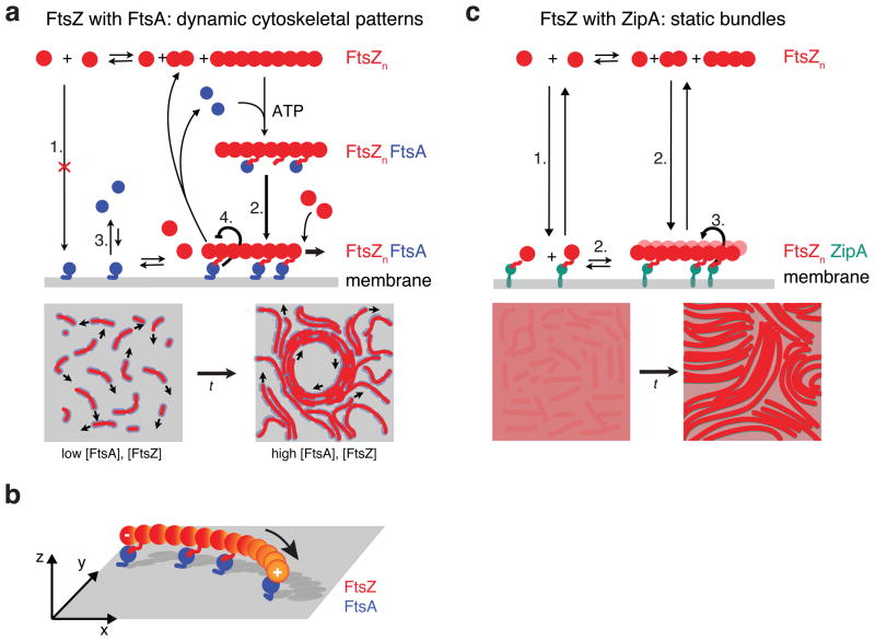
Bacterial cytokinesis is commonly initiated by the Z-ring, a cytoskeletal structure that assembles at the site of division. Its primary component is FtsZ, a tubulin superfamily GTPase, which is recruited to the membrane by the actin-related protein FtsA. Both proteins are required for the formation of the Z-ring, but if and how they influence each other's assembly dynamics is not known. Here, we reconstituted FtsA-dependent recruitment of FtsZ polymers to supported membranes, where both proteins self-organize into complex patterns, such as fast-moving filament bundles and chirally rotating rings. Using fluorescence microscopy and biochemical perturbations, we found that these large-scale rearrangements of FtsZ emerge from its polymerization dynamics and a dual, antagonistic role of FtsA: recruitment of FtsZ filaments to the membrane and negative regulation of FtsZ organization. Our findings provide a model for the initial steps of bacterial cell division and illustrate how dynamic polymers can self-organize into large-scale structures.


Figures

Figure 1
Figure 1. FtsZ and FtsA self-organize into a rapidly reorganizing filament network
(a) Snapshots showing typical cytoskeletal patterns of FtsZ emerging from its interaction with FtsA on a supported membrane. Similar results were obtained in more than 100 experiments. After adding GTP (to 3 mM at t = 0 min), short filaments of FtsZ start attaching to the membrane. While their density increases, they self-organize into a rapidly reorganizing filament network, forming traveling streams and rotating swirls (orange arrows) (FtsZ, 1.5 μM with 10 % FtsZ-Alexa488; FtsA, 0.5 μM). (b) Representative time-intensity curve corresponding to the amount of FtsZ polymerizing on the membrane after adding GTP (c) Overlay of two individual frames from Supplementary Video 2 separated by 50s. While FtsZ bundles outside of the ring are constantly rearranging, the bundles inside of the vortex are more persistent. (d) Representative kymograph along the circumference of a vortex shown in (a), see yellow dashed arrow at 22.5 min. The slope of the orange line corresponds to the velocity (x/t) of the vortex. Kymographs were obtained and analyzed for 40 different vortices. (e) Array of dynamic rings of FtsZ. (f) Ring diameters were determined by measuring the peak-to-peak distance in the intensity profile (red line shown in micrograph). Right, scatterplot of ring diameters (red dots), average value (solid red line, n = 132), standard deviation (dashed line and grey background) and corresponding histogram. Source data is given in Supplementary Table 1.
Figure 2
Figure 2. Reorganization of the FtsZ filament network emerges from FtsZ polymerization dynamics and not from filament sliding
(a) Typical micrograph of a single FtsZ molecule (white, FtsZ-Cy5) in an FtsZ vortex (red, FtsZ-Alexa488) (FtsZ, 1.25 μM with 30 % FtsZ-Alexa488 and 0.4 % FtsZ-Cy5; FtsA, 0.4 μM). (b) Kymograph along the circumference of the vortex shown in (a). The slope of the white dashed line shows corresponds to the velocity of the vortex. While the FtsZ ring is rotating, individual FtsZ proteins appear for one to three frames (3 to 9 sec) without moving (Supplementary Videos 6–8). Similar results were obtained in more than 20 experiments. (c) Distribution of individual FtsZ lifetimes (red circles) in a linear-log plot and linear fit (black line). See text for details. (d) FtsZ filaments can show different kinds of polymerization dynamics when recruited to the membrane by FtsA. Representative snapshots, maximum intensity projections (MIP) and kymographs of (i) filament polymerization and delayed depolymerization, (ii) treadmilling and (iii) fragmentation. Cyan arrowhead in MIPs indicate start of FtsZ polymerization on the membrane, yellow arrows indicate the direction of polymerization. The slopes of the yellow dashed lines correspond to the polymerization (1.) and depolymerization rate (2.) of the FtsZ filament. All scale bars correspond to 500 nm or 5 s. (38 filaments from 5 independent experiments were analyzed, see also Supplementary Fig. 3). (FtsZ, 0.4 μM with 10 % FtsZ-Alexa488; FtsA, 0.2 μM) (e) Illustration of a FtsZ filament recruited to the membrane by FtsA. After binding and unipolar polymerization, the filament detaches either after polymerization from the opposite end (i,ii) or after fragmentation (iii).
Figure 3
Figure 3. FtsZ organizes into static bundles of dynamic filaments with ZipA as membrane anchor
(a) Representative snapshots showing formation of FtsZ bundles with ZipA as membrane anchor. Similar results were obtained in more than 70 experiments. First, FtsZ intensity increases homogeneously, followed by condensation of FtsZ into long thick filament bundles (FtsZ, 1.5 μM with 30% FtsZ- Alexa488; His-Δ22-ZipA, 0.5 μM; supported membrane contained 2% Ni-chelating lipids). Different concentrations of Ni-chelating lipids (1% to 8%) gave similar results (Supplementary Figure 4). (b) Representative time-intensity curve corresponding to the amount of FtsZ bound to the membrane. (c) SDS-PAGE gel from co-pelleting assay with either FtsA and ATP (top row), ZipA (bottom row) or without membrane anchor (far right lanes). In contrast to ZipA, FtsA only recruits FtsZ to the membrane in the presence of GTP (left lanes versus middle lanes); S: supernatant, P: pellet. Result shown represents eight independent experiments.
Figure 4
Figure 4. FtsZ and FtsA co-assemble on the membrane
(a) Typical micrograph of reorganizing filament patterns with fluorescently labeled FtsZ and FtsA (FtsZ, 1.1 μM with 30% FtsZ-Alexa488; FtsA, 0.4 μM with 10 % Cy5-GG-FtsA). (b and c) Representative intensity traces corresponding to the amount of FtsZ (red) and FtsA (blue). (b) After adding GTP (orange arrowhead), FtsZ and FtsA assemble simultaneously on the membrane. Similar micrographs and intensity curves were obtained in more than 50 experiments. (c) Binding of FtsA is slow at a low concentration of FtsZ (< c.c. = critical concentration of FtsZ allowing for filament network formation), but facilitated at higher FtsZ concentration (> c.c.). Orange triangle indicates time of FtsZ addition. Similar intensity traces were obtained in five independent experiments. (d) Bar plot representing the amount of FtsA co-pelleting with vesicles (P) or remaining in the supernatant (SN) after 30 min of incubation (see also Supplementary Fig. 7b). The presence of FtsZ filaments (+FtsZ, GTP), increases the amount of FtsA bound to the membrane (p=0.0391). Error bars represent s.d. from n=3 independent experiments. Source data is given in Supplementary Table 1.
Figure 5
Figure 5. FtsA destabilizes the FtsZ filaments network
(a and b) Representative intensity traces corresponding to the amount of FtsZ (red) and FtsA (blue) co-assembling on the membrane. Prepolymerized FtsZ filaments are disassembled after addition of FtsA (a), but remain stable after addition of ZipA (b) (Supplementary Video 16) (FtsZ, 1.2 μM; FtsA or His-Δ22-ZipA, 0.3 μM). Intensity traces correspond to four independent experiments respectively. Orange triangle indicates the time of addition of either protein. (c) Left: Mean intensity traces for FtsZ depolymerization upon rapid dilution (FtsZ and FtsA (purple); FtsZ and ZipA (turquoise). Inset: mean disassembly times obtained from single-exponential (FtsZ with FtsA) and double-exponential fits (FtsZ with ZipA). Thin lines and error bars illustrate s.e.m (n=8 (FtsZ with FtsA) and n=6 (FtsZ with ZipA)). Right: Representative snapshots showing dilution-induced FtsZ disassembly. After adding buffer (at time point indicated by orange arrowhead and dashed line), FtsZ filaments rapidly disassembled when recruited by FtsA. In contrast, with ZipA FtsZ bundles can persist for more than 5 min. Intensities of micrographs have been normalized to have constant overall intensity. Raw data is shown in Supplementary Video 17.
Figure 6
Figure 6. Model for membrane-based FtsZ polymerization
(a) Model for FtsZ-FtsA co-assembly on the membrane starting from top left: FtsA does not recruit monomeric FtsZ to the membrane (1.). After FtsZ polymerization, an FtsA-FtsZ filament complex forms and attaches to the membrane, which facilitates binding of FtsA to the membrane (2. versus 3.). Here, FtsZ filaments can further polymerize or disassemble into short filaments or monomers, which are not longer recruited to the membrane by FtsA leading to their detachment. The dual, antagonistic role of FtsA is highlighted by two thick arrows: FtsA co-assembles with FtsZ (2.), but allows for the rapid disassembly of FtsZ filaments (4.). Bottom: With increasing density, lateral interactions between short, dynamic filaments give rise to a rapidly reorganizing filament network, i.e. streams and vortices. (b) Illustration of a polar, curved FtsZ filament recruited to the membrane by FtsA. Anchoring a polar filament with a curved rigid conformation to a surface creates chiral asymmetries of the system, i.e. the membrane-bound filament is not identical to its mirror image. (c) Model for the formation of static bundles of dynamic FtsZ filaments mediated by ZipA. In contrast to FtsA, ZipA can recruit polymerized and non-polymerized FtsZ to the membrane (1.). Membrane-recruited FtsZ initiates FtsZ polymerization (2). Lateral interactions allow FtsZ filaments to organize into thick bundles, which is further enhanced by ZipA (3.). Short filaments and monomeric FtsZ can remain on the membrane after depolymerization of fragmentation. Illustration of GTP hydrolysis by FtsZ has been omitted for clarity.

Comment in

Similar articles

Cited by

References

    1. Cabeen MT, Jacobs-Wagner C. The bacterial cytoskeleton. Annu Rev Genet. 2010;44:365–392. - PubMed
    1. Ingerson-Mahar M, Gitai Z. A growing family: the expanding universe of the bacterial cytoskeleton. FEMS Microbiol Rev. 2012;36:256–266. - PMC - PubMed
    1. Margolin W. FtsZ and the division of prokaryotic cells and organelles. Nat Rev Mol Cell Biol. 2005;6:862–871. - PMC - PubMed
    1. Adams DW, Errington J. Bacterial cell division: assembly, maintenance and disassembly of the Z ring. Nat Rev Micro. 2009;7:642–653. - PubMed
    1. Erickson HP, Taylor DW, Taylor KA, Bramhill D. Bacterial cell division protein FtsZ assembles into protofilament sheets and minirings, structural homologs of tubulin polymers. Proceedings of the National Academy of Sciences. 1996;93:519–523. - PMC - PubMed

Publication types

MeSH terms