Comparative Study
. 2013 Oct 31:2013:790343.
doi: 10.1155/2013/790343. eCollection 2013.

Deliberations on the external morphology and modification of the labial segments in the Nepomorpha (Heteroptera: Insecta) with notes on the phylogenetic characteristics

Affiliations
Comparative Study

Deliberations on the external morphology and modification of the labial segments in the Nepomorpha (Heteroptera: Insecta) with notes on the phylogenetic characteristics

Jolanta Brożek. ScientificWorldJournal. .

Abstract

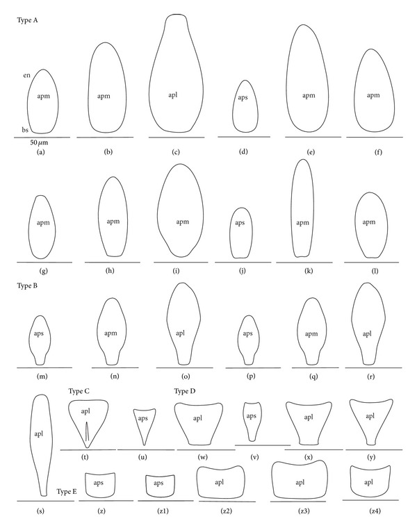
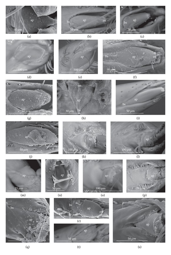
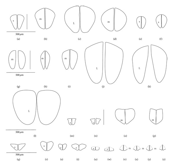
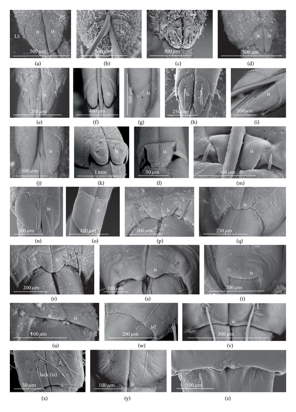
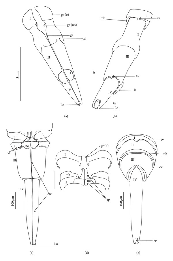
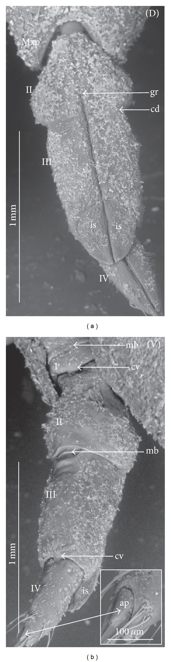
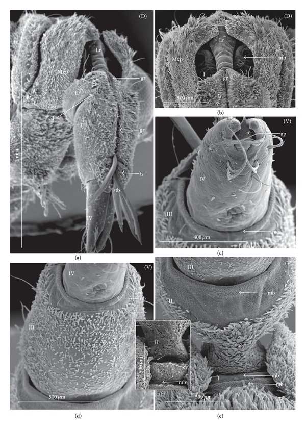
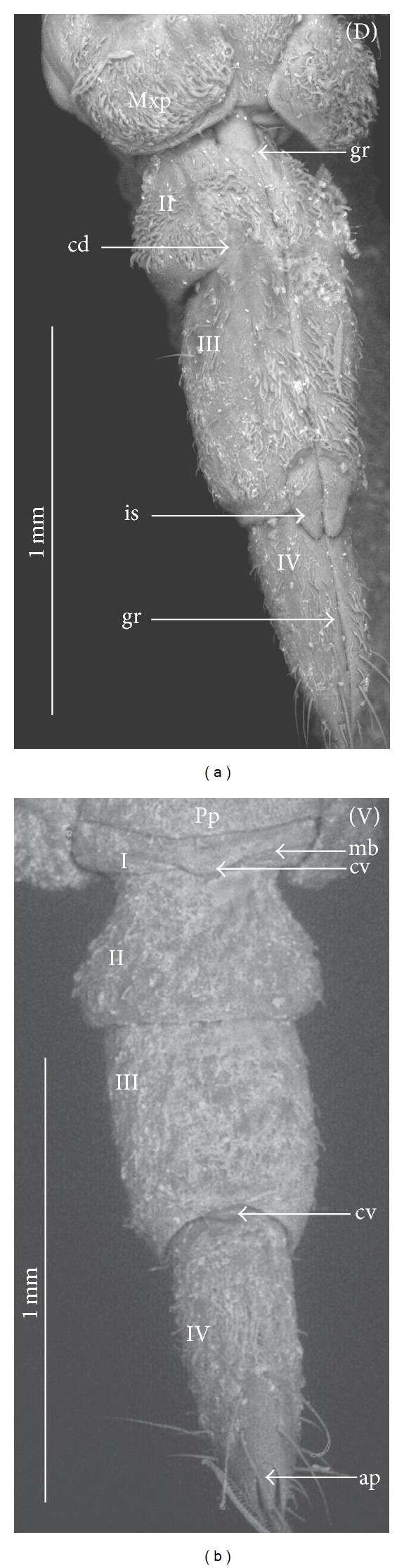
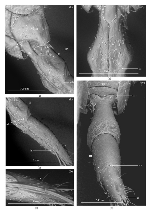
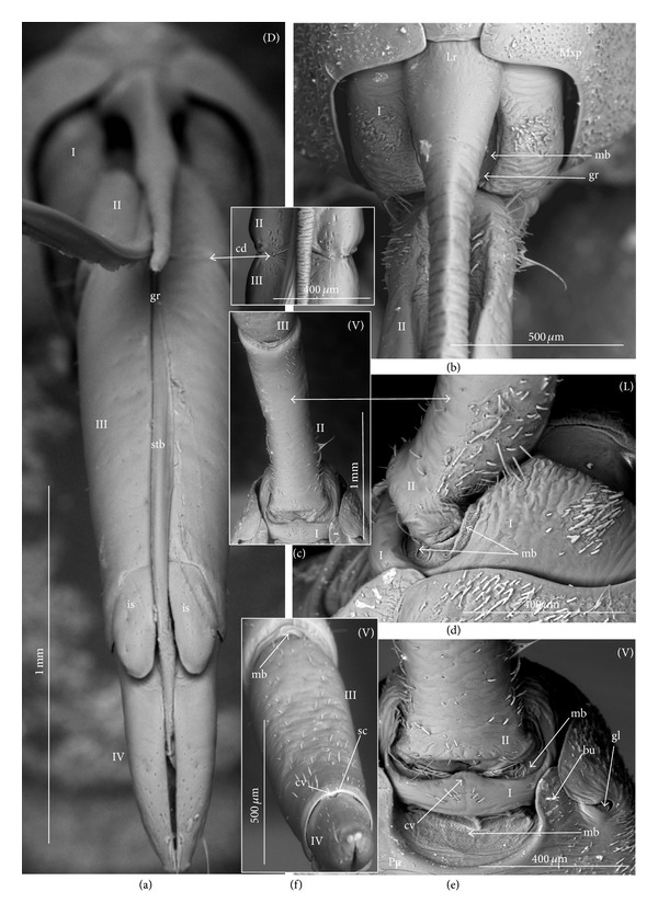
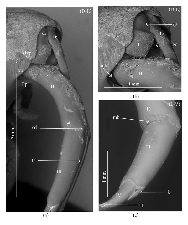
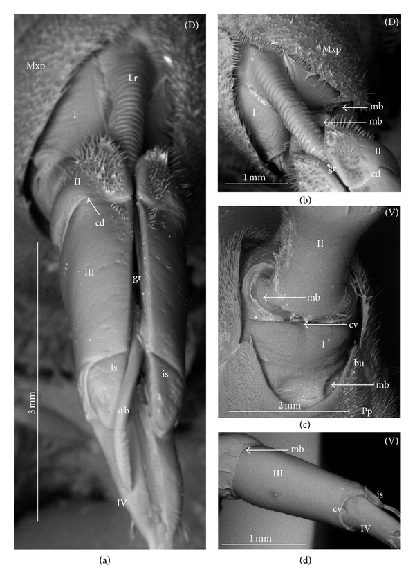
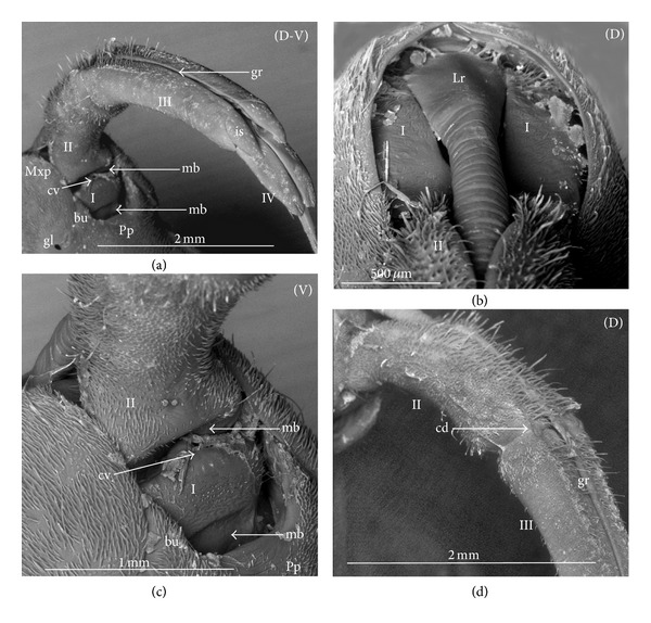
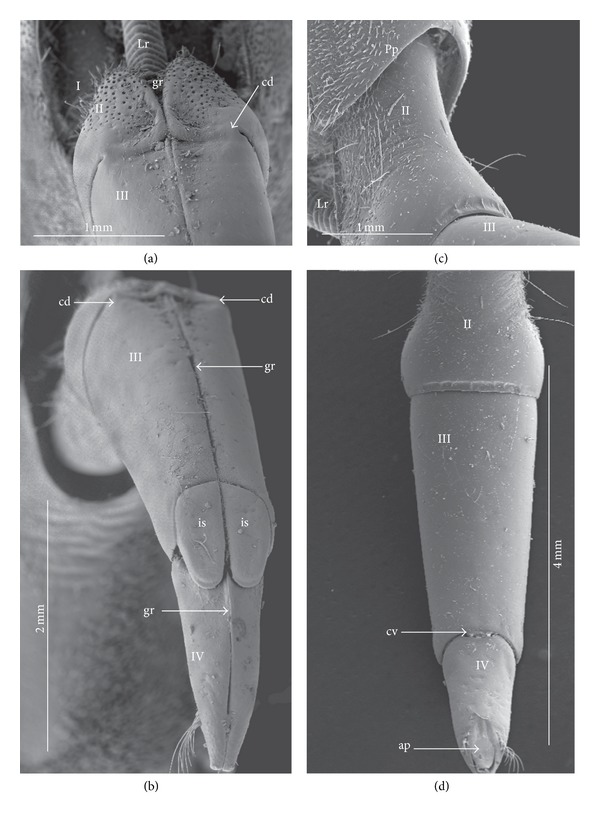
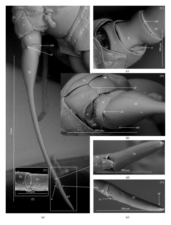
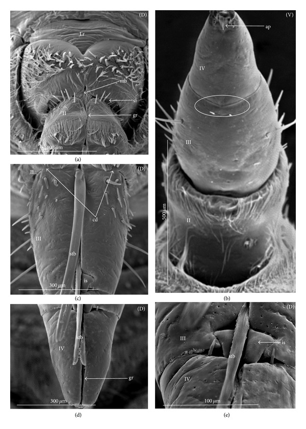
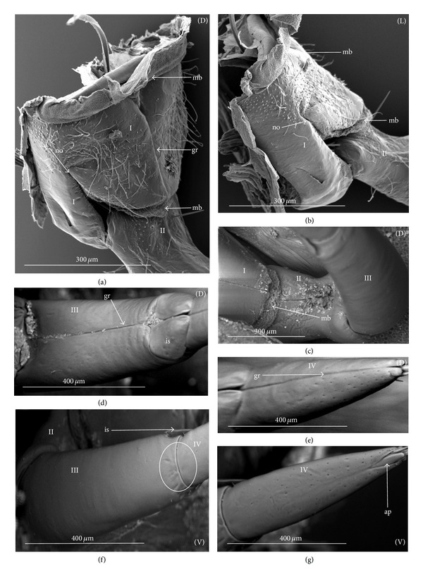
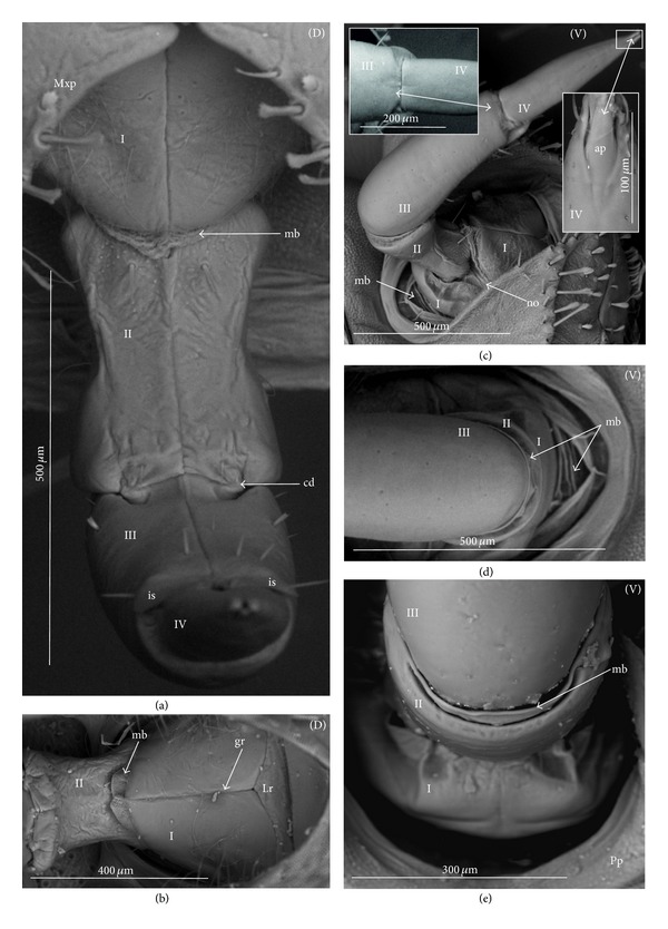
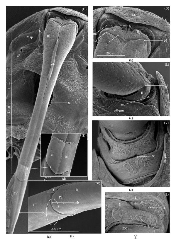
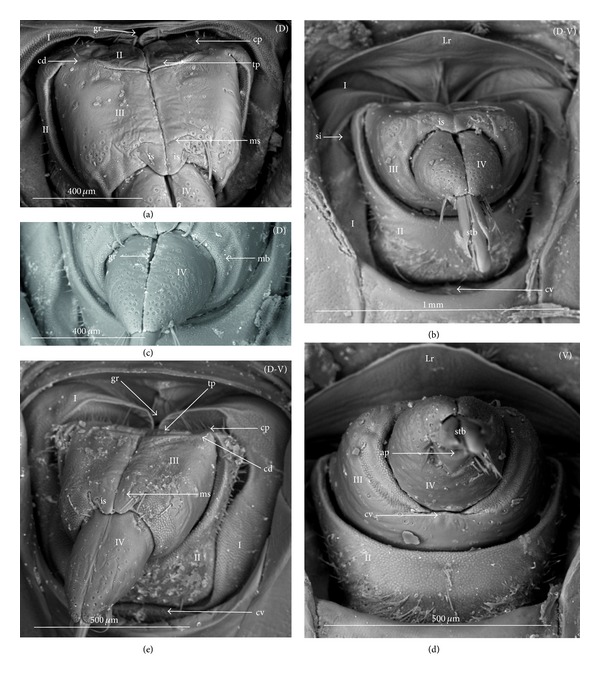
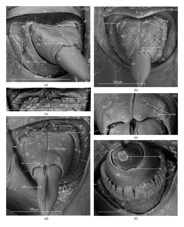
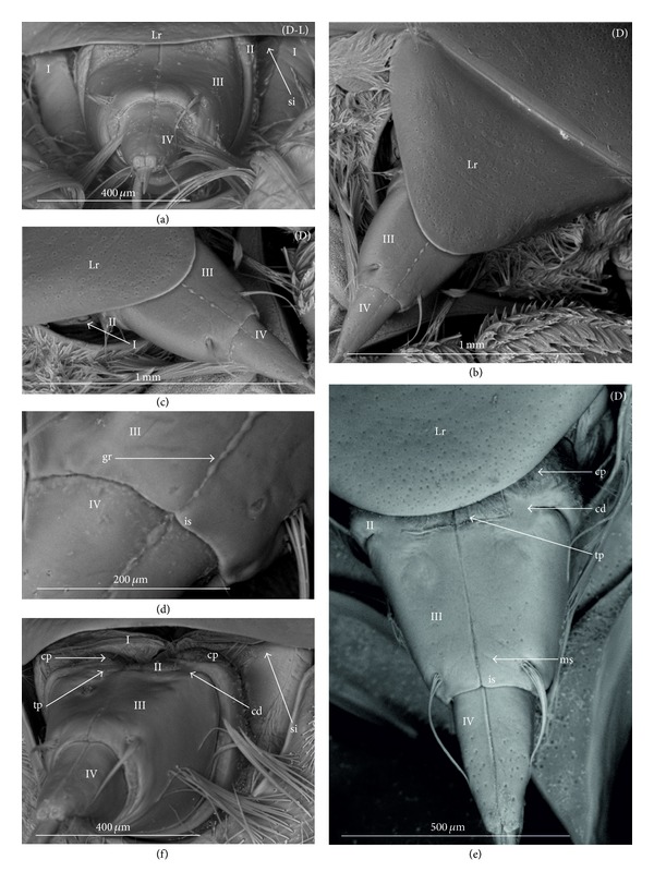
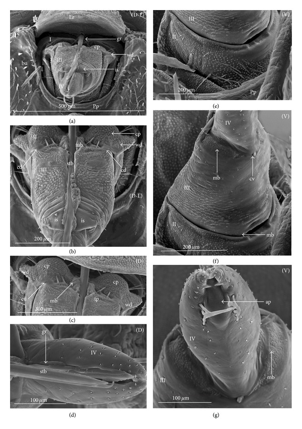
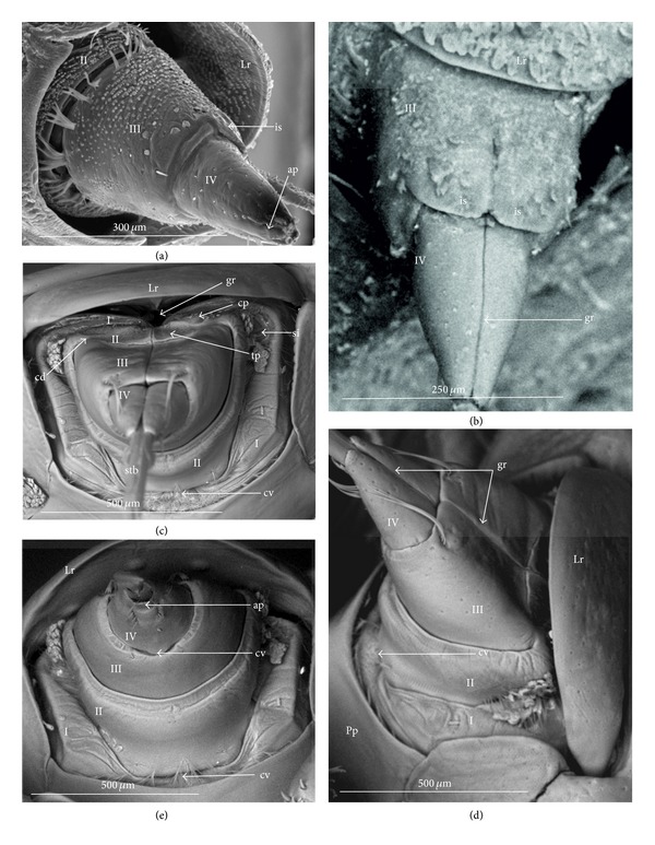
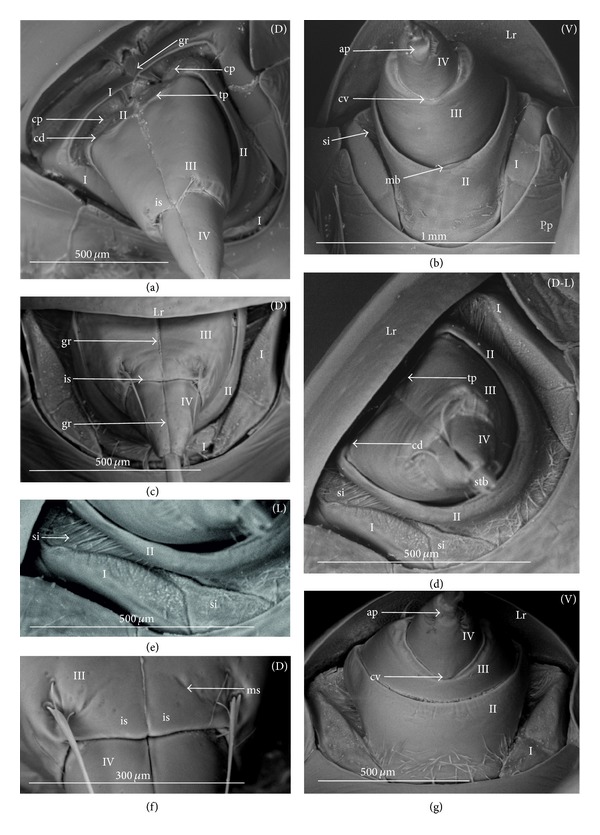
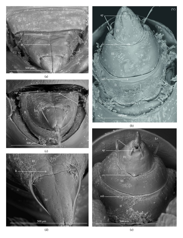
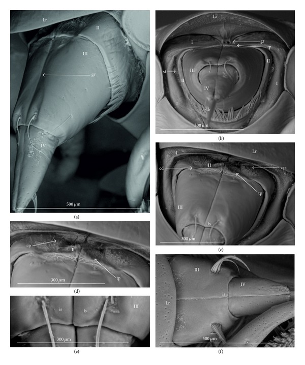
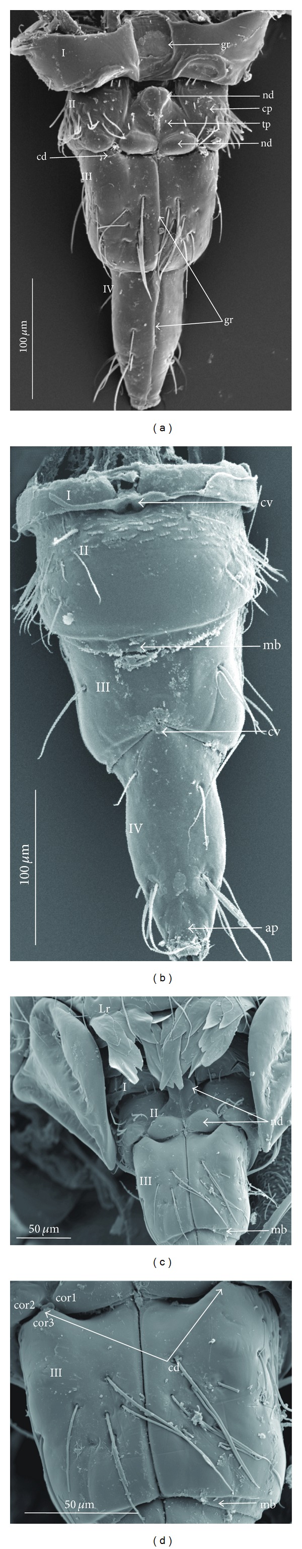
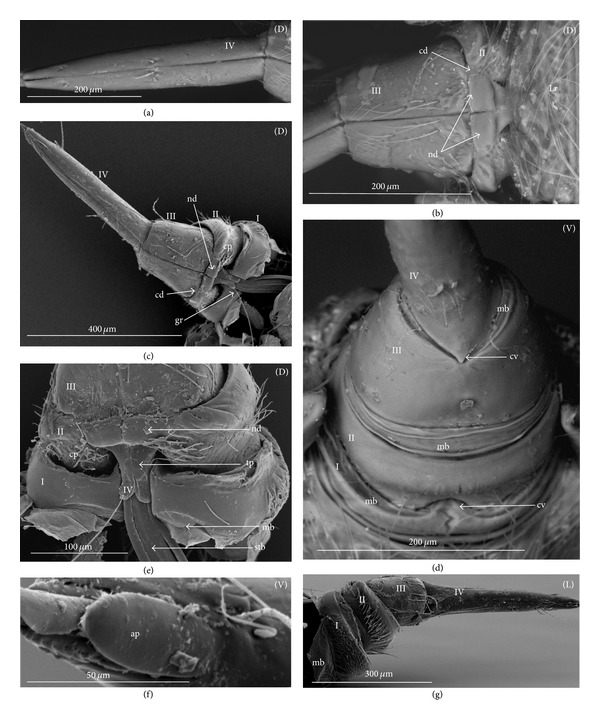
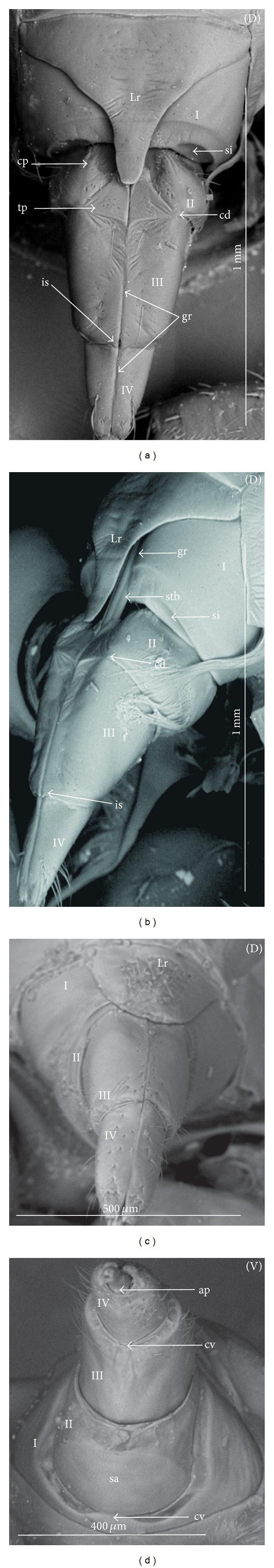
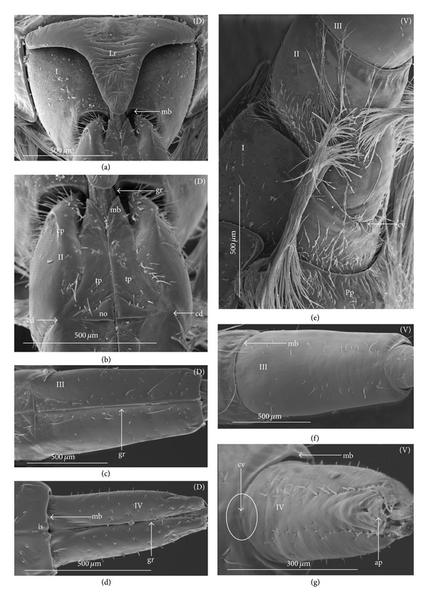
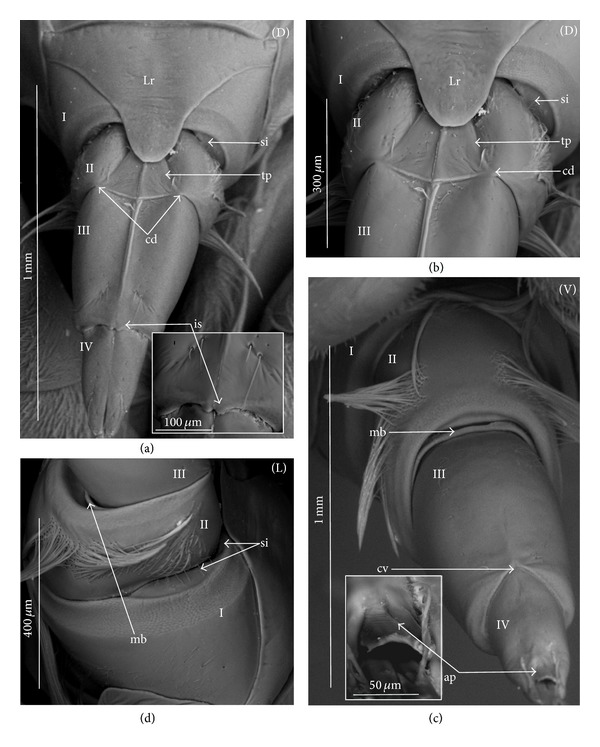
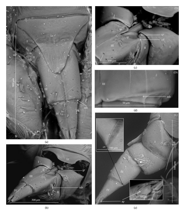
The present study provides new data concerning the external morphology of the labial segments of 46 species from nine Nepomorpha families using the scanning electron microscope. The labial segments are described in detail and images of their structures are presented for the systematic groups. Subsequent segments of the labium (I, II, III, and IV) are shaped similarly in all investigated taxa but carry individual characters in some (sub-)families. Five morphologically distinct forms of the apical plate and five intercalary sclerites have been identified. Additionally, three types of the articulation on the dorsal side between the third and second segments are interpreted as the new characters. The presence of the midventral condyle on the distal edge of the first segment and the third segment has been reanalyzed. New position of the midventral condyle on the proximal edge of the fourth labial segment has been distinguished in several groups. The new set of characters has been estimated from the plesiomorphic taxa of the Nepoidea (Nepidae and Belostomatidae) and subsequently through the more advanced taxa in the relation to the outgroup (Gerromorpha). The evaluation of these characters has revealed twenty-seven new apomorphies for the labium in the Nepomorpha.


Figures

Figure 1
Figure 1
Various shapes of the apical plate in the Nepomorpha: Nepinae: (a) Curicta granulosa, (b) Borborophyes mayri, (c) Laccotrephes japonensis, (d) Nepa cinerea, Ranatrinae: (e) Cercotmetus asiaticus, (f) Ranatra chinensis, Ochteridae; (g) Ochterus marginatus, Aphelocheiridae: (h) Aphelocheirus aestivalis, Naucoridae: Limnocorinae: (i) Limnocoris lutzi, Helotrephidae: (j) Tiphotrephes indicus, (k) Hydrotrephes visayasensis, (l) Hydrotrephes balnearius, Belostomatidae: Belostomatinae: (m) Belostoma flumineum, (n) Deinostoma dilatatum, (o) Appasus major, (p) Hydrocyrius colombiae, (q) Limnogeton fieberi, Lethocerinae: (r) Lethocerus deyrollei, Gelastocoridae: Nerthrinae: (s) Nerthra nepaeformis, Gelastocorinae: (t) Gelastocoris oculatus, Pleidae: (u) Paraplea frontalis, Notonectidae: Anisopinae: (w) Anisops camaroonensis, Notonectinae: (v) Nychia sappho, (x) Enithares bergrothi, (y) Notonecta glauca. Naucoridae: Cheirochelinae: (z) Cheirochela feana, (z1). Coptocatus oblongulus, Cryphocricinae: (z2) Cryphocricos hungerfordi. Laccocorinae: (z3) Laccocoris hoogstraali, Naucorinae: (z4) Naucoris maculatus. The same scale bar has been applied for all images (50 μm), bs: base of the apical plate, en: tip of the apical plate.
Figure 2
Figure 2
Various shapes of the apical plate in the Nepomorpha: Nepinae: (a) Laccotrephes japonensis, Ranatrinae: (b) Cercotmetus asiaticus, (c) Ranatra chinensis, Belostomatidae: Belostomatinae: (d) Belostoma flumineum, (e) Hydrocyrius colombiae, Lethocerinae: (f) Lethocerus deyrollei, Ochteridae: (g) Ochterus marginatus, Gelastocoridae: Gelastocorinae: (h) Gelastocoris oculatus, Nerthrinae: (i) Nerthra nepaeformis, Aphelocheiridae: (j) Aphelocheirus aestivalis, Naucoridae: Cheirochelinae: (k) Cheirochela feana, Cryphocricinae: (l) Cryphocricos hungerfordi. Laccocorinae: (m) Laccocoris hoogstraali, Limnocorinae: (n) Limnocoris lutzi, Naucorinae: (o) Pelocoris femoratus, (p) Naucoris maculatus, Pleidae: (q) Paraplea frontalis, Helotrephidae: (r) Hydrotrephes visayasensis, Notonectidae: Notonectinae: (s) Notonecta glauca, (t) Nychia sappho.
Figure 3
Figure 3
Various shapes of intercalary sclerites in the Nepomorpha: Nepinae: (a) Curicta granulosa, (b) Borborophyes mayri, (c) Laccotrephes japonensis, (d) Nepa cinerea, Ranatrinae: (e) Cercotmetus asiaticus, (f) Ranatra chinensis. Belostomatidae: Belostomatinae: (g) Belostoma flumineum, (h) Deinostoma dilatatum, (i) Appasus major, (j) Hydrocyrius colombiae, (k) Limnogeton fieberi, Lethocerinae: (l) Lethocerus deyrollei, Ochteridae: (m) Ochterus marginatus, Gelastocoridae: Gelastocorinae: (n) Gelastocoris oculatus, Nerthrinae: (o) Nerthra nepaeformis, Aphelocheiridae: (p) Aphelocheirus aestivalis, Naucoridae: Cheirochelinae: (q) Cheirochela feana, (r) Gestroiella limnocoroides, (s) Coptocatus oblongulus, (t) Tanycricos longiceps, Limnocorinae: (u) Limnocoris lutzi, Cryphocricinae: (w) Cryphocricos hungerfordi, Laccocorinae: (v) Laccocoris hoogstraali, Naucorinae, (x) Naucoris maculatus, Notonectidae: Anisopinae: (y) Anisops camaroonensis, Notonectinae: (z) Notonecta glauca. The dotted line indicates a border between the third labial segment and the intercalary sclerites. The same scale bar has been applied for all images (500 μm).
Figure 4
Figure 4
Examples of diversified intercalary sclerites in the Nepomorpha. Nepinae: (a) Borborophyes mayri, (b) Laccotrephes japonensis, (c) Nepa cinerea, Ranatrinae: (d) Cercotmetus asiaticus, (e) Ranatra chinensis. Belostomatidae: Belostomatinae: (f) Belostoma flumineum, (g) Deinostoma dilatatum, (h) Appasus major, (i) Hydrocyrius colombiae, (j) Limnogeton fieberi, Lethocerinae: (k) Lethocerus deyrollei, Ochteridae; (l) Ochterus marginatus, Gelastocoridae: Gelastocorinae: (m) Gelastocoris oculatus, Nerthrinae: (n) Nerthra nepaeformis, Aphelocheiridae: (o) Aphelocheirus aestivalis, Naucoridae: Cheirochelinae: (p) Cheirochela feana, (q) Gestroiella limnocoroides, (r) Coptocatus oblongulus, (s) Tanycricos longiceps, Limnocorinae: (t) Limnocoris lutzi, Cryphocricinae: (u) Cryphocricos hungerfordi, Laccocorinae: (w) Laccocoris hoogstraali, Naucorinae: (v) Naucoris maculatus, Pleidae: (x) Paraplea frontalis, Notonectidae: Anisopinae: (y) Anisops camaroonensis, Notonectinae: (z) Notonecta glauca.
Figure 5
Figure 5
General model of the labium focusing on the character of the segment in the Belostomatidae and Helotrephidae. (a) dorsal side of the labial segments in Hydrocyrius colombiae (Belostomatidae), (b) ventral side of the labial segments in Hydrocyrius colombiae (Belostomatidae), (c) dorsal side of the labial segment in Hydrotrephes balnearius (Helotrephidae), (d) detailed shape of the I and II segments, dorsal view, (e) ventral side of the labial segments. ap: apical plate, cd: dorsal condyle, cv: midventral condyle, gr (o): open stylet groove, gr: closed stylet groove, gr (no): stylet groove has no definite lips, is: intercalary sclerites, lo: lateral lobe of apex, mb: intersegmental membrane, no: nodule of triangular plate, tp: triangular plate, I, II, III, IV numbers of the labial segments.
Figure 6
Figure 6
Shapes of the labial segments of the Nepidae (Nepinae). (a)-(b). Borborophyes mayri. (a) Dorsal view (D) of the labium, shapes of the II, III, and IV segments are visible, the stylet groove (gr) is opened, the articulation of the dorsal condyle (cd) between the II and III segments on the dorsal side, intercalary sclerites (is) on the III segment. (b) The shape of the first segment is visible, the base of the second (II) segment in narrower than the base of the first (I) segment, the midventral condyles (cv) on the distal edge of the first segment (I) and third segments are present, the oval apical plate and the intersegmental membranes are present. Mxp: maxillary plates.
Figure 7
Figure 7
Shapes of the labial segments of the Nepidae (Nepinae). (a)–(e) Laccotrephes japonensis. (a) Dorsal view (D) of the labium, the II, III and IV segments are visible, the articulation of the dorsal condyle (cd) between the II and III segments on the dorsal side, intercalary sclerites (is) on the III, segment. (b) The shape of the first segment slightly visible in the dorsal position, the base of the second (II) segment is almost as wide as the end of the first (I) segment, the stylet groove (gr) is opened, (c) ventral view of the IV segment, the oval apical plate is visible, (d) ventral view (V), the connection between the third and fourth segments on the ventral side, the midventral condyle (cv) on the third segment is slightly visible, (e) the ventral view of the first and second segments (cuplike), the intersegmental membranes can be observed, (f) the midventral condyle (cv) on the distal edge of the first segment (I). Lr: labrum, Mxp: maxillary plates.
Figure 8
Figure 8
Shapes of the labial segments of the Nepidae (Ranatrinae). (a)-(b) Cercotmetus asiaticus. (a) Dorsal view (D) of the labium, the II, III and IV segments are visible, the articulation of the dorsal condyle (cd) between the II and III segments on the dorsal side, intercalary sclerites (is) on the III segment, the stylet groove (gr) is partly opened at the base of the second segment, only the remaining segment of the stylet groove is closed. (b) Ventral view (V), shape of the first segment is slightly visible with the midventral condyle (cv), the base of the second (II) segment in narrower than the end of the first (I) segment, the distal margin is wide, the connection between the third and fourth segment with the midventral condyle (cv) on the distal edge of the third segment, on the IV segment the oval apical plate is visible. Mxp: maxillary plates.
Figure 9
Figure 9
Shapes of the labial segments of the Nepidae (Ranatrinae). (a)–(e) Ranatra chinensis. (a) Lateral view (L) of the II segment, the stylet groove (gr) is partly opened at the base of the second segment, (b) the shape of the second segment, dorsal view (narrow the proximal part and a wider distal edge), the articulation of the dorsal condyle (cd) between the II and III segments on the dorsal side, (c) intercalary sclerites (is) on the III segment, the stylet groove is closed on the distal part of the second segment (II) and in the remaining segments (II and IV), (d) ventral view (V), the shape of the first segment with the midventral condyle (cv), the base of the second (II) segment in narrower than the end of the first (I) segment, the distal margin is wide, the connection between the third and fourth segments with the midventral condyle (cv) on the distal edge of the third segment, on the IV segment the oval apical plate is visible, and (e) the shape of the IV segment. Mxp: maxillary plates, Pp: posterior plate of the cranium, stb: stylets bundle.
Figure 10
Figure 10
Shapes of the labial segments of the Belostomatidae. (a)–(f) Belostoma flumineum. (a) Dorsal view (D) of the labium, all segments are visible, the articulation of the dorsal condyle (cd) between the II and III segments on the dorsal side, (b) the shape of the first segment (I), the stylet groove is opened, the shape of the proximal part of the second segment, the stylet groove is opened, (c) the elongated second segment, ventral view (V), (d) the shape of the first segment (I) in the lateral view (L), the stylet groove is opened, the intersegmental membrane is visible, (e) the midventral condyle (cv) on the distal edge of the first segment, ventral view (V), the intersegmental membrane is distinctly visible, (f) the connection between the third and fourth segments on the ventral side, the midventral condyle (cv) on the third segment is clearly visible. Bu: buccula of the posterior plate, gl: orifice of the maxillary gland, Lr: labrum, Mxp: maxillary plate, Pp: posterior plate of the cranium, stb: stylet bundle.
Figure 11
Figure 11
Shapes of the labial segments of the Belostomatidae. (a)–(c). Deinostoma dilatatum. (a) Dorso-lateral view (D-L) of the labium, the articulation of the dorsal condyle (cd) between the II and III segments on the dorsal side, the second has the same width proximally and distally, the second segment is slightly longer than the third (b) the shape of the first segment (I) the dorsal side is clearly visible in a slightly rectangular form and the stylet groove is opened, (c) the shapes of the III and IV segments, intercalary sclerites (is) and the apical plate can be observed. Bu: buccula of the posterior plate, gl: orifice of the maxillary gland, Lr: labrum, Mxp: maxillary plate, Pp: posterior plate of the cranium, sp: suspensory plate.
Figure 12
Figure 12
Shapes of the labial segments of the Belostomatidae. (a)–(d) Hydrocyrius colombiae. (a) Dorsal view (D) of the labium, all segments are visible, the articulation of the dorsal condyle (cd) between the II and III segments on the dorsal side, the intercalary sclerites (is) and the stylet groove are visible, (b) the shape of the first segment (I) dorsal view of the triangular shape with the distal margin that is curved inside, the stylet groove is opened, the shape of the proximal part of the second stylet groove is opened, the intersegmental membrane is visible, (c) ventral view (V), the first segment with the midventral condyle, the second segment is elongated, (d) the shape of the third segment (III), ventral view (V), the midventral condyle (cv) on the distal edge of the third segment is visible, the intersegmental membranes are distinctly visible. Bu: buccula of posterior plate. Lr: labrum, Mxp: maxillary plate, Pp: posterior plate of the cranium, stb: stylet bundle.
Figure 13
Figure 13
Shapes of the labial segments of the Belostomatidae. (a)–(d) Limnogeton fieberi. (a) Dorso-ventral view (D-V) of the labium, all segments and intercalary sclerites are visible (is) (b) the shape of the first segment (I) the dorsal side is clearly visible in a slightly rectangular form and the stylet groove is opened, (c) the shape of the first (I) segment and the proximal part of the second (II) segment, the midventral condyle on the distal edge of the first segment and the intersegmental membrane (mb) are visible, (d) the articulation of the dorsal condyle (cd) between the II and III segments on the dorsal side, the elongated second segment, the second segment has the same width proximally and distally, it is slightly longer than the third. Bu: buccula of posterior plate, gl: orifice of the maxillary gland, Lr: labrum, Mxp: maxillary plate, Pp: posterior plate of the cranium.
Figure 14
Figure 14
Shapes of the labial segments of the Belostomatidae (Lethocerinae). (a)–(d) Lethocerus deyrollei. (a) The shape of the first (I) and the second (II) segments, the stylet groove (gr) is opened, the shape of the proximal part of the second stylet groove is opened, the articulation of the dorsal condyle (cd) between the II and III segments on the dorsal side, (b) dorsal view (D) of the labium, the III and IV segments are visible, the articulation of the dorsal condyle (cd) between the II and III segments on the dorsal side, intercalary sclerites (is) on the III segment, (c) lateral view on the second segment. (d) Ventral view (V), the second segment is short, the third segment is the longest, the connection between the third and fourth segments on the ventral side, the midventral condyle (cv) on the third segment is clearly visible, and the shape of the apical plate. Lr: labrum, Pp: posterior plate of the cranium.
Figure 15
Figure 15
Shapes of the labial segments of the Ochteridae. (a)–(f) Ochterus piliferus. (a) Lateral view of the labium, all segments are visible, (b) the square shape of the first segment (I), one intersegmental sclerite (si), the shape of the second segment (II), the dorsal condyle (cd) between the II and III segments on the dorsal side, a wider proximal part of the third segment, ventral view of the intersegmental membrane (mb), (c) the shape of the first (I) and second (II) segments, ventral view (V), lack of the midventral condyle (cv) on the distal edge, one intersegmental sclerite (si), (d) pairs of small intercalary sclerites, the shape of the fourth segment, (e) ventral view, the apical plate (ap) is visible, and (f) the connection between the third and fourth segments on the ventral side, the midventral condyle (cv) is absent (circle). Lr: labrum.
Figure 16
Figure 16
Shapes of the labial segments of the Gelastocoridae. (a)–(e) Gelastocoris oculatus. (a) The shape of the first and second segment, dorsal view, the stylet groove is closed, one intersegmental sclerit (si) and intersegmental membrane (mb) are visible, (b) ventral view of the I, II and III segments, the midventral condyle (cv) on the distal edge of III and IV segments is invisible (circle), triangular shape of the apical plate, (c) the shape of the second segment (II), the dorsal condyle (cd) between the II and III segments on the dorsal side, small intercalary sclerites (is), (d) the shape of the IV segment, ventral view (V), the stylet groove is closed, and (e) pairs of the small intercalary sclerites, magnified. Lr: labrum.
Figure 17
Figure 17
Shapes of the labial segments of the Gelastocoridae. (a)–(g) Nerthra nepaeformis. (a) Dorsal view (D) of the labium, the shapes of the I and II segments, a deep notch on the lateral slide, the stylet groove is closed, (b) the shape of the first segment (I), lateral and ventral view, intersegmental membrane (mb) is visible, (c) the shape of the second segment (II)—dorsal view, and the third (III) segment—ventral view, (d) the third segment, dorsal view with the intercalary sclerites, (e) dorsal view of the fourth (IV) segment, (f) no midventral condyle (cv) on the distal edge of the third segment (circle), and (g) ventral view of the fourth segment, the apical plate is visible. Lr: labrum.
Figure 18
Figure 18
Shapes of the labial segments of the Gelastocoridae. (a)–(e) Nerthra macrothorax. (a) Dorsal view (D) of the labium, all segments are visible, the dorsal condyle (cd) between the II and III segments on the dorsal side, intercalary sclerites, (b) slightly triangular shape of the first segment (I), intersegmental membrane (mb), dorsal view, (c) the shape of the first (I) segment with a notch (no), the cuplike second (II) segment, lateral view, no midventral condyle (cv) on the distal edge of the third segment, the shape of the apical plate (ap), (d) the intersegmental membranes connecting the I, II and III segments, the second segment is retracted into the first one, and (e) ventral view, a connection between the first (I) and second (II) segments, no midventral condyle (cv) on the distal edge of the first segment, Lr: labrum, Mxp: maxillary plate, Pp: posterior plate of the cranium.
Figure 19
Figure 19
Shapes of the labial segments of the Aphelocheiridae. (a)–(g) Aphelocheirus aestivalis (a) Dorsal view (D) of the labium, all segments (I, II, III, IV) are visible, (b) the shape of the first segment (I), the stylet groove (gr) is opened in the proximal part, the dorsal surface of the second segment (II) is divided into the triangular plate (tp) and the convex plate (cp), the second segment (II)—the stylet groove is opened, the articulation of the dorsal condyle (cd) between the II and III segments on the dorsal side, (c) lateral view, the proximal part of the third segment, the intersegmental membrane (mb) is visible, the second segment, ventral (V) view, (d) shapes of the intercalary sclerites (is), (e) the midventral condyle (cv) on the distal edge of the first segment, ventral view (V), the intersegmental membrane is distinctly visible, the shape of the first (I) and second (II) segments, viewed from the ventral side, (f) the connection between third and fourth segments on the ventral side, the midventral condyle (cv) on the third segment is not visible (circle), only the membrane (mb) is present, and (g) an intersegmental sclerit (si) between the first and second segment, lateral view. bu: buccula of the posterior plate, gl: orifice of the maxillary gland, Lr: labrum, Mxp: maxillary plate, Pp: posterior plate of the cranium, stb: stylet bundle.
Figure 20
Figure 20
Shapes of the labial segments of the Naucoridae. Cheirochelinae, (a)–(d) Cheirochela feana, (e) Gestroiella limnocoroides. (a) Dorsal view (D) of the labium, the shape of the first segment (I), the stylet groove (gr) is opened, the dorsal surface of the second segment (II) is divided into the triangular plate (tp) and the convex plate (cp), in the second segment (II) the stylet groove is closed, the articulation of the dorsal condyle (cd) between the II and III segments, the shape of the intercalary sclerites (is), (b) the shape of the segment on the dorsal and ventral side (D-V), the lateral membrane with the intersegmental sclerit (si), the midventral condyle (cv) on the proximal edge of the first segment is visible, (c) the dorsal view, the conical shape of the fourth segment, the intersegmental membrane (mb), the stylet groove is closed (gr), (d) ventral view of the segments, the connection between the third and fourth segments, the midventral condyle (cv) and the apical plate (ap) are visible, and (e) the view from the dorsal and ventral sides, the first segment with the open stylet groove (gr) and with the midventral condyle (cv) on the distal edge, dorsally the surface of the second segment is divided into the triangular plate (tp) and the concave plate (cp), the articulation of the dorsal condyle (cd), the groove is closed in the II, III, and IV segments, the intercalary sclerites are distinct. Lr: labrum, stb: stylet bundle.
Figure 21
Figure 21
Shapes of the labial segments of the Naucoridae. Cheirochelinae, (a) Coptocatus oblongulus, (b)-(c) Coptocatus kinabalu, (d)–(f) Tanycricos longiceps. (a)-(b) Dorsal and lateral views (D-L) of the labium, the shape of the first segment (I) and intersegmental sclerites (si), the dorsal surface of the second segment (II) is divided into the triangular plate (tp) and the convex plate (cp), in the second segment (II) the stylet groove is closed, the articulation of the dorsal condyle (cd) between the II and III, segments, the shape of the intercalary sclerites (is) with the visible membrane (ms), the conical shape of the IV segment, (c) the dorsal surface of the second segment (II) is divided into the triangular plate (tp) and the convex plate (cp), the lateral membrane with the intersegmental sclerit (si) viewed in detail, (d) the shape of the segment on the dorsal side (D), dorsal view of the first segment with the open stylet groove (gr), the second segment is divided into the triangular plate (tp) and the convex plate (cp), the articulation of the dorsal condyle is distinctly visible, the stylet groove is closed in the II, III and IV segments, the intercalary sclerites are present, the conical shape of the fourth segment, (e) the intercalary sclerites magnified, the membranes of these sclerites are marked, and (f) ventral view on the segments, the connection between the third and fourth segments, the midventral condyle (cv) and the apical plate (ap) can be observed. Lr: labrum, stb: stylet bundle.
Figure 22
Figure 22
Shapes of the labial segments of the Naucoridae. Laccocorinae, (a)–(d) Laccocoris hoogstraali, (e)-(f) Heleocoris humeralis. (a)–(c) Dorsal and lateral views (D-L) of the labium, the shape of the first segment (I) and intersegmental sclerites (si), the tubular shape of the third segment (II) and the conical shape of the IV segment, (d) the intercalary sclerites (is) are slightly visible (reduced), (e) dorsal surface of the second segment (II) is divided into the triangular plate (tp) and the convex plate (cp), the intercalary sclerites (is) are slightly visible (reduced), and (f) the shape of the segment on the dorsal side (D), dorsal view of the first segment with the open stylet groove (gr), the second segment is divided into the triangular plate (tp) and the concave plate (cp), the articulation of the dorsal condyle is distinctly visible, the stylet groove is closed in the II, III, and IV segments. Lr: labrum.
Figure 23
Figure 23
Shapes of the labial segments of the Naucoridae. Limnocorinae, (a)–(g) Limnocoris lutzi, (a)-(b) Dorsal and lateral view (D-L) of the labium, the shape of the first segment (I) with the open stylet groove (gr), the second segment dorsally divided into the triangular plate (tp) and the strong convex plate (cp), the wing plate laterally extended (wd), the intercalary sclerites are distinctly visible, the articulation of condyles and the intersegmental membrane are visible, (c) the dorsal surface of the second segment in detailed, (d) the conical shape of the fourth segment, the tubular shape of the third segment (II) and the conical shape of the IV segment, the stylet groove is opened, (e) the connection between the second and third segments on the ventral side (V), (f) the shape of the third segment and its connection with the fourth segment, the midventral condyle is probably situated on the fourth segment, and (g) the shape of the IV segment and the apical plate from the ventral side (V). bu: buccula, Lr: labrum, Pp: posterior plate of the cranium, stb: stylet bundle.
Figure 24
Figure 24
Shapes of the labial segments of the Naucoridae. Cryphocricinae, (a)-(b) Cryphocricos hungerfordi, (c)–(e) Ambrysus occidentalis. (a) Lateral view (L) of the labium, the first segment (I) is invisible, the part of the second segment is visible, the tubular third and the conical fourth segments are visible, (b) intercalary sclerites (is) are distinctly visible, the stylet groove (gr) is closed, (c) dorsal and lateral view (D-L), in the first segment viewed from the dorsal side the stylet groove (gr) is opened, the second segment is divided into the triangular plate (tp) and the convex plate (cp), the intercalary sclerites are not distinctly visible (reduced), the articulation of condyles and the intersegmental membrane with the intersegmental sclerit (si) are visible, on the ventral side of the first segment the midventral condyle (cv) is present, (d) the shape of the segment in the dorso-lateral view (D-L), the stylet groove is closed in the III and IV segments, the midventral condyle on the first segment is visible, and (e) the shape of the segments, ventral view (V),the midventral condyle situated on the proximal edge of the fourth segment, the apical plate viewed from the ventral side (V). bu: buccula, Lr: labrum, Pp: posterior plate of the cranium.
Figure 25
Figure 25
Shapes of the labial segments of the Naucoridae: Naucorinae. (a)-(b) Ilyocoris cimicoides, (c)–(g) Pelocoris femoratus. (a) Dorsal view (V) of the labium, the first segment (I) with the open stylet groove (gr), the second segment is divided into the triangular plate (tr) and the convex plate (cp), intercalary sclerites (is) are distinctly visible, the stylet groove (gr) is closed in the II, III and IV segments, the articulation between the second and third segments (cd), (b) ventral view of the shape of the segments (V), the midventral condyle is situated on the proximal edge of the fourth segment, the apical plate viewed from the ventral side (V), the intersegmental sclerit (si) can be observed, (c) the shape of the segments in the dorsal view (D), intercalary sclerites are visible, the conical shape of the IV segment, the elements of the first segment (lateral and ventral sides), (d) dorsal and lateral views (D-L), the second segment is divided into the triangular plate (tp) and the convex plate, the articulation of condyles and the intersegmental membrane with two intersegmental sclerites (si) are visible, on the ventral side of the first segment the midventral condyle (cv) is present, (e) intersegmental sclerites (si) magnified, (f) the intercalary sclerites are distinctly visible, the membrane (ms) is slightly visible, and (g) the shape of the segments, ventral view (V), the midventral condyle is situated on the proximal edge of the fourth segment, ventral view of the apical plate (V). Lr: labrum, Pp: posterior plate of the cranium.
Figure 26
Figure 26
Shapes of the labial segments of the Naucoridae. Naucorinae, (a)-(b) Macrocoris rhantoides, (c)–(e) Neomacrocoris handlirschi. (a) Dorsal view (D) of the labium, the first segment (I) invisible, the second segment with the triangular plate partly visible, the stylet groove in the II, III and IV segments is closed, (b) ventral view (V), the first segment with the midventral condyle (cv) and the proximal edge of the fourth segment, the apical plate is present, (c) the shape of the segments, dorsal view (D), the second segment is divided into the triangular plate (tr) and the convex plate (cp), the articulation between the second and third segments (cd), (d) the intercalary sclerites (is) are slightly visible, and (e) ventral view (V), the proximal edge of the fourth segment possess the midventral condyle (cv), the apical plate can be observed. Lr: labrum, stb: stylet bundle.
Figure 27
Figure 27
Shapes of the labial segments of the Naucoridae. Naucorinae, (a)–(e) Naucoris maculatus, (f) Namtokocoris siamensis. (a) Lateral view on the labium, the II segment has the shape of a narrow strap, the III segment is tubular, the IV segment is conical, the stylet groove is closed in these segments, (b) dorsal view (D) of the labium, the first segment (I) with the open stylet groove (gr), in the second segment the triangular plate is visible, laterally, the intersegmental sclerit (si) can be observed, (c) the shape of the segments in the dorsal view (D), the second segment is divided into the triangular plate (tr) and the convex plate (cp), intercalary sclerites are slightly visible, the articulation between the second and third segments (cd), (d) dorsal view (D), the second segment is divided into the triangular plate (tp) and the concave plate, detailed view, (e) the intercalary sclerites are slightly visible, and (f) dorsal view of the labium, the III and IV segment are visible, the intercalary sclerites (is) are almost reduced. Lr: labrum, stb: stylet bundle.
Figure 28
Figure 28
Shapes of the labial segments of the Pleidae. (a)–(d) Paraplea frontalis. (a) Dorsal view of the labium, the first segment is a narrow strap with the open stylet groove (gr), the second segment is divided into the triangular plate (tp) and the convex plate (cp), the triangular plate possess two pairs of nodules (no), the III segment is tubular, the IV segment is conical, the stylet groove is closed in these segments, (b) ventral view (V) of the labium, on the distal edge of the first segment (I) and the proximal edge of the fourth segment there is the midventral condyle (cv), the triangular apical plate can be observed, (c) the shape of the II segment, detailed view (D), the proximal part (no) of the triangular plate is slightly raised, and (d) the articulation between the second and third segments (cd) consists of three corners (condyles), the cor3 is distinctly protruding, there are no intercalary sclerites on the third segment (loss).
Figure 29
Figure 29
Shapes of the labial segments of the Helotrephidae. (a)–(g) Hydrotrephes balnearius. (a) Dorsal view of the labium, the shape and size of the IV segment (this segment is longer than the remaining three), (b) the shape of the second segment (a triangular plate with evidently raised nodules in the distal part of the triangular plate) and the tubular third segment, (c) dorsal view of the labium, the first segment (I) is a narrow strap with the open stylet groove (gr), the second segment is divided into the triangular plate (tp) and the concave plate (cp), the triangular plate features pairs of nodules (no), the tubular III segment, the articulation between the second and third segments (cd), the IV segment has a conical shape, the stylet groove is closed in these segments, (d) ventral view (V) of the labium, on the distal edge of the first segment (I) and the proximal edge of the fourth segment there is the midventral condyle (cv), (e) the I and II segments magnified, (f) the oval apical plate can be observed, and (g) lateral view of all segments (the fourth segment is the longest). Lr: labrum, stb: stylet bundle.
Figure 30
Figure 30
Shapes of the labial segments of the Notonectidae. Anisopinae. (a)-(b) Anisops camaroonensis, (c)-(d) Buenoa uhleri. (a) Dorsal view of the labium, the shape of the second segment—the planar triangular plate (tp) and the slightly convex plate (cp), the third segment is tubular, the fourth segment is conical, the articulation between the second and third segments (cd), the intersegmental sclerit (si) is visible on the membrane of the first segment, the stylet groove (gr) in the III and IV segments is closed, (b) lateral view of the segments, the first segment and the base of the second segment each have an open a stylet groove (gr), (c) dorsal view of the labium, the first segment (I), the second (II) segment is invisible (covered by the labrum), the III segment is tubular, the IV segment is conical, the stylet groove is closed in these segments, and (d) ventral view (V) of the labium, on the distal edge of the first segment (I) and proximal edge of the fourth (IV) segment there are midventral condyles (cv), the apical plate can be observed, the second segment on the ventral side is evidently modified—the convex sack (sa) can be observed. Lr: labrum, stb: stylet bundle.
Figure 31
Figure 31
Shapes of the labial segments of the Notonectidae. Notonectinae. (a)–(d). Notonecta glauca. (a) Dorsal view on the segments: the first segment is wide, the stylet groove is covered by the labrum, the base of the second segment possesses the open stylet groove (gr), (b) dorsal view of the labium, the shape of the second segment is that of a triangular plate (tp) with a distinctly visible nodule (no) and a convex plate (cp), the articulation between the second and third segments (cd), (c) the third segment is tubular, the stylet groove (gr) is closed, (d) the fourth segment is conical, the stylet groove (gr) is closed, (e) ventral view (V) of the segments, the first segment on the distal edge has a midventral condyle (cv), this segment is narrower ventrally than dorsally, (f) ventral view (V), the III segment is tubular, and (g) the IV segment is conical, the stylet groove is closed in this segment, on the fourth segment there is situated the midventral condyle (cv) but it is only slightly visible (circle), the apical plate can be observed. Lr: labrum, Pp: posterior plate of the cranium.
Figure 32
Figure 32
Shapes of the labial segments of the Notonectidae. Notonectinae. (a)–(d) Enithares bergrothi. (a) Dorsal view of the shape of the segments (I, II, III and IV), the first segment is wide, the stylet groove is covered by the labrum, the shape and size of the reduced intercalary sclerites, the stylet groove (gr) in the III and IV segments is closed, (b) dorsal view (V) of the segments in detail, the second segment is divided dorsally into the triangular plate (tp) and the convex plate (cp), the articulation between the II and III segments is visible, (c) ventral view (V), the shape of the segments, the midventral condyle on the proximal edge of the fourth segment is visible, the trapezoidal apical plate can be observed, the membranes (mb) are visible, and (d) lateral view, the first segment is narrower ventrally than dorsally, the second segment is narrower dorsally than ventrally, the intersegmental sclerites can be observed. Lr: labrum.
Figure 33
Figure 33
Shapes of the labial segments of the Notonectidae. Notonectinae. (a)–(e) Nychia sappho. (a) Dorsal view of the shape of the segments (I, II, III, and IV), the first segment is wide, the stylet groove is covered by the labrum, (b) dorsal view of the labium, the first segment with the open t stylet groove (gr), the second segment, at the base of this segment the stylet groove is opened, the third segment is tubular, he fourth segment is conical, the stylet groove (gr) in III and IV segments is closed, (c) dorsal view (V) of the segments in detail, the second segment is divided dorsally into the triangular plate (tp) and the convex plate (cp), the articulation between the II and III segments is visible, (d) the shapes and sizes of the reduced intercalary sclerites, and (e) lateral view, the first segment has a midventral condyle (cv) on the distal edge, this segment is narrower ventrally than dorsally, the second segment is narrower dorsally than ventrally, the III segment is tubular, the IV segment is conical, on the fourth segment there is the midventral condyle (cv) but it is only slightly visible, the trapezoidal apical plate (ap) can be observed. Lr: labrum.

Similar articles

Cited by

References

    1. Weirauch C, Schuh RT. Systematics and evolution of heteroptera: 25 years of progress. Annual Review of Entomology. 2011;56:487–510. - PubMed
    1. Schuh RT. (Systematic Zoology).Evolutionary Trends in Heteroptera. Part II. Mouthpart-Structures and Feeding Strategies. 1979;28 Edited by R. H. Cobben.
    1. Hamilton KGA. Morphology and evolution of the rhynchotan head (Insecta: Hemiptera, Homoptera) Canadian Entomologist. 1981;113:953–974.
    1. Schuh RT, Slater JA. True Bugs of the World (Hemiptera: Heteroptera). Classification and Natural History. New York, NY, USA: Cornell University Press; 1995.
    1. Sweet MH. (Landesmuseen Neue Serie).Justification for the Aradimorpha as an Infraor Der of the Subor Der Heteroptera (Hemiptera, Prosorrhyncha) with Special Reference to the Pregenital Abdominal Structure1. Denisia 19, Zugleich Kataloge Der OÖ. 2006;50

Publication types

LinkOut - more resources