

doi: 10.1152/advan.00066.2013.
T cell responses: naive to memory and everything in between
Affiliations
- PMID: 24292902
- PMCID: PMC4089090
- DOI: 10.1152/advan.00066.2013
Item in Clipboard
T cell responses: naive to memory and everything in between
Adv Physiol Educ.
2013 Dec.
No abstract available
Figures
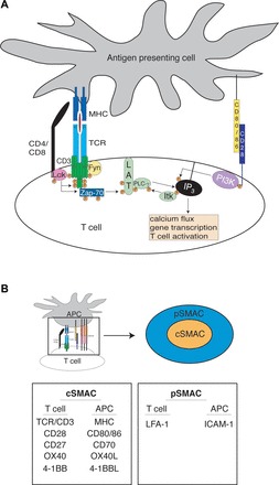
T cell activation. A: simplified model of early events in T cell signaling. In the absence of CD28 costimulation, canonical T cell signaling is mostly stalled at the linker for activation of T cells (LAT) signalosome stage, where phospholipase C (PLC)-γ remains unactivated. MHC, major histocompatibility complex; TCR, T cell receptor; P, phosphorylation; ZAP-70, ζ-chain-associated protein kinase 70; IP3, inositol 1,4,5-trisphosphate; PI3K, phosphatidylinositol 3-kinase; B: sustained interaction between a T cell and an antigen-presenting cell (APC) results in the formation of an immunological synapse. Membrane reorganization creates a greater apposition of surface areas into which signaling and adhesion molecules are ordered into a “bulls-eye” arrangement. The relevant molecules are listed according to location. SMAC, supramolecular activation complex; L, ligand; LFA-1, lymphocyte function-associated antigen 1.
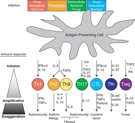
T cell differentiation. Top: APC [i.e., dendritic cell (DC)] recognition of a spectrum of pathogens through various pathogen-associated molecular pattern receptors results in cytokine release from the APC. Along with TCR engagement, milieu cytokines initiate (top middle) differentiation to one of a variety T cell subsets programmed by transcription factors to specifically respond to the spectrum of the instigating pathogen [pathogen and T helper (Th) subset color coordinated]. Upon differentiation, T cells themselves produce cytokines, which feed back into the cellular milieu, amplifying and balancing the immune response to promote specific pathogen clearance (bottom middle) and host survival. Finally, sustained, ill-timed, or otherwise exaggerated T cell immune responses from any of the T cell subsets results in a range of immunopathologies from autoimmunity to allergy and cancer (bottom). IFN, interferon; TGF-β, transforming growth factor-β; Gz, granzyme.
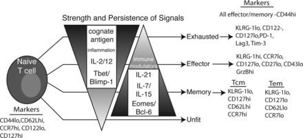
T cell memory. T cells may assume many phenotypes in response to stimulation. The eventual fate of a T cell depends on many environmental queues, including, but not limited to, cytokines, inflammatory and immune-modulatory products, and tissue-specific factors. These signals, in turn, influence the transcriptional profile of the T cell, leading to developmental choices. Generally, IL-7/15/21 and Eomes/Bcl-6 are considered to tip the scale toward memory. In contrast, IL-2/12, inflammatory products, and T-bet/Blimp-1 weigh toward terminal differentiation and effectors. Exhaustion can result when T cells experience these factors too intensely or for too long. A proper balancing of all these factors will lead to long-lived, protective memory. Markers commonly used to identify cells within a particular functional grouping are given on the right. KLRG-1, killer cell lectin-like receptor subfamily G member 1; PD-1, programmed cell death-1; Lag3, lymphocyte activation gene 3; Tim-3, T cell Ig mucin-3.
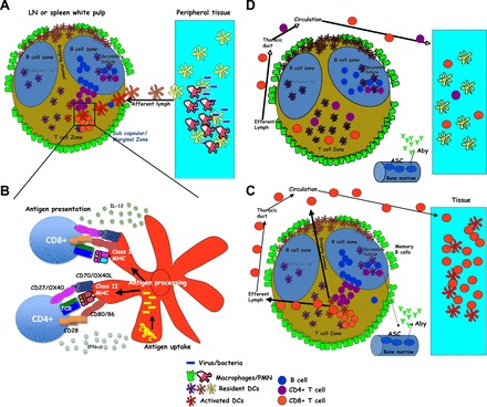
The anatomy of a T cell response. A: microbe invasion and proliferation at the site of infection, leading to the initial recruitment of phagocytes and containment of the infection. Tissue-resident APCs acquire antigen and migrate into the local secondary lymphoid organ (SLO) after being activated by the local inflammatory processes. LN, lymph node. B: during transit, APCs process and present pathogen-derived antigens in the context of class I and class II MHC. APCs also upregulate various cell surface molecules and cytokines important in providing the necessary costimulation to T cells within the SLO. C: antigen-stimulated CD4 T cells collaborate with B cells to promote antibody production, class switching, and memory B cell formation. Both CD4 and CD8 T cells clonally expand and migrate out of the SLO and into the infection site, where their effector functions facilitates the elimination of the pathogen. Aby, antibody; ASC, Antibody secreting cell; D: most T cells die off, leaving a memory pool with precursor frequency, antigen sensitivity, and trafficking capacity optimized for initiating rapid secondary responses in situ. PMN, polymorphonuclear leukocytes.
Similar articles
-
SnapShot: effector and memory T cell differentiation.Cell. 2009 Aug 7;138(3):606.e1-2. doi: 10.1016/j.cell.2009.07.020. Cell. 2009. PMID: 19665979 Free PMC article. No abstract available.
-
New insights into lymphocyte activation and differentiation.Curr Opin Immunol. 2013 Jun;25(3):297-9. doi: 10.1016/j.coi.2013.05.013. Epub 2013 Jun 5. Curr Opin Immunol. 2013. PMID: 23746790 No abstract available.
-
T cell memory.Annu Rev Immunol. 2002;20:551-79. doi: 10.1146/annurev.immunol.20.100101.151926. Epub 2001 Oct 4. Annu Rev Immunol. 2002. PMID: 11861612 Review.
-
T cell proliferation, differentiation, and restoration in lymphopenic individuals: for contributed volumes.Adv Exp Med Biol. 2002;512:135-9. doi: 10.1007/978-1-4615-0757-4_18. Adv Exp Med Biol. 2002. PMID: 12405197 Review. No abstract available.
-
Lifespan of lymphocytes.Immunol Res. 1995;14(1):1-12. doi: 10.1007/BF02918494. Immunol Res. 1995. PMID: 7561338 Review.
Cited by
-
Infection and Immune Memory: Variables in Robust Protection by Vaccines Against SARS-CoV-2.Front Immunol. 2021 May 11;12:660019. doi: 10.3389/fimmu.2021.660019. eCollection 2021. Front Immunol. 2021. PMID: 34046033 Free PMC article. Review.
-
Human CD8+ T Cells Exhibit a Shared Antigen Threshold for Different Effector Responses.J Immunol. 2020 Sep 15;205(6):1503-1512. doi: 10.4049/jimmunol.2000525. Epub 2020 Aug 17. J Immunol. 2020. PMID: 32817332 Free PMC article.
-
Analysis of Immune and Inflammation Characteristics of Atherosclerosis from Different Sample Sources.Oxid Med Cell Longev. 2022 Apr 25;2022:5491038. doi: 10.1155/2022/5491038. eCollection 2022. Oxid Med Cell Longev. 2022. PMID: 35509837 Free PMC article.
-
Paracrine costimulation of IFN-γ signaling by integrins modulates CD8 T cell differentiation.Proc Natl Acad Sci U S A. 2018 Nov 6;115(45):11585-11590. doi: 10.1073/pnas.1804556115. Epub 2018 Oct 22. Proc Natl Acad Sci U S A. 2018. PMID: 30348790 Free PMC article.
-
Advances in modular control of CAR-T therapy with adapter-mediated CARs.Adv Drug Deliv Rev. 2022 Aug;187:114358. doi: 10.1016/j.addr.2022.114358. Epub 2022 May 23. Adv Drug Deliv Rev. 2022. PMID: 35618140 Free PMC article. Review.
References
-
- Appleby MW, Gross JA, Cooke MP, Levin SD, Qian X, Perlmutter RM. Defective T cell receptor signaling in mice lacking the thymic isoform of p59fyn. Cell 70: 751–763, 1992 - PubMed
-
- Bacchetta R, Passerini L, Gambineri E, Dai M, Allan SE, Perroni L, Dagna-Bricarelli F, Sartirana C, Matthes-Martin S, Lawitschka A, Azzari C, Ziegler SF, Levings MK, Roncarolo MG. Defective regulatory and effector T cell functions in patients with FOXP3 mutations. J Clin Invest 116: 1713–1722, 2006 - PMC - PubMed
-
- Bachmann MF, Wolint P, Walton S, Schwarz K, Oxenius A. Differential role of IL-2R signaling for CD8+ T cell responses in acute and chronic viral infections. Eur J Immunol 37: 1502–1512, 2007 - PubMed
MeSH terms
Grants and funding
LinkOut - more resources
Full Text Sources
Other Literature Sources