. 2014 Jan;71(1):61-78.
doi: 10.1002/cm.21159. Epub 2013 Dec 10.

CLIC5 stabilizes membrane-actin filament linkages at the base of hair cell stereocilia in a molecular complex with radixin, taperin, and myosin VI

Affiliations

CLIC5 stabilizes membrane-actin filament linkages at the base of hair cell stereocilia in a molecular complex with radixin, taperin, and myosin VI

Felipe T Salles et al. Cytoskeleton (Hoboken). 2014 Jan.

Abstract

Chloride intracellular channel 5 protein (CLIC5) was originally isolated from microvilli in complex with actin binding proteins including ezrin, a member of the Ezrin-Radixin-Moesin (ERM) family of membrane-cytoskeletal linkers. CLIC5 concentrates at the base of hair cell stereocilia and is required for normal hearing and balance in mice, but its functional significance is poorly understood. This study investigated the role of CLIC5 in postnatal development and maintenance of hair bundles. Confocal and scanning electron microscopy of CLIC5-deficient jitterbug (jbg) mice revealed progressive fusion of stereocilia as early as postnatal day 10. Radixin (RDX), protein tyrosine phosphatase receptor Q (PTPRQ), and taperin (TPRN), deafness-associated proteins that also concentrate at the base of stereocilia, were mislocalized in fused stereocilia of jbg mice. TPRQ and RDX were dispersed even prior to stereocilia fusion. Biochemical assays showed interaction of CLIC5 with ERM proteins, TPRN, and possibly myosin VI (MYO6). In addition, CLIC5 and RDX failed to localize normally in fused stereocilia of MYO6 mutant mice. Based on these findings, we propose a model in which these proteins work together as a complex to stabilize linkages between the plasma membrane and subjacent actin cytoskeleton at the base of stereocilia.

Keywords: PTPRQ; chloride intracellular channel 5 (CLIC5); cytoskeleton; deafness; ezrin-radixin-moesin (ERM); hair cell; myosin VI (MYO6); radixin; stereocilia; taperin.


Conflict of interest statement

The authors declare no conflict of interest.

Figures
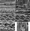
Fig. 1
Fig. 1
Morphological defects of hair cells in jitterbug mutant. Auditory (A–G) and vestibular (H–J) hair cells from P17 and P10 (inset) mice. (A) Inner hair cells (IHC) from a heterozygous control (jbg/+) viewed from lateral aspect near the apex of the cochlear duct. (B) IHC near the apex of a cochlea from a jitterbug mutant (jbg/jbg) showing elongated and fused stereocilia lying parallel to the surface of the epithelium (arrows), and fused stereocilia with lifted membranes (arrowhead). Inset shows IHC with fused stereocilia (arrow) at P10. (C) Control IHC at the base with staircase arrangement of stereocilia. (D) Mutant IHC at the base lack the shortest row of stereocilia and exhibit fusion of several stereocilia (arrowheads) as well as mass fusion indicated by the lifted plasma membrane (*). (E) Outer hair cells (OHC) at the base of control cochlear duct showing regular arrangement of stereocilia arrays. (F) OHC at the base of a mutant cochlear duct devoid of individual, non-fused stereocilia. (G) OHC from mid-turn of mutant cochlear duct with varying degrees of stereocilia fusion, including entire arm of V-shaped bundle (arrows) and fusion at the ends of bundles (*). (H) Utricular hair cells (UHC) from heterozygous control showing stereocilia arranged in rows of increasing height and uniform diameter within each row. (I) Mutant UHC bundle lacks a regular staircase array and displays thickened stereocilia. (J) High magnification view of proximal end of mutant UHC bundle with a morphology reflecting apparent fusion of multiple parallel actin bundles covered by a common membrane. Scale bars: 10 µm (A, B), 5 µm (inset, C, D, E, F, G), 2 µm (H–J).
Fig. 2
Fig. 2
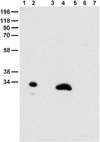
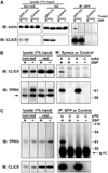
Characterization of monospecific CLIC5 antibody (APB56) on Western blot. Blot probed with CLIC5 polyclonal antibody following cross-adsorption against CLIC4 and subsequent affinity-purification on CLIC5 (APB56). (Lanes 1, 2), bacterial extract containing 50 ng untagged recombinant CLIC4 and CLIC5, respectively; (Lanes 3–7), 10 mg SDS-soluble extract of mouse stomach, lung, pancreas, small intestine, and liver, respectively. APB56 does not cross-react with CLIC4; a single major band is detected in lung extract.
Fig. 3
Fig. 3
CLIC5 concentrates at the base of the hair bundle. (A, B) Fluorescence confocal micrographs of adult rat whole mount organ of Corti preparations stained with specific antibody (APB56) against CLIC5 (green); actin filaments were counterstained with phal-loidin (red). Specimens were gently squashed to facilitate visualization of staining along the proximal-distal axis of stereocilia; note that requisite use of heat-induced antigen retrieval compromised the morphological preservation of bundles as well as the quality of F-actin staining. In both inner (A) and outer (B) hair cells, CLIC5 is concentrated at the base of stereocilia (arrows). (C–E) Expression of GFP-CLIC5 fusion protein in vestibular hair cell. The transfected cell (C) exhibits accumulation of CLIC5 towards the proximal end of the stereocilia. (D) Actin filaments were counterstained with phalloidin (red). (E) Merged images of C and D. Scale bars: 5 µm. [Color figure can be viewed in the online issue, which is available at wileyonlinelibrary.com.]
Fig. 4
Fig. 4
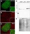
Characterization of GFP-CLIC5 fusion protein. (A–C) LLC-PK1-CL4 epithelial cell transfected with GFP-CLIC5 (A) and counterstained with phalloidin (B). Merged images (C) with boxed area to indicate region shown in insets. GFP-CLIC5 concentrates in microvilli-like surface structures rich in F-actin. Scale bar: 20 µm. (D) Western blot of LLC-PK1-CL4 extracts probed with whole antiserum (B132) against CLIC5. Blot contains two-fold serial dilutions (lanes 8, 4, 2, 1) of extracts from untransfected (untransf), mock-transfected cells treated with transfection reagent with no DNA and GFP-CLIC5 transfected cultures. Bacterial lysate containing 50 ng untagged human CLIC5 (R) was loaded on the same gel as a positive control. A band migrating between ~55 and 60 kDa and corresponding to the expected size of the fusion protein is detected only in the GFP-CLIC5 culture; a faster migrating background band (*) is seen in all cultures. Note that no endogenous CLIC5 (~32 kDa) was detected. (E) Western blot stained with Coomassie Blue as a loading control. [Color figure can be viewed in the online issue, which is available at wileyonlinelibrary.com.]
Fig. 5
Fig. 5
Immunogold localization of CLIC5 at the apical surface of inner hair cells from adult rat. (A) Low magnification overview showing high density of gold particles at the bases of ster-eocilia (brackets) as well as the inter-stereocilia membrane domain (arrows); gold labeling is sparse at the tips and shafts of stereocilia as well as core actin rootlets projecting into the cutic-ular plate (arrowheads). (B) High magnification of CLIC5 staining at the base of stereocilia and inter-stereocilia region. Scale bars: 500 nm.
Fig. 6
Fig. 6
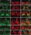
Localization of PTPRQ, RDX, and TPRN in mature hair cells of jitterbug mutant. Inner hair cells near the apex of the cochlea stained with antibodies (green) against PTPRQ at P40 (A–F), RDX at P17 (G–L), and TPRN at P21 (M–R) and counterstained with phalloidin (red). (A-F) In control cells (jbg/+), intense PTPRQ staining is restricted to the bases of stereocilia (A, C, arrow) as well as the intervening apical plasma membrane (A, C, arrowhead); in mutant cells (jbg/jbg), PTPRQ is distributed in a diffused pattern extending along the membrane covering the shaft of the still recognizable stereocilia (D, F, arrows). (G-L) Control cells exhibit enriched RDX staining at the base and a lower level of staining along the stereocilia shaft towards the tip (G, I, arrows); mutant cells exhibit a diffuse pattern of RDX staining extending along the membrane covering the shaft of the still recognizable, but severely malformed stereocilia (J, L, arrows). (M–R) In control cells, TPRN staining is enriched at the base of stereocilia (M, O, arrows); in mutant cells, TPRN staining is distributed in a diffused pattern extending along the shaft of the malformed stereocilia (P, R, arrows). TPRN staining is also dispersed along the shaft of a single individual stereocilium (P, R, arrowhead). Scale bars: 5 µm.
Fig. 7
Fig. 7
Localization of PTPRQ, RDX, and TPRN in immature hair cells of jitterbug mutant. Inner hair cells near the apex of the cochlea stained with antibodies (green) against PTPRQ at P10 (A–F), RDX at P7 (G–L), and TPRN at P10 (M–R) and counter-stained with phalloidin (red). (A–F) In control cells (jbg/+), PTPRQ is enriched at the bases of stereocilia (A, C, arrows); in mutant cells (jbg/jbg), PTPRQ labeling extends along the stereocilia shaft and is not restricted to the base (D, F, arrow). (G–L) Control cells exhibit RDX staining enriched at the bases of stereocilia (G, I, arrows); in mutant cells a small amount of RDX extends along the shaft towards the tip of a single stereocilium (J, L, arrow). (M–R) TPRN staining is restricted to the bases of stereocilia in control cells (M, O, arrows) as well as mutant cells (P, R, arrows). Kinocilia are stained positive with the TPRN antibody (P, R, arrowheads). Scale bars: 5 µm.
Fig. 8
Fig. 8
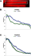
PTPRQ and RDX redistribute along the lengths of stereocilia at P10 in jitterbug mutant mice. Analysis of PTPRQ (B) and RDX (C) distributions in control (jbg/+) and mutant (jbg/jbg) stereocilia. (A) For measurement, a rectangular selection was drawn along the length of stereocilia from base (left) to tip (right), and intensity profiles obtained in ImageJ (NIH). Plots show mean intensities (AU, arbitrary units) ± S.D. (n = 10 stereocilia from the tallest row of inner hair cells). In control stereocilia, PTPRQ is very concentrated at the base with relatively low expression towards the tips (B, lower green line), similar to the distribution of RDX (C, lower green line). In mutant stereocilia, the proteins show an altered distribution, no longer as restricted to a high expression at the base and with redistribution along the shaft (B and C, upper blue lines). [Color figure can be viewed in the online issue, which is available at wileyonlinelibrary.com.]
Fig. 9
Fig. 9
CLIC5 associates with Ezrin and TPRN in LLC-PK1 epithelial cells. (A) Endogenous ezrin coimmunoprecipitates with GFP-CLIC5. Cells transfected with GFP-CLIC5 (GFP-C5) or mock-transfected (Control) were treated with DSP (+) or DMSO alone (−), followed by immunoprecipitation with GFP antibody. Immunoblots of starting lysates (nonreduced and reduced) and immunoprecipitates were probed with ezrin polyclonal antibody and then reprobed with CLIC5 polyclonal antibody. (B) GFP-CLIC5 coimmunoprecipitates with the C-terminal region of TPRN. Cells double transfected with GFP-CLIC5 and Xpress-TPRN308–711 were treated with DSP (+) or DMSO alone (−), followed by immunoprecipitation with antibody against the Xpress epitope (X) or an isotype matched control (C) monoclonal antibody. Immunoblots were probed with CLIC5 polyclonal antibody and then reprobed with TPRN polyclonal antibody (TPRN); band indicated by the arrow corresponds to CLIC5. (C) Xpress-TPRN308–711 coimmunoprecipitates with GFP-CLIC5. Double transfected cells were treated with DSP (+) or DMSO (−) followed by immunoprecipitation with antibodies against GFP (G) or a control (C) polyclonal antibody. Immunoblots were probed with TPRN polyclonal antibody and then reprobed with whole antiserum against CLIC5. IgG heavy chain (Ig HC) is indicated.
Fig. 10
Fig. 10
CLIC5 associates tightly with the detergent-insoluble actin cytoskeleton and interacts with RDX, ezrin, and MYO6 in an affinity pull-down assay using mouse lung extract. (A) Summary of scheme used to fractionate lung tissue. (B) Blots containing fractions were probed with antibodies that recognize both CLIC5 (arrowhead, 5) and CLIC4 (arrowhead, 4), antibodies against α-actinin and β-actin, and the lectin concanavalin A to detect glycosylated membrane proteins. Equal volumes were loaded for H, S1, and S2; 10 times more sample was loaded for P2 and S3. CLIC5 was highly insoluble and resistant to extraction with detergent (S3), indicating a tight association with the cytoskeleton; α-actinin and β-actin, both of which are cytoskeletal proteins, show relatively poor recovery after detergent treatment in comparison to glycosylated membrane proteins detected by concanavalin A (compare P2 and S3). (C) Blots containing equivalent volumes of fractions as well as starting material (S) and eluates from BSA (B), GST (G), or 6His-CLIC5 (C) beads used in the pull-down assay. RDX antibody detects a major band migrating at ~82 kDa (arrowheads) and several additional faster migrating bands that may represent RDX degradation products; RDX is highly resistant to detergent extraction (compare P2 and S3). In the pull-down assay, RDX is present only in the CLIC5 bead eluate (arrowhead). Two potential degradation products (*) are enriched in the CLIC5 eluate relative to the starting extract or fractionation samples, suggesting that these fragments have greater affinity for CLIC5 than intact RDX. Ezrin antibody recognizes a major band at ~82 kDa. In the pull-down assay, ezrin is detected only in the CLIC5 eluate. MYO6 antibody recognizes a prominent band migrating at ~200 kDa and a faster migrating band at ~160 kDa (*); MYO6 is highly resistant to detergent extraction (compare P2 and S3). The similar fractionation profiles of the prominent and faster migrating band (*) suggests that the latter is a MYO6 degradation product. The faster migrating species is enriched in the CLIC5 eluant. With increased contrast, a faint band (arrowhead) corresponding to the ~200 kDa migrating intact MYO6 band is detected in the CLIC5 eluate.
Fig. 11
Fig. 11
Localization of CLIC5 and RDX in hair cells from Myo6 mutant Snell’s waltzer 2 Jackson (sv-2J) mice. Inner hair cells from the apex of P15 cochleae from heterozygous control (sv-2J/+) mice (A, B) and homozygous mutant (sv-2J/sv-2J) mice (C, D). Hair cells were stained with antibodies (green) against CLIC5 (A, C) or RDX (B, D) and counterstained with phalloidin (red). Both proteins are enriched at the base of stereocilia in control cells (arrows). However, in Myo6 mutant cells, both proteins exhibit diffused staining along the shaft of giant fused stereocilia. Staining of CLIC5 and RDX is enriched at the surface of shafts (arrowheads), indicating preferential association with the membrane; accumulation of CLIC5 or RDX is no longer seen at the bases of fused stereocilia (asterisks). Scale bar: 10 µm.
Fig. 12
Fig. 12
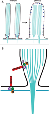
Simplified model for membrane-cytoskeletal linking complex at the base of stereocilia. (A) In stereocilia of wild-type hair cell (left panel) the cytoplasmic domain of PTPRQ (red lines) associates with a juxtamembrane protein complex (blue spheres) consisting of CLIC5, RDX, MYO6, and/or TPRN, thereby linking PTPRQ to actin filaments at the tapered base of stereocilia as well as linking actin filaments of the cuticular plate with the interstereocilia membrane domain. In the absence of CLIC5 (right panel), the protein complex is altered (blue squares), thereby compromising function and possibly dissociation of interacting partners, leading to destabilized membrane-cytoskeletal attachments, consequent membrane lifting, and loss of stereocilia individuality. (B) Hypothetical molecular complexes linking PTPRQ to submembranous actin: CLIC5 (C), RDX (R), MYO6 (M), and TPRN (T). Based on homology with the minus end-capping protein phostensin [Lai et al., 2009; Rehman et al., 2010; Wang et al., 2012], TPRN may associate with the minus ends of actin filaments that may terminate at the tapered base. MYO6 minus-end directed motor activity could help establish and dynamically maintain the localization of the protein complex at the base of stereocilia and simultaneously act on its own as an independent membrane-cytoskeletal linker. [Color figure can be viewed in the online issue, which is available at wileyonlinelibrary.com.]

Similar articles

Cited by

References

    1. Ahmed ZM, Morell RJ, Riazuddin S, Gropman A, Shaukat S, Ahmad MM, Mohiddin SA, Fananapazir L, Caruso RC, Husnain T, et al. Mutations of MYO6 are associated with recessive deafness, DFNB37. Am J Hum Genet. 2003;72:1315–1322. - PMC - PubMed
    1. Ahmed ZM, Goodyear R, Riazuddin S, Lagziel A, Legan PK, Behra M, Burgess SM, Lilley KS, Wilcox ER, Griffith AJ, et al. The tip-link antigen, a protein associated with the transduction complex of sensory hair cells, is protocadherin-15. J Neurosci. 2006;26:7022–7034. - PMC - PubMed
    1. Alagramam KN, Goodyear RJ, Geng R, Furness DN, van Aken AF, Marcotti W, Kros CJ, Richardson GP. Mutations in protocadherin 15 and cadherin 23 affect tip links and mechanotransduction in mammalian sensory hair cells. PLoS One. 2011;6:e19183. - PMC - PubMed
    1. Avraham KB, Hasson T, Sobe T, Balsara B, Testa JR, Skvorak AB, Morton CC, Copeland NG, Jenkins NA. Characterization of unconventional MYO6, the human homologue of the gene responsible for deafness in Snell’s waltzer mice. Hum Mol Genet. 1997;6:1225–1231. - PubMed
    1. Belyantseva IA, Boger ET, Naz S, Frolenkov GI, Sellers JR, Ahmed ZM, Griffith AJ, Friedman TB. Myosin-XVa is required for tip localization of whirlin and differential elongation of hair-cell stereocilia. Nat Cell Biol. 2005;7:148–156. - PubMed

Publication types

LinkOut - more resources